 Movie
Movie Controller


+ Open data

- Basic information
| Entry | Database: EMDB / ID: EMD-9948 | |||||||||
|---|---|---|---|---|---|---|---|---|---|---|
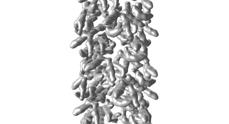
| Title | Structure of unknow protein 4 | |||||||||
 Map data Map data | ||||||||||
 Sample Sample |
| |||||||||
 Keywords Keywords | filament / SIGNALING PROTEIN | |||||||||
| Function / homology |  Function and homology information Function and homology informationCARD8 inflammasome complex assembly / CARD8 inflammasome complex / NACHT domain binding / Formation of apoptosome / CARD domain binding / self proteolysis / NLRP3 inflammasome complex / negative regulation of lipopolysaccharide-mediated signaling pathway / negative regulation of NLRP3 inflammasome complex assembly / cysteine-type endopeptidase activator activity ...CARD8 inflammasome complex assembly / CARD8 inflammasome complex / NACHT domain binding / Formation of apoptosome / CARD domain binding / self proteolysis / NLRP3 inflammasome complex / negative regulation of lipopolysaccharide-mediated signaling pathway / negative regulation of NLRP3 inflammasome complex assembly / cysteine-type endopeptidase activator activity / Regulation of the apoptosome activity / Hydrolases; Acting on peptide bonds (peptidases) / negative regulation of interleukin-1 beta production / pattern recognition receptor activity / negative regulation of tumor necrosis factor-mediated signaling pathway / activation of innate immune response / intrinsic apoptotic signaling pathway / antiviral innate immune response / positive regulation of interleukin-1 beta production / negative regulation of canonical NF-kappaB signal transduction / molecular condensate scaffold activity / protein destabilization / positive regulation of inflammatory response / peptidase activity / regulation of apoptotic process / defense response to virus / inflammatory response / protein homodimerization activity / protein-containing complex / nucleoplasm / nucleus / cytoplasm / cytosol Similarity search - Function | |||||||||
| Biological species |  Homo sapiens (human) Homo sapiens (human) | |||||||||
| Method | helical reconstruction / cryo EM / Resolution: 3.7 Å | |||||||||
 Authors Authors | Gong Q / Xu C | |||||||||
| Funding support |  Singapore, 2 items Singapore, 2 items
| |||||||||
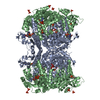
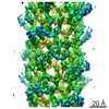
 Citation Citation | Journal: Nat Commun / Year: 2021 Title: Structural basis for distinct inflammasome complex assembly by human NLRP1 and CARD8. Authors: Qin Gong / Kim Robinson / Chenrui Xu / Phuong Thao Huynh / Kelvin Han Chung Chong / Eddie Yong Jun Tan / Jiawen Zhang / Zhao Zhi Boo / Daniel Eng Thiam Teo / Kenneth Lay / Yaming Zhang / ...Authors: Qin Gong / Kim Robinson / Chenrui Xu / Phuong Thao Huynh / Kelvin Han Chung Chong / Eddie Yong Jun Tan / Jiawen Zhang / Zhao Zhi Boo / Daniel Eng Thiam Teo / Kenneth Lay / Yaming Zhang / John Soon Yew Lim / Wah Ing Goh / Graham Wright / Franklin L Zhong / Bruno Reversade / Bin Wu /  Abstract: Nod-like receptor (NLR) proteins activate pyroptotic cell death and IL-1 driven inflammation by assembling and activating the inflammasome complex. Closely related sensor proteins NLRP1 and CARD8 ...Nod-like receptor (NLR) proteins activate pyroptotic cell death and IL-1 driven inflammation by assembling and activating the inflammasome complex. Closely related sensor proteins NLRP1 and CARD8 undergo unique auto-proteolysis-dependent activation and are implicated in auto-inflammatory diseases; however, their mechanisms of activation are not understood. Here we report the structural basis of how the activating domains (FIIND-CARD) of NLRP1 and CARD8 self-oligomerize to assemble distinct inflammasome complexes. Recombinant FIIND-CARD of NLRP1 forms a two-layered filament, with an inner core of oligomerized CARD surrounded by an outer ring of FIIND. Biochemically, self-assembled NLRP1-CARD filaments are sufficient to drive ASC speck formation in cultured human cells-a process that is greatly enhanced by NLRP1-FIIND which forms oligomers in vitro. The cryo-EM structures of NLRP1-CARD and CARD8-CARD filaments, solved here at 3.7 Å, uncover unique structural features that enable NLRP1 and CARD8 to discriminate between ASC and pro-caspase-1. In summary, our findings provide structural insight into the mechanisms of activation for human NLRP1 and CARD8 and reveal how highly specific signaling can be achieved by heterotypic CARD interactions within the inflammasome complexes. | |||||||||
| History |
|
- Structure visualization
| Movie |
Movie viewer |
|---|---|
| Structure viewer | EM map: SurfViewMolmilJmol/JSmol |
| Supplemental images |
- Downloads & links
-EMDB archive
| Map data | emd_9948.map.gz | 4.4 MB | EMDB map data format | |
|---|---|---|---|---|
| Header (meta data) | emd-9948-v30.xmlemd-9948.xml | 19.9 KB 19.9 KB | Display Display | EMDB header |
| FSC (resolution estimation) | emd_9948_fsc.xml | 4.7 KB | Display | FSC data file |
| Images |  emd_9948.png emd_9948.png | 82 KB | ||
| Filedesc metadata | emd-9948.cif.gz | 5.7 KB | ||
| Others | emd_9948_additional_1.map.gzemd_9948_additional_2.map.gzemd_9948_half_map_1.map.gzemd_9948_half_map_2.map.gz | 2.9 MB 515.1 KB 4.4 MB 4.4 MB | ||
| Archive directory |  http://ftp.pdbj.org/pub/emdb/structures/EMD-9948ftp://ftp.pdbj.org/pub/emdb/structures/EMD-9948 http://ftp.pdbj.org/pub/emdb/structures/EMD-9948ftp://ftp.pdbj.org/pub/emdb/structures/EMD-9948 | HTTPS FTP |
-Related structure data
| Related structure data |  6k9fMC  9943C  9946C  9947C  6k7vC  6k8jC  6k99C M: atomic model generated by this map C: citing same article ( |
|---|---|
| Similar structure data |


-Links
| EMDB pages | EMDB (EBI/PDBe) / EMDataResource |
|---|---|
| Related items in Molecule of the Month |


-Map
| File | Download / File: emd_9948.map.gz / Format: CCP4 / Size: 3.6 MB / Type: IMAGE STORED AS FLOATING POINT NUMBER (4 BYTES) | ||||||||||||||||||||||||||||||||||||||||||||||||||||||||||||||||||||
|---|---|---|---|---|---|---|---|---|---|---|---|---|---|---|---|---|---|---|---|---|---|---|---|---|---|---|---|---|---|---|---|---|---|---|---|---|---|---|---|---|---|---|---|---|---|---|---|---|---|---|---|---|---|---|---|---|---|---|---|---|---|---|---|---|---|---|---|---|---|
| Projections & slices | Image control
Images are generated by Spider. generated in cubic-lattice coordinate | ||||||||||||||||||||||||||||||||||||||||||||||||||||||||||||||||||||
| Voxel size | X=Y=Z: 1.1 Å | ||||||||||||||||||||||||||||||||||||||||||||||||||||||||||||||||||||
| Density |
| ||||||||||||||||||||||||||||||||||||||||||||||||||||||||||||||||||||
| Symmetry | Space group: 1 | ||||||||||||||||||||||||||||||||||||||||||||||||||||||||||||||||||||
| Details | EMDB XML:
CCP4 map header:
| ||||||||||||||||||||||||||||||||||||||||||||||||||||||||||||||||||||
 Z (Sec.)
 Y (Row.)
 X (Col.)


-Supplemental data
-Additional map: new additional map with ccp4 format, for model map fitting
| File | emd_9948_additional_1.map | ||||||||||||
|---|---|---|---|---|---|---|---|---|---|---|---|---|---|
| Annotation | new additional map with ccp4 format, for model map fitting | ||||||||||||
| Projections & Slices |
| ||||||||||||
| Density Histograms |


-Additional map: #1
| File | emd_9948_additional_2.map | ||||||||||||
|---|---|---|---|---|---|---|---|---|---|---|---|---|---|
| Projections & Slices |
| ||||||||||||
| Density Histograms |


-Half map: #2
| File | emd_9948_half_map_1.map | ||||||||||||
|---|---|---|---|---|---|---|---|---|---|---|---|---|---|
| Projections & Slices |
| ||||||||||||
| Density Histograms |


-Half map: #1
| File | emd_9948_half_map_2.map | ||||||||||||
|---|---|---|---|---|---|---|---|---|---|---|---|---|---|
| Projections & Slices |
| ||||||||||||
| Density Histograms |


- Sample components
-Entire : caspase recruitment domain containing=protein 8
| Entire | Name: caspase recruitment domain containing=protein 8 |
|---|---|
| Components |
|
-Supramolecule #1: caspase recruitment domain containing=protein 8
| Supramolecule | Name: caspase recruitment domain containing=protein 8 / type: complex / ID: 1 / Parent: 0 / Macromolecule list: all |
|---|---|
| Source (natural) | Organism: Homo sapiens (human) |
-Macromolecule #1: Caspase recruitment domain-containing protein 8
| Macromolecule | Name: Caspase recruitment domain-containing protein 8 / type: protein_or_peptide / ID: 1 / Number of copies: 12 / Enantiomer: LEVO |
|---|---|
| Source (natural) | Organism: Homo sapiens (human) |
| Molecular weight | Theoretical: 10.445787 KDa |
| Recombinant expression | Organism:  |
| Sequence | String: GPGMAAFVKE NHRQLQARMG DLKGVLDDLQ DNEVLTENEK ELVEQEKTRQ SKNEALLSMV EKKGDLALDV LFRSISERDP YLVSYLRQQ NL UniProtKB: Caspase recruitment domain-containing protein 8 |
-Experimental details
-Structure determination
| Method | cryo EM |
|---|---|
 Processing Processing | helical reconstruction |
| Aggregation state | filament |
-Sample preparation
| Buffer | pH: 8 |
|---|---|
| Grid | Model: Quantifoil R1.2/1.3 / Material: COPPER / Mesh: 300 / Pretreatment - Type: GLOW DISCHARGE / Pretreatment - Time: 60 sec. / Pretreatment - Atmosphere: AIR |
| Vitrification | Cryogen name: ETHANE / Chamber humidity: 100 % / Chamber temperature: 277.15 K / Instrument: FEI VITROBOT MARK III |
- Electron microscopy
| Microscope | FEI TITAN KRIOS |
|---|---|
| Image recording | Film or detector model: GATAN K2 SUMMIT (4k x 4k) / Detector mode: COUNTING / Number real images: 1424 / Average electron dose: 80.0 e/Å2 |
| Electron beam | Acceleration voltage: 300 kV / Electron source:  FIELD EMISSION GUN FIELD EMISSION GUN |
| Electron optics | Illumination mode: FLOOD BEAM / Imaging mode: DARK FIELD / Cs: 2.7 mm / Nominal magnification: 115000 |
| Sample stage | Specimen holder model: FEI TITAN KRIOS AUTOGRID HOLDER / Cooling holder cryogen: NITROGEN |
| Experimental equipment |  Model: Titan Krios / Image courtesy: FEI Company |
+Image processing

-Atomic model buiding 1
| Refinement | Space: REAL / Protocol: RIGID BODY FIT |
|---|---|
| Output model | PDB-6k9f: |
