Movie
Movie Controller

[English] 日本語
 Yorodumi
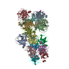
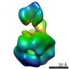
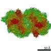
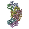
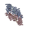
- EMDB-8090: Cryo-EM structure of GluA2/3 AMPA receptor heterotetramer (model I) -
+ Open data

- Basic information
| Entry | Database: EMDB / ID: EMD-8090 | |||||||||
|---|---|---|---|---|---|---|---|---|---|---|
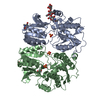
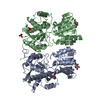
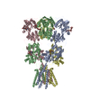
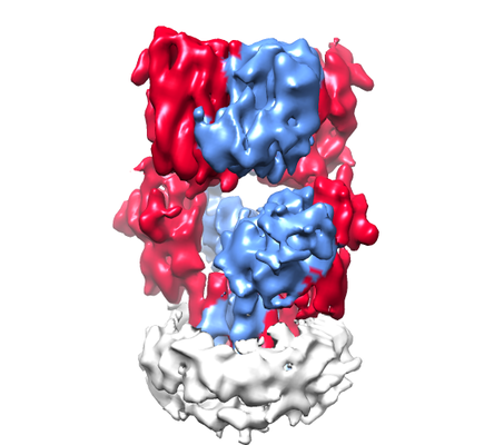
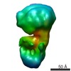
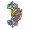
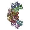
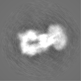
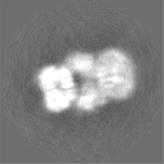
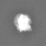
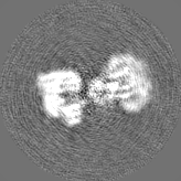
| Title | Cryo-EM structure of GluA2/3 AMPA receptor heterotetramer (model I) | |||||||||
 Map data Map data | None | |||||||||
 Sample Sample |
| |||||||||
 Keywords Keywords | AMPA glutamate receptor / Signaling protein | |||||||||
| Function / homology |  Function and homology information Function and homology informationTrafficking of AMPA receptors / ligand-gated calcium channel activity / Synaptic adhesion-like molecules / spine synapse / dendritic spine neck / dendritic spine cytoplasm / dendritic spine head / cellular response to amine stimulus / Activation of AMPA receptors / ligand-gated monoatomic cation channel activity ...Trafficking of AMPA receptors / ligand-gated calcium channel activity / Synaptic adhesion-like molecules / spine synapse / dendritic spine neck / dendritic spine cytoplasm / dendritic spine head / cellular response to amine stimulus / Activation of AMPA receptors / ligand-gated monoatomic cation channel activity / protein heterotetramerization / perisynaptic space / Trafficking of GluR2-containing AMPA receptors / response to lithium ion / AMPA glutamate receptor activity / AMPA glutamate receptor clustering / parallel fiber to Purkinje cell synapse / regulation of receptor recycling / kainate selective glutamate receptor activity / immunoglobulin binding / AMPA glutamate receptor complex / extracellularly glutamate-gated ion channel activity / cellular response to glycine / ionotropic glutamate receptor complex / asymmetric synapse / Unblocking of NMDA receptors, glutamate binding and activation / glutamate receptor binding / positive regulation of synaptic transmission / conditioned place preference / synaptic cleft / regulation of synaptic transmission, glutamatergic / response to fungicide / cytoskeletal protein binding / extracellular ligand-gated monoatomic ion channel activity / glutamate-gated receptor activity / cellular response to brain-derived neurotrophic factor stimulus / regulation of long-term synaptic depression / somatodendritic compartment / glutamate-gated calcium ion channel activity / presynaptic active zone membrane / ionotropic glutamate receptor signaling pathway / ionotropic glutamate receptor binding / dendrite cytoplasm / excitatory synapse / dendrite membrane / ligand-gated monoatomic ion channel activity involved in regulation of presynaptic membrane potential / positive regulation of excitatory postsynaptic potential / dendritic shaft / SNARE binding / synaptic membrane / PDZ domain binding / protein tetramerization / establishment of protein localization / synaptic transmission, glutamatergic / transmitter-gated monoatomic ion channel activity involved in regulation of postsynaptic membrane potential / receptor internalization / cerebral cortex development / postsynaptic density membrane / modulation of chemical synaptic transmission / Schaffer collateral - CA1 synapse / long-term synaptic potentiation / terminal bouton / synaptic vesicle / synaptic vesicle membrane / amyloid-beta binding / presynapse / growth cone / signaling receptor activity / presynaptic membrane / scaffold protein binding / chemical synaptic transmission / protein homotetramerization / dendritic spine / perikaryon / postsynaptic membrane / neuron projection / postsynaptic density / external side of plasma membrane / axon / neuronal cell body / synapse / dendrite / protein kinase binding / protein-containing complex binding / glutamatergic synapse / cell surface / endoplasmic reticulum / protein-containing complex / membrane / identical protein binding / plasma membrane Similarity search - Function | |||||||||
| Biological species |  | |||||||||
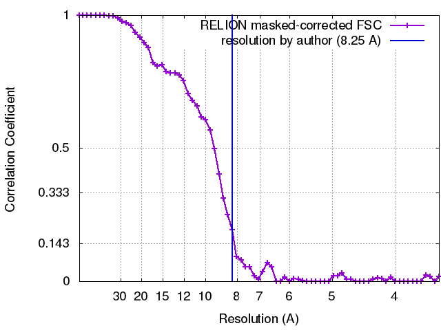
| Method | single particle reconstruction / cryo EM / Resolution: 8.25 Å | |||||||||
 Authors Authors | Herguedas B / Garcia-Nafria J | |||||||||
 Citation Citation | Journal: Science / Year: 2016 Title: Structure and organization of heteromeric AMPA-type glutamate receptors. Authors: Beatriz Herguedas / Javier García-Nafría / Ondrej Cais / Rafael Fernández-Leiro / James Krieger / Hinze Ho / Ingo H Greger /  Abstract: AMPA-type glutamate receptors (AMPARs), which are central mediators of rapid neurotransmission and synaptic plasticity, predominantly exist as heteromers of the subunits GluA1 to GluA4. Here we ...AMPA-type glutamate receptors (AMPARs), which are central mediators of rapid neurotransmission and synaptic plasticity, predominantly exist as heteromers of the subunits GluA1 to GluA4. Here we report the first AMPAR heteromer structures, which deviate substantially from existing GluA2 homomer structures. Crystal structures of the GluA2/3 and GluA2/4 N-terminal domains reveal a novel compact conformation with an alternating arrangement of the four subunits around a central axis. This organization is confirmed by cysteine cross-linking in full-length receptors, and it permitted us to determine the structure of an intact GluA2/3 receptor by cryogenic electron microscopy. Two models in the ligand-free state, at resolutions of 8.25 and 10.3 angstroms, exhibit substantial vertical compression and close associations between domain layers, reminiscent of N-methyl-D-aspartate receptors. Model 1 resembles a resting state and model 2 a desensitized state, thus providing snapshots of gating transitions in the nominal absence of ligand. Our data reveal organizational features of heteromeric AMPARs and provide a framework to decipher AMPAR architecture and signaling. | |||||||||
| History |
|
- Structure visualization
| Movie |
Movie viewer |
|---|---|
| Structure viewer | EM map: SurfViewMolmilJmol/JSmol |
| Supplemental images |
- Downloads & links
-EMDB archive
| Map data | emd_8090.map.gz | 2.5 MB | EMDB map data format | |
|---|---|---|---|---|
| Header (meta data) | emd-8090-v30.xmlemd-8090.xml | 22.4 KB 22.4 KB | Display Display | EMDB header |
| FSC (resolution estimation) | emd_8090_fsc.xml | 5.8 KB | Display | FSC data file |
| Images |  emd_8090.png emd_8090.png | 122.2 KB | ||
| Masks | emd_8090_msk_1.mapemd_8090_msk_2.map | 16.8 MB 16.8 MB | Mask map | |
| Filedesc metadata | emd-8090.cif.gz | 7.5 KB | ||
| Others | emd_8090_half_map_1.map.gzemd_8090_half_map_2.map.gz | 12.8 MB 12.8 MB | ||
| Archive directory |  http://ftp.pdbj.org/pub/emdb/structures/EMD-8090ftp://ftp.pdbj.org/pub/emdb/structures/EMD-8090 http://ftp.pdbj.org/pub/emdb/structures/EMD-8090ftp://ftp.pdbj.org/pub/emdb/structures/EMD-8090 | HTTPS FTP |
-Related structure data
| Related structure data | 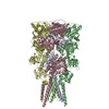 5ideMC 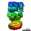 8091C 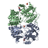 5fwxC 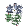 5fwyC 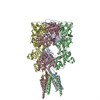 5idfC M: atomic model generated by this map C: citing same article ( |
|---|---|
| Similar structure data |
-Links
| EMDB pages | EMDB (EBI/PDBe) / EMDataResource |
|---|---|
| Related items in Molecule of the Month |


-Map
| File | Download / File: emd_8090.map.gz / Format: CCP4 / Size: 16.8 MB / Type: IMAGE STORED AS FLOATING POINT NUMBER (4 BYTES) | ||||||||||||||||||||||||||||||||||||||||||||||||||||||||||||||||||||
|---|---|---|---|---|---|---|---|---|---|---|---|---|---|---|---|---|---|---|---|---|---|---|---|---|---|---|---|---|---|---|---|---|---|---|---|---|---|---|---|---|---|---|---|---|---|---|---|---|---|---|---|---|---|---|---|---|---|---|---|---|---|---|---|---|---|---|---|---|---|
| Annotation | None | ||||||||||||||||||||||||||||||||||||||||||||||||||||||||||||||||||||
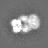
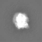
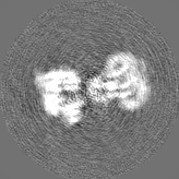
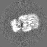
| Projections & slices | Image control
Images are generated by Spider. | ||||||||||||||||||||||||||||||||||||||||||||||||||||||||||||||||||||
| Voxel size | X=Y=Z: 1.76 Å | ||||||||||||||||||||||||||||||||||||||||||||||||||||||||||||||||||||
| Density |
| ||||||||||||||||||||||||||||||||||||||||||||||||||||||||||||||||||||
| Symmetry | Space group: 1 | ||||||||||||||||||||||||||||||||||||||||||||||||||||||||||||||||||||
| Details | EMDB XML:
CCP4 map header:
| ||||||||||||||||||||||||||||||||||||||||||||||||||||||||||||||||||||
 Z (Sec.)
 Y (Row.)
 X (Col.)


-Supplemental data
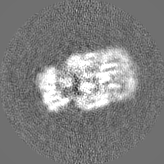
-Mask #1
| File | emd_8090_msk_1.map | ||||||||||||
|---|---|---|---|---|---|---|---|---|---|---|---|---|---|
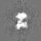
| Projections & Slices |
| ||||||||||||
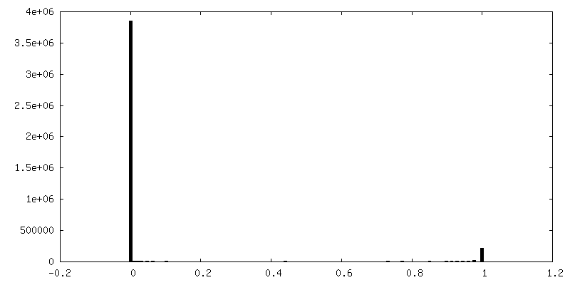
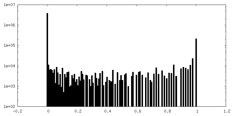
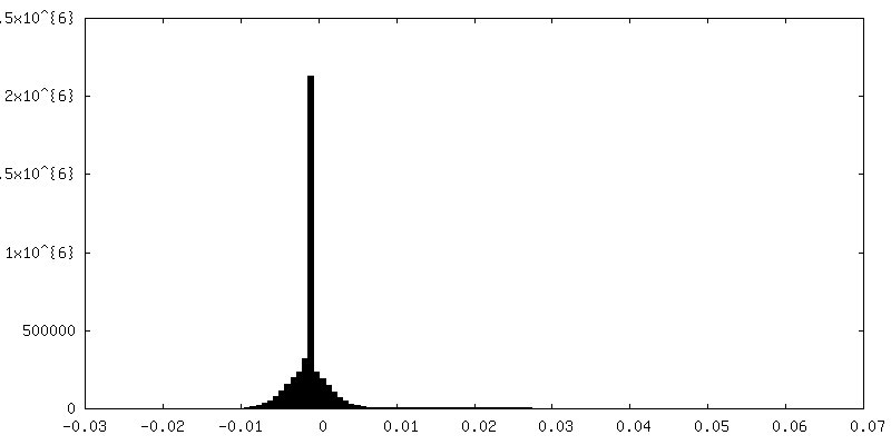
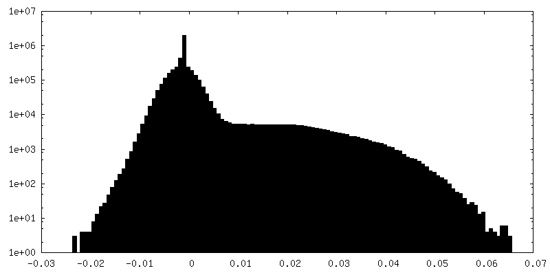
| Density Histograms |


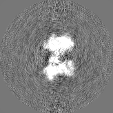
-Mask #2
| File | emd_8090_msk_2.map | ||||||||||||
|---|---|---|---|---|---|---|---|---|---|---|---|---|---|
| Projections & Slices |
| ||||||||||||
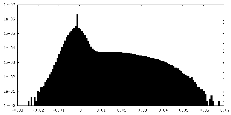
| Density Histograms |


-Half map: None
| File | emd_8090_half_map_1.map | ||||||||||||
|---|---|---|---|---|---|---|---|---|---|---|---|---|---|
| Annotation | None | ||||||||||||
| Projections & Slices |
| ||||||||||||
| Density Histograms |

-Half map: None
| File | emd_8090_half_map_2.map | ||||||||||||
|---|---|---|---|---|---|---|---|---|---|---|---|---|---|
| Annotation | None | ||||||||||||
| Projections & Slices |
| ||||||||||||
| Density Histograms |

- Sample components
-Entire : AMPA GluA2/3 heterotetramer
| Entire | Name: AMPA GluA2/3 heterotetramer |
|---|---|
| Components |
|
-Supramolecule #1: AMPA GluA2/3 heterotetramer
| Supramolecule | Name: AMPA GluA2/3 heterotetramer / type: complex / ID: 1 / Parent: 0 / Macromolecule list: all Details: Full-length GluA2/3 heterotetramer containing the A2_N292C and A3_265C mutations |
|---|---|
| Source (natural) | Organism: |
| Molecular weight | Theoretical: 400 KDa |
-Macromolecule #1: Glutamate receptor 2
| Macromolecule | Name: Glutamate receptor 2 / type: protein_or_peptide / ID: 1 Details: The sequence corresponds to mature rat GluA2 (residues 22-883, isoform Flip, edited at R/G and Q/R sites) with a Myc tag after the first residue and the N292C mutation Number of copies: 2 / Enantiomer: LEVO |
|---|---|
| Source (natural) | Organism: |
| Molecular weight | Theoretical: 97.663188 KDa |
| Recombinant expression | Organism:  Homo sapiens (human) Homo sapiens (human) |
| Sequence | String: VEQKLISEED LSSNSIQIGG LFPRGADQEY SAFRVGMVQF STSEFRLTPH IDNLEVANSF AVTNAFCSQF SRGVYAIFGF YDKKSVNTI TSFCGTLHVS FITPSFPTDG THPFVIQMRP DLKGALLSLI EYYQWDKFAY LYDSDRGLST LQAVLDSAAE K KWQVTAIN ...String: VEQKLISEED LSSNSIQIGG LFPRGADQEY SAFRVGMVQF STSEFRLTPH IDNLEVANSF AVTNAFCSQF SRGVYAIFGF YDKKSVNTI TSFCGTLHVS FITPSFPTDG THPFVIQMRP DLKGALLSLI EYYQWDKFAY LYDSDRGLST LQAVLDSAAE K KWQVTAIN VGNINNDKKD ETYRSLFQDL ELKKERRVIL DCERDKVNDI VDQVITIGKH VKGYHYIIAN LGFTDGDLLK IQ FGGANVS GFQIVDYDDS LVSKFIERWS TLEEKEYPGA HTATIKYTSA LTYDAVQVMT EAFRCLRKQR IEISRRGNAG DCL ANPAVP WGQGVEIERA LKQVQVEGLS GNIKFDQNGK RINYTINIME LKTNGPRKIG YWSEVDKMVV TLTELPSGND TSGL ENKTV VVTTILESPY VMMKKNHEML EGNERYEGYC VDLAAEIAKH CGFKYKLTIV GDGKYGARDA DTKIWNGMVG ELVYG KADI AIAPLTITLV REEVIDFSKP FMSLGISIMI KKPQKSKPGV FSFLDPLAYE IWMCIVFAYI GVSVVLFLVS RFSPYE WHT EEFEDGRETQ SSESTNEFGI FNSLWFSLGA FMQQGCDISP RSLSGRIVGG VWWFFTLIII SSYTANLAAF LTVERMV SP IESAEDLSKQ TEIAYGTLDS GSTKEFFRRS KIAVFDKMWT YMRSAEPSVF VRTTAEGVAR VRKSKGKYAY LLESTMNE Y IEQRKPCDTM KVGGNLDSKG YGIATPKGSS LGTPVNLAVL KLSEQGVLDK LKNKWWYDKG ECGAKDSGSK EKTSALSLS NVAGVFYILV GGLGLAMLVA LIEFCYKSRA EAKRMKVAKN PQNINPSSSQ NSQNFATYKE GYNVYGIESV KI UniProtKB: Glutamate receptor 2 |
-Macromolecule #2: Glutamate receptor 3
| Macromolecule | Name: Glutamate receptor 3 / type: protein_or_peptide / ID: 2 Details: The sequence corresponds to the mature rat GluA3 subunit (residues 23-888, Flip isoform) with a Flag tag after the first residue and mutated at R439G and R265C Number of copies: 2 / Enantiomer: LEVO |
|---|---|
| Source (natural) | Organism: |
| Molecular weight | Theoretical: 99.075664 KDa |
| Recombinant expression | Organism: Homo sapiens (human) |
| Sequence | String: GDYKDDDDKF PNTISIGGLF MRNTVQEHSA FRFAVQLYNT NQNTTEKPFH LNYHVDHLDS SNSFSVTNAF CSQFSRGVYA IFGFYDQMS MNTLTSFCGA LHTSFVTPSF PTDADVQFVI QMRPALKGAI LSLLSYYKWE KFVYLYDTER GFSVLQAIME A AVQNNWQV ...String: GDYKDDDDKF PNTISIGGLF MRNTVQEHSA FRFAVQLYNT NQNTTEKPFH LNYHVDHLDS SNSFSVTNAF CSQFSRGVYA IFGFYDQMS MNTLTSFCGA LHTSFVTPSF PTDADVQFVI QMRPALKGAI LSLLSYYKWE KFVYLYDTER GFSVLQAIME A AVQNNWQV TARSVGNIKD VQEFRRIIEE MDRRQEKRYL IDCEVERINT ILEQVVILGK HSRGYHYMLA NLGFTDILLE RV MHGGANI TGFQIVNNEN PMVQQFIQRW VRLDECEFPE AKNAPLKYTS ALTHDAILVI AEAFRYLRRQ RVDVSRRGSA GDC LANPAV PWSQGIDIER ALKMVQVQGM TGNIQFDTYG RRTNYTIDVY EMKVSGSRKA GYWNEYERFV PFSDQQISND SSSS ENRTI VVTTILESPY VMYKKNHEQL EGNERYEGYC VDLAYEIAKH VGIKYKLSIV GDGKYGARDP ETKIWNGMVG ELVYG RADI AVAPLTITLV REEVIDFSKP FMSLGISIMI KKPQKSKPGV FSFLDPLAYE IWMCIVFAYI GVSVVLFLVS RFSPYE WHL EDNNEEPRDP QSPPDPPNEF GIFNSLWFSL GAFMQQGCDI SPRSLSGRIV GGVWWFFTLI IISSYTANLA AFLTVER MV SPIESAEDLA KQTEIAYGTL DSGSTKEFFR RSKIAVYEKM WSYMKSAEPS VFTKTTADGV ARVRKSKGKF AFLLESTM N EYIEQRKPCD TMKVGGNLDS KGYGVATPKG SALGTPVNLA VLKLSEQGIL DKLKNKWWYD KGECGAKDSG SKDKTSALS LSNVAGVFYI LVGGLGLAMM VALIEFCYKS RAESKRMKLT KNTQNFKPAP ATNTQNYATY REGYNVYGTE SVKI UniProtKB: Glutamate receptor 3 |
-Experimental details
-Structure determination
| Method | cryo EM |
|---|---|
 Processing Processing | single particle reconstruction |
| Aggregation state | particle |
-Sample preparation
| Concentration | 0.03 mg/mL |
|---|---|
| Buffer | pH: 7.4 / Details: 25 mM Tris pH 7.4, 0.25 % DDM, 150 mM NaCl |
| Grid | Model: Quantifoil R1.2/1.3 / Material: COPPER / Mesh: 300 / Support film - Material: CARBON / Support film - topology: CONTINUOUS / Pretreatment - Type: GLOW DISCHARGE |
| Vitrification | Cryogen name: ETHANE / Chamber humidity: 100 % / Chamber temperature: 277 K / Instrument: FEI VITROBOT MARK IV / Details: Incubated for 1 minute, blotted for 3 seconds. |
- Electron microscopy
| Microscope | FEI TITAN KRIOS |
|---|---|
| Specialist optics | Energy filter - Name: GIF Quantum / Energy filter - Lower energy threshold: 0 eV / Energy filter - Upper energy threshold: 20 eV |
| Image recording | Film or detector model: GATAN K2 SUMMIT (4k x 4k) / Detector mode: COUNTING / Digitization - Frames/image: 1-20 / Number grids imaged: 2 / Number real images: 980 / Average exposure time: 25.0 sec. / Average electron dose: 40.0 e/Å2 |
| Electron beam | Acceleration voltage: 300 kV / Electron source:  FIELD EMISSION GUN FIELD EMISSION GUN |
| Electron optics | C2 aperture diameter: 70.0 µm / Calibrated magnification: 28409 / Illumination mode: OTHER / Imaging mode: BRIGHT FIELD / Cs: 2.7 mm / Nominal defocus max: 4.0 µm / Nominal defocus min: 1.0 µm |
| Sample stage | Specimen holder model: FEI TITAN KRIOS AUTOGRID HOLDER / Cooling holder cryogen: NITROGEN |
| Experimental equipment |  Model: Titan Krios / Image courtesy: FEI Company |
+Image processing
-Atomic model buiding 1
| Initial model |
| ||||||||||||
|---|---|---|---|---|---|---|---|---|---|---|---|---|---|
| Details | For GluA2 chains (A,C), 2 copies of GluA2NTD (3HSY) and two copies of GluA2 LBD (1FTO) were fitted. For GluA3 chains (B,D), 2 copies of GluA3NTD (3O21) and two copies of GluA2 LBD (3UA8) were fitted.For the TMD region, the 4 chains of 3KG2 TMD(residues 509-544 594-629 784-817) were fitted as a rigid body. After fitting the sequence was corrected including mutations and side chains were removed. | ||||||||||||
| Refinement | Space: REAL / Protocol: RIGID BODY FIT | ||||||||||||
| Output model | PDB-5ide: |