 Movie
Movie Controller


+ Open data

- Basic information
| Entry | Database: EMDB / ID: EMD-1851 | |||||||||
|---|---|---|---|---|---|---|---|---|---|---|
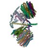
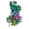
| Title | EM map of the negative stained SMG-1-9 complex. | |||||||||
 Map data Map data | This is the EM map of the negative stained SMG-1-9 complex. | |||||||||
 Sample Sample |
| |||||||||
 Keywords Keywords | Nonsense-mediated mRNA decay / NMD / SMG-1 / SMG-8 / SMG-9 / SMG1C / 3D-Electron Microscopy | |||||||||
| Function / homology | nuclear-transcribed mRNA catabolic process, nonsense-mediated decay Function and homology information Function and homology information | |||||||||
| Biological species |  Homo sapiens (human) Homo sapiens (human) | |||||||||
| Method | single particle reconstruction / negative staining / Resolution: 24.0 Å | |||||||||
 Authors Authors | Arias-Palomo E / Yamashita A / Fernandez IS / Nunez-Ramirez R / Bamba Y / Izumi N / Ohno S / Llorca O | |||||||||
 Citation Citation | Journal: Genes Dev / Year: 2011 Title: The nonsense-mediated mRNA decay SMG-1 kinase is regulated by large-scale conformational changes controlled by SMG-8. Authors: Ernesto Arias-Palomo / Akio Yamashita / Israel S Fernández / Rafael Núñez-Ramírez / Yumi Bamba / Natsuko Izumi / Shigeo Ohno / Oscar Llorca /  Abstract: Nonsense-mediated mRNA decay (NMD) is a eukaryotic surveillance pathway that regulates the degradation of mRNAs harboring premature translation termination codons. NMD also influences the expression ...Nonsense-mediated mRNA decay (NMD) is a eukaryotic surveillance pathway that regulates the degradation of mRNAs harboring premature translation termination codons. NMD also influences the expression of many physiological transcripts. SMG-1 is a large kinase essential to NMD that phosphorylates Upf1, which seems to be the definitive signal triggering mRNA decay. However, the regulation of the kinase activity of SMG-1 remains poorly understood. Here, we reveal the three-dimensional architecture of SMG-1 in complex with SMG-8 and SMG-9, and the structural mechanisms regulating SMG-1 kinase. A bent arm comprising a long region of HEAT (huntington, elongation factor 3, a subunit of PP2A and TOR1) repeats at the N terminus of SMG-1 functions as a scaffold for SMG-8 and SMG-9, and projects from the C-terminal core containing the phosphatidylinositol 3-kinase domain. SMG-9 seems to control the activity of SMG-1 indirectly through the recruitment of SMG-8 to the N-terminal HEAT repeat region of SMG-1. Notably, SMG-8 binding to the SMG-1:SMG-9 complex specifically down-regulates the kinase activity of SMG-1 on Upf1 without contacting the catalytic domain. Assembly of the SMG-1:SMG-8:SMG-9 complex induces a significant motion of the HEAT repeats that is signaled to the kinase domain. Thus, large-scale conformational changes induced by SMG-8 after SMG-9-mediated recruitment tune SMG-1 kinase activity to modulate NMD. | |||||||||
| History |
|
- Structure visualization
| Movie |
Movie viewer |
|---|---|
| Structure viewer | EM map: SurfViewMolmilJmol/JSmol |
| Supplemental images |
 UCSF Chimera
- Downloads & links
-EMDB archive
| Map data | emd_1851.map.gz | 181.2 KB | EMDB map data format | |
|---|---|---|---|---|
| Header (meta data) | emd-1851-v30.xmlemd-1851.xml | 10.4 KB 10.4 KB | Display Display | EMDB header |
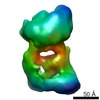
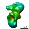
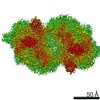
| Images | 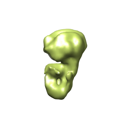 EMD1851.png EMD1851.png | 46.1 KB | ||
| Archive directory |  http://ftp.pdbj.org/pub/emdb/structures/EMD-1851ftp://ftp.pdbj.org/pub/emdb/structures/EMD-1851 http://ftp.pdbj.org/pub/emdb/structures/EMD-1851ftp://ftp.pdbj.org/pub/emdb/structures/EMD-1851 | HTTPS FTP |
-Related structure data


-Links
| EMDB pages | EMDB (EBI/PDBe) / EMDataResource |
|---|
-Map
| File | Download / File: emd_1851.map.gz / Format: CCP4 / Size: 1.9 MB / Type: IMAGE STORED AS FLOATING POINT NUMBER (4 BYTES) | ||||||||||||||||||||||||||||||||||||||||||||||||||||||||||||||||||||
|---|---|---|---|---|---|---|---|---|---|---|---|---|---|---|---|---|---|---|---|---|---|---|---|---|---|---|---|---|---|---|---|---|---|---|---|---|---|---|---|---|---|---|---|---|---|---|---|---|---|---|---|---|---|---|---|---|---|---|---|---|---|---|---|---|---|---|---|---|---|
| Annotation | This is the EM map of the negative stained SMG-1-9 complex. | ||||||||||||||||||||||||||||||||||||||||||||||||||||||||||||||||||||
| Projections & slices | Image control
Images are generated by Spider. | ||||||||||||||||||||||||||||||||||||||||||||||||||||||||||||||||||||
| Voxel size | X=Y=Z: 4.2 Å | ||||||||||||||||||||||||||||||||||||||||||||||||||||||||||||||||||||
| Density |
| ||||||||||||||||||||||||||||||||||||||||||||||||||||||||||||||||||||
| Symmetry | Space group: 1 | ||||||||||||||||||||||||||||||||||||||||||||||||||||||||||||||||||||
| Details | EMDB XML:
CCP4 map header:
| ||||||||||||||||||||||||||||||||||||||||||||||||||||||||||||||||||||
 Z (Sec.)
 Y (Row.)
 X (Col.)


-Supplemental data
- Sample components
-Entire : Human SMG-1 kinase bound to SMG-9
| Entire | Name: Human SMG-1 kinase bound to SMG-9 |
|---|---|
| Components |
|
-Supramolecule #1000: Human SMG-1 kinase bound to SMG-9
| Supramolecule | Name: Human SMG-1 kinase bound to SMG-9 / type: sample / ID: 1000 Oligomeric state: One molecule of SMG-1 binds to one molecule of SMG-9 Number unique components: 2 |
|---|---|
| Molecular weight | Theoretical: 470 KDa |
-Macromolecule #1: SMG-1
| Macromolecule | Name: SMG-1 / type: protein_or_peptide / ID: 1 / Name.synonym: SMG-1 / Number of copies: 1 / Oligomeric state: Monomer / Recombinant expression: Yes |
|---|---|
| Source (natural) | Organism: Homo sapiens (human) / synonym: Human |
| Molecular weight | Theoretical: 410 KDa |
| Recombinant expression | Organism: 293T cells / Recombinant plasmid: pEF_Flag-HA-SBP |
| Sequence | GO: nuclear-transcribed mRNA catabolic process, nonsense-mediated decay |
-Macromolecule #2: SMG-9
| Macromolecule | Name: SMG-9 / type: protein_or_peptide / ID: 2 / Name.synonym: SMG-9 / Number of copies: 1 / Oligomeric state: Monomer / Recombinant expression: Yes |
|---|---|
| Source (natural) | Organism: Homo sapiens (human) / synonym: Human |
| Molecular weight | Theoretical: 60 KDa |
| Recombinant expression | Organism: 293T Cells / Recombinant plasmid: pSR_Strep-HA |
| Sequence | GO: nuclear-transcribed mRNA catabolic process, nonsense-mediated decay |
-Experimental details
-Structure determination
| Method | negative staining |
|---|---|
 Processing Processing | single particle reconstruction |
| Aggregation state | particle |
-Sample preparation
| Buffer | pH: 7.5 Details: 10 mM HEPES-KOH at pH7.5, 150 mM NaCl, 20% glycerol, 10 mM MgCl2 |
|---|---|
| Staining | Type: NEGATIVE Details: Protein was adsorbed for 1 minute, washed in two water drops and stained with 2% w/v uranyl formate for 1 minute. |
| Grid | Details: 400 mesh copper grid |
| Vitrification | Cryogen name: NONE / Instrument: OTHER |
- Electron microscopy
| Microscope | JEOL 1230 |
|---|---|
| Temperature | Average: 294 K |
| Alignment procedure | Legacy - Astigmatism: Objective lens astigmatism was corrected at 80,000 times magnification |
| Details | Microscope JEOL JEM-1230 |
| Image recording | Category: FILM / Film or detector model: KODAK SO-163 FILM / Digitization - Scanner: OTHER / Digitization - Sampling interval: 10.5 µm / Details: Scanner MINOLTA Dimage Scan Multi PRO / Bits/pixel: 16 |
| Tilt angle min | 0 |
| Electron beam | Acceleration voltage: 100 kV / Electron source: TUNGSTEN HAIRPIN |
| Electron optics | Illumination mode: FLOOD BEAM / Imaging mode: BRIGHT FIELD / Cs: 2.9 mm / Nominal defocus max: 1.2 µm / Nominal defocus min: 0.5 µm / Nominal magnification: 50000 |
| Sample stage | Specimen holder: Side entry single tilt holder / Specimen holder model: JEOL / Tilt angle max: 20 |
-Image processing
| Details | Particles were collected manually. |
|---|---|
| CTF correction | Details: Each micrograph |
| Final reconstruction | Applied symmetry - Point group: C1 (asymmetric) / Algorithm: OTHER / Resolution.type: BY AUTHOR / Resolution: 24.0 Å / Resolution method: FSC 0.5 CUT-OFF / Software - Name: EMAN, Xmipp / Number images used: 15608 |
