 Movie
Movie Controller

[English] 日本語
 Yorodumi
- EMDB-76717: In situ cryo-EM structure of axonal microtubules from ghost neuro... -
+ Open data

- Basic information
| Entry |  | |||||||||
|---|---|---|---|---|---|---|---|---|---|---|
| Title | In situ cryo-EM structure of axonal microtubules from ghost neurons (expanded lattice) | |||||||||
 Map data Map data | sharpened map from local refinement | |||||||||
 Sample Sample |
| |||||||||
 Keywords Keywords | cytoskeleton / neuron / microtubules / axon / STRUCTURAL PROTEIN | |||||||||
| Function / homology |  Function and homology information Function and homology informationMicrotubule-dependent trafficking of connexons from Golgi to the plasma membrane / Cargo trafficking to the periciliary membrane / Sealing of the nuclear envelope (NE) by ESCRT-III / netrin-activated signaling pathway / Carboxyterminal post-translational modifications of tubulin / Intraflagellar transport / netrin receptor binding / COPI-independent Golgi-to-ER retrograde traffic / HSP90 chaperone cycle for steroid hormone receptors (SHR) in the presence of ligand / pyramidal neuron differentiation ...Microtubule-dependent trafficking of connexons from Golgi to the plasma membrane / Cargo trafficking to the periciliary membrane / Sealing of the nuclear envelope (NE) by ESCRT-III / netrin-activated signaling pathway / Carboxyterminal post-translational modifications of tubulin / Intraflagellar transport / netrin receptor binding / COPI-independent Golgi-to-ER retrograde traffic / HSP90 chaperone cycle for steroid hormone receptors (SHR) in the presence of ligand / pyramidal neuron differentiation / COPI-mediated anterograde transport / Kinesins / PKR-mediated signaling / Aggrephagy / RHO GTPases activate IQGAPs / Mitotic Prometaphase / EML4 and NUDC in mitotic spindle formation / COPI-dependent Golgi-to-ER retrograde traffic / glial cell differentiation / Resolution of Sister Chromatid Cohesion / dorsal root ganglion development / The role of GTSE1 in G2/M progression after G2 checkpoint / Recycling pathway of L1 / dentate gyrus development / axonemal microtubule / organelle transport along microtubule / Hedgehog 'off' state / RHO GTPases Activate Formins / Loss of Nlp from mitotic centrosomes / Recruitment of mitotic centrosome proteins and complexes / Loss of proteins required for interphase microtubule organization from the centrosome / Separation of Sister Chromatids / Anchoring of the basal body to the plasma membrane / forebrain morphogenesis / Recruitment of NuMA to mitotic centrosomes / AURKA Activation by TPX2 / cerebellar cortex morphogenesis / Regulation of PLK1 Activity at G2/M Transition / MHC class II antigen presentation / neuron projection arborization / smoothened signaling pathway / homeostasis of number of cells within a tissue / motor behavior / adult behavior / response to L-glutamate / centrosome cycle / sperm principal piece / startle response / 'de novo' protein folding / intercellular bridge / flagellated sperm motility / regulation of synapse organization / sperm end piece / locomotory exploration behavior / microtubule polymerization / ciliary tip / response to tumor necrosis factor / response to mechanical stimulus / neuron apoptotic process / neurogenesis / sperm flagellum / adult locomotory behavior / cytoplasmic microtubule / condensed chromosome / peptide binding / cellular response to calcium ion / gene expression / visual learning / axon guidance / locomotory behavior / hippocampus development / cell periphery / filopodium / neuromuscular junction / myelin sheath / neuron migration / memory / cerebral cortex development / intracellular protein transport / synapse organization / microtubule cytoskeleton organization / recycling endosome / mitotic spindle / structural constituent of cytoskeleton / cytoplasmic ribonucleoprotein granule / microtubule cytoskeleton / cilium / neuron differentiation / mitotic cell cycle / lamellipodium / growth cone / protein-folding chaperone binding / microtubule binding / microtubule / Hydrolases; Acting on acid anhydrides; Acting on GTP to facilitate cellular and subcellular movement / protein stabilization / membrane raft / protein heterodimerization activity / protein domain specific binding / axon Similarity search - Function | |||||||||
| Biological species |   Mus musculus (house mouse) Mus musculus (house mouse) | |||||||||
| Method | helical reconstruction / cryo EM / Resolution: 3.66 Å | |||||||||
 Authors Authors | Bodakuntla S / Marelli J / Vasquez-Montes V / Biertumpfel C / Mizuno N | |||||||||
| Funding support |  United States, 1 items United States, 1 items
| |||||||||
 Citation Citation | Journal: To Be Published Title: Ghost neuron unlocks in situ high-resolution structural mapping of intracellular architecture in neurons Authors: Bodakuntla S / Marelli J / Vasquez-Montes V / Biertumpfel C / Mizuno N | |||||||||
| History |
|
- Structure visualization
| Supplemental images |
|---|

- Downloads & links
-EMDB archive
| Map data | emd_76717.map.gz | 418.2 MB | EMDB map data format | |
|---|---|---|---|---|
| Header (meta data) | emd-76717-v30.xmlemd-76717.xml | 22.5 KB 22.5 KB | Display Display | EMDB header |
| FSC (resolution estimation) | emd_76717_fsc.xml | 19.9 KB | Display | FSC data file |
| Images |  emd_76717.png emd_76717.png | 76.2 KB | ||
| Masks | emd_76717_msk_1.map | 824 MB | Mask map | |
| Filedesc metadata | emd-76717.cif.gz | 7.3 KB | ||
| Others | emd_76717_half_map_1.map.gzemd_76717_half_map_2.map.gz | 765.5 MB 765.5 MB | ||
| Archive directory |  http://ftp.pdbj.org/pub/emdb/structures/EMD-76717ftp://ftp.pdbj.org/pub/emdb/structures/EMD-76717 http://ftp.pdbj.org/pub/emdb/structures/EMD-76717ftp://ftp.pdbj.org/pub/emdb/structures/EMD-76717 | HTTPS FTP |
-Related structure data
| Related structure data |  12ruMC  12rtC  12rvC M: atomic model generated by this map C: citing same article ( |
|---|---|
| Similar structure data |


-Links
| EMDB pages | EMDB (EBI/PDBe) / EMDataResource |
|---|---|
| Related items in Molecule of the Month |


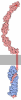
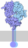

-Map
| File | Download / File: emd_76717.map.gz / Format: CCP4 / Size: 824 MB / Type: IMAGE STORED AS FLOATING POINT NUMBER (4 BYTES) | ||||||||||||||||||||||||||||||||||||
|---|---|---|---|---|---|---|---|---|---|---|---|---|---|---|---|---|---|---|---|---|---|---|---|---|---|---|---|---|---|---|---|---|---|---|---|---|---|
| Annotation | sharpened map from local refinement | ||||||||||||||||||||||||||||||||||||
| Projections & slices | Image control
Images are generated by Spider. | ||||||||||||||||||||||||||||||||||||
| Voxel size | X=Y=Z: 0.824 Å | ||||||||||||||||||||||||||||||||||||
| Density |
| ||||||||||||||||||||||||||||||||||||
| Symmetry | Space group: 1 | ||||||||||||||||||||||||||||||||||||
| Details | EMDB XML:
|
 Z (Sec.)
 Y (Row.)
 X (Col.)


-Supplemental data
-Mask #1
| File | emd_76717_msk_1.map | ||||||||||||
|---|---|---|---|---|---|---|---|---|---|---|---|---|---|
| Projections & Slices |
| ||||||||||||
| Density Histograms |


-Half map: half map from local refinement
| File | emd_76717_half_map_1.map | ||||||||||||
|---|---|---|---|---|---|---|---|---|---|---|---|---|---|
| Annotation | half map from local refinement | ||||||||||||
| Projections & Slices |
| ||||||||||||
| Density Histograms |


-Half map: half map from local refinement
| File | emd_76717_half_map_2.map | ||||||||||||
|---|---|---|---|---|---|---|---|---|---|---|---|---|---|
| Annotation | half map from local refinement | ||||||||||||
| Projections & Slices |
| ||||||||||||
| Density Histograms |


- Sample components
-Entire : Microtubules, axonal
| Entire | Name: Microtubules, axonal |
|---|---|
| Components |
|
-Supramolecule #1: Microtubules, axonal
| Supramolecule | Name: Microtubules, axonal / type: organelle_or_cellular_component / ID: 1 / Parent: 0 / Macromolecule list: #1-#2 Details: explant axon from thalamus primary mouse embryo E15.5 tissue after ghost preparation (hypotonic treatment and mechanical plasma membrane removal) |
|---|---|
| Source (natural) | Organism: Mus musculus (house mouse) / Strain: CD-1 / Organ: Brain / Tissue: Thalamus / Location in cell: Axon |
-Macromolecule #1: Detyrosinated tubulin alpha-1A chain
| Macromolecule | Name: Detyrosinated tubulin alpha-1A chain / type: protein_or_peptide / ID: 1 / Details: TUBA1A component of microtubules / Number of copies: 2 / Enantiomer: LEVO |
|---|---|
| Source (natural) | Organism: Mus musculus (house mouse) / Strain: CD-1 / Organ: Brain / Tissue: Thalamus |
| Molecular weight | Theoretical: 50.188441 KDa |
| Sequence | String: MRECISIHVG QAGVQIGNAC WELYCLEHGI QPDGQMPSDK TIGGGDDSFN TFFSETGAGK HVPRAVFVDL EPTVIDEVRT GTYRQLFHP EQLITGKEDA ANNYARGHYT IGKEIIDLVL DRIRKLADQC TGLQGFLVFH SFGGGTGSGF TSLLMERLSV D YGKKSKLE ...String: MRECISIHVG QAGVQIGNAC WELYCLEHGI QPDGQMPSDK TIGGGDDSFN TFFSETGAGK HVPRAVFVDL EPTVIDEVRT GTYRQLFHP EQLITGKEDA ANNYARGHYT IGKEIIDLVL DRIRKLADQC TGLQGFLVFH SFGGGTGSGF TSLLMERLSV D YGKKSKLE FSIYPAPQVS TAVVEPYNSI LTTHTTLEHS DCAFMVDNEA IYDICRRNLD IERPTYTNLN RLIGQIVSSI TA SLRFDGA LNVDLTEFQT NLVPYPRIHF PLATYAPVIS AEKAYHEQLS VAEITNACFE PANQMVKCDP RHGKYMACCL LYR GDVVPK DVNAAIATIK TKRTIQFVDW CPTGFKVGIN YQPPTVVPGG DLAKVQRAVC MLSNTTAIAE AWARLDHKFD LMYA KRAFV HWYVGEGMEE GEFSEAREDM AALEKDYEEV GVDSVEGEGE EEGEEY UniProtKB: Tubulin alpha-1A chain |
-Macromolecule #2: Tubulin beta-3 chain
| Macromolecule | Name: Tubulin beta-3 chain / type: protein_or_peptide / ID: 2 / Details: TUBB3 component of microtubules / Number of copies: 2 / Enantiomer: LEVO |
|---|---|
| Source (natural) | Organism: Mus musculus (house mouse) / Strain: CD-1 / Organ: Brain / Tissue: Thalamus |
| Molecular weight | Theoretical: 50.467492 KDa |
| Sequence | String: MREIVHIQAG QCGNQIGAKF WEVISDEHGI DPSGNYVGDS DLQLERISVY YNEASSHKYV PRAILVDLEP GTMDSVRSGA FGHLFRPDN FIFGQSGAGN NWAKGHYTEG AELVDSVLDV VRKECENCDC LQGFQLTHSL GGGTGSGMGT LLISKVREEY P DRIMNTFS ...String: MREIVHIQAG QCGNQIGAKF WEVISDEHGI DPSGNYVGDS DLQLERISVY YNEASSHKYV PRAILVDLEP GTMDSVRSGA FGHLFRPDN FIFGQSGAGN NWAKGHYTEG AELVDSVLDV VRKECENCDC LQGFQLTHSL GGGTGSGMGT LLISKVREEY P DRIMNTFS VVPSPKVSDT VVEPYNATLS IHQLVENTDE TYCIDNEALY DICFRTLKLA TPTYGDLNHL VSATMSGVTT SL RFPGQLN ADLRKLAVNM VPFPRLHFFM PGFAPLTARG SQQYRALTVP ELTQQMFDAK NMMAACDPRH GRYLTVATVF RGR MSMKEV DEQMLAIQSK NSSYFVEWIP NNVKVAVCDI PPRGLKMSST FIGNSTAIQE LFKRISEQFT AMFRRKAFLH WYTG EGMDE MEFTEAESNM NDLVSEYQQY QDATAEEEGE MYEDDDEESE AQGPK UniProtKB: Tubulin beta-3 chain |
-Macromolecule #3: GUANOSINE-5'-TRIPHOSPHATE
| Macromolecule | Name: GUANOSINE-5'-TRIPHOSPHATE / type: ligand / ID: 3 / Number of copies: 2 / Formula: GTP |
|---|---|
| Molecular weight | Theoretical: 523.18 Da |
| Chemical component information | 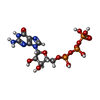 ChemComp-GTP: |
-Macromolecule #4: MAGNESIUM ION
| Macromolecule | Name: MAGNESIUM ION / type: ligand / ID: 4 / Number of copies: 2 / Formula: MG |
|---|---|
| Molecular weight | Theoretical: 24.305 Da |
-Macromolecule #5: GUANOSINE-5'-DIPHOSPHATE
| Macromolecule | Name: GUANOSINE-5'-DIPHOSPHATE / type: ligand / ID: 5 / Number of copies: 2 / Formula: GDP |
|---|---|
| Molecular weight | Theoretical: 443.201 Da |
| Chemical component information | 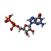 ChemComp-GDP: |
-Experimental details
-Structure determination
| Method | cryo EM |
|---|---|
 Processing Processing | helical reconstruction |
| Aggregation state | filament |
-Sample preparation
| Buffer | pH: 7.2 / Details: Gibco Neurobasal Media (diluted 1:3 to 160 mOsm) |
|---|---|
| Grid | Model: Quantifoil R2/4 / Material: GOLD / Mesh: 200 / Support film - Material: CARBON / Support film - topology: HOLEY / Pretreatment - Type: GLOW DISCHARGE / Pretreatment - Time: 60 sec. / Pretreatment - Atmosphere: AIR / Pretreatment - Pressure: 3.8000000000000003 kPa / Details: coated with poly-L-lysine and laminin |
| Vitrification | Cryogen name: ETHANE / Chamber humidity: 100 % / Chamber temperature: 303 K / Instrument: FEI VITROBOT MARK IV / Details: blot force 4, blot time 4 s. |
| Details | ghost neurons prepared by hypotonic and mechanical treatment |
- Electron microscopy
| Microscope | TFS KRIOS |
|---|---|
| Specialist optics | Energy filter - Name: GIF Bioquantum |
| Image recording | Film or detector model: GATAN K3 BIOQUANTUM (6k x 4k) / Digitization - Dimensions - Width: 11520 pixel / Digitization - Dimensions - Height: 8184 pixel / Number grids imaged: 10 / Number real images: 4172 / Average electron dose: 52.2 e/Å2 / Details: curated image number |
| Electron beam | Acceleration voltage: 300 kV / Electron source:  FIELD EMISSION GUN FIELD EMISSION GUN |
| Electron optics | Illumination mode: FLOOD BEAM / Imaging mode: BRIGHT FIELD / Nominal defocus max: 3.0 µm / Nominal defocus min: 0.8 µm / Nominal magnification: 105000 |
| Sample stage | Specimen holder model: FEI TITAN KRIOS AUTOGRID HOLDER / Cooling holder cryogen: NITROGEN |
| Experimental equipment |  Model: Titan Krios / Image courtesy: FEI Company |
+Image processing

-Atomic model buiding 1
| Initial model | PDB ID: Chain - Source name: PDB / Chain - Initial model type: experimental model |
|---|---|
| Details | iterative rounds of manual building and automated real-space refinement |
| Refinement | Space: REAL / Protocol: FLEXIBLE FIT |
| Output model | PDB-12ru: |

