 Movie
Movie Controller

[English] 日本語
 Yorodumi
- PDB-12rt: In situ cryo-EM structure of axonal microtubules from ghost neuro... -
+ Open data

- Basic information
| Entry | Database: PDB / ID: 12rt | |||||||||
|---|---|---|---|---|---|---|---|---|---|---|
| Title | In situ cryo-EM structure of axonal microtubules from ghost neurons (compact lattice) | |||||||||
 Components Components |
| |||||||||
 Keywords Keywords | STRUCTURAL PROTEIN / cytoskeleton / microtubules / neuron / axon | |||||||||
| Function / homology |  Function and homology information Function and homology informationMicrotubule-dependent trafficking of connexons from Golgi to the plasma membrane / Cargo trafficking to the periciliary membrane / Sealing of the nuclear envelope (NE) by ESCRT-III / netrin-activated signaling pathway / Carboxyterminal post-translational modifications of tubulin / Intraflagellar transport / netrin receptor binding / COPI-independent Golgi-to-ER retrograde traffic / HSP90 chaperone cycle for steroid hormone receptors (SHR) in the presence of ligand / pyramidal neuron differentiation ...Microtubule-dependent trafficking of connexons from Golgi to the plasma membrane / Cargo trafficking to the periciliary membrane / Sealing of the nuclear envelope (NE) by ESCRT-III / netrin-activated signaling pathway / Carboxyterminal post-translational modifications of tubulin / Intraflagellar transport / netrin receptor binding / COPI-independent Golgi-to-ER retrograde traffic / HSP90 chaperone cycle for steroid hormone receptors (SHR) in the presence of ligand / pyramidal neuron differentiation / COPI-mediated anterograde transport / Kinesins / PKR-mediated signaling / RHO GTPases activate IQGAPs / Aggrephagy / Mitotic Prometaphase / EML4 and NUDC in mitotic spindle formation / Resolution of Sister Chromatid Cohesion / COPI-dependent Golgi-to-ER retrograde traffic / glial cell differentiation / dorsal root ganglion development / Recycling pathway of L1 / The role of GTSE1 in G2/M progression after G2 checkpoint / dentate gyrus development / axonemal microtubule / forebrain morphogenesis / Hedgehog 'off' state / RHO GTPases Activate Formins / Loss of Nlp from mitotic centrosomes / Recruitment of mitotic centrosome proteins and complexes / Loss of proteins required for interphase microtubule organization from the centrosome / organelle transport along microtubule / Anchoring of the basal body to the plasma membrane / Recruitment of NuMA to mitotic centrosomes / AURKA Activation by TPX2 / Separation of Sister Chromatids / cerebellar cortex morphogenesis / Regulation of PLK1 Activity at G2/M Transition / MHC class II antigen presentation / microtubule plus-end / neuron projection arborization / smoothened signaling pathway / homeostasis of number of cells within a tissue / motor behavior / adult behavior / response to L-glutamate / centrosome cycle / sperm principal piece / startle response / intercellular bridge / flagellated sperm motility / regulation of synapse organization / sperm end piece / 'de novo' protein folding / locomotory exploration behavior / microtubule polymerization / response to tumor necrosis factor / sperm flagellum / response to mechanical stimulus / neuron apoptotic process / neurogenesis / adult locomotory behavior / cytoplasmic microtubule / ciliary tip / condensed chromosome / peptide binding / visual learning / hippocampus development / cellular response to calcium ion / axon guidance / locomotory behavior / gene expression / cell periphery / filopodium / intracellular protein transport / neuromuscular junction / cerebral cortex development / neuron migration / microtubule cytoskeleton organization / memory / synapse organization / recycling endosome / mitotic spindle / structural constituent of cytoskeleton / cytoplasmic ribonucleoprotein granule / microtubule cytoskeleton / mitotic cell cycle / neuron differentiation / cilium / myelin sheath / microtubule / lamellipodium / protein folding / protein-folding chaperone binding / growth cone / microtubule binding / Hydrolases; Acting on acid anhydrides; Acting on GTP to facilitate cellular and subcellular movement / protein stabilization / membrane raft / protein heterodimerization activity Similarity search - Function | |||||||||
| Biological species |   Mus musculus (house mouse) Mus musculus (house mouse) | |||||||||
| Method | ELECTRON MICROSCOPY / helical reconstruction / cryo EM / Resolution: 3.96 Å | |||||||||
 Authors Authors | Bodakuntla, S. / Marelli, J. / Vasquez-Montes, V. / Biertumpfel, C. / Mizuno, N. | |||||||||
| Funding support |  United States, 1items United States, 1items
| |||||||||
 Citation Citation | Journal: To Be Published Title: Ghost neuron unlocks in situ high-resolution structural mapping of intracellular architecture in neurons Authors: Bodakuntla, S. / Marelli, J. / Vasquez-Montes, V. / Biertumpfel, C. / Mizuno, N. | |||||||||
| History |
|
- Structure visualization
| Structure viewer | Molecule: MolmilJmol/JSmol |
|---|
- Downloads & links
-Download
| PDBx/mmCIF format | 12rt.cif.gz | 311.7 KB | Display | PDBx/mmCIF format |
|---|---|---|---|---|
| PDB format | pdb12rt.ent.gz | 250.2 KB | Display | PDB format |
| PDBx/mmJSON format | 12rt.json.gz | Tree view | PDBx/mmJSON format | |
| Others |  Other downloads Other downloads |
-Validation report
| Arichive directory | https://data.pdbj.org/pub/pdb/validation_reports/2r/12rtftp://data.pdbj.org/pub/pdb/validation_reports/2r/12rt | HTTPS FTP |
|---|
-Related structure data
| Related structure data |  76716MC  12ruC  12rvC C: citing same article ( M: map data used to model this data |
|---|---|
| Similar structure data |


-Links
 PDBj
PDBj


- Assembly
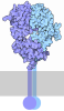
| Deposited unit | 
|
|---|---|
| 1 |
|
-Components
| #1: Protein | Mass: 50188.441 Da / Num. of mol.: 2 / Source method: isolated from a natural source / Details: TUBA1A component of microtubules / Source: (natural) Mus musculus (house mouse) / Organ: Brain / Plasmid details: Axon / Strain: CD-1 / Tissue: Thalamus / References: UniProt: P68369#2: Protein | Mass: 50467.492 Da / Num. of mol.: 2 / Source method: isolated from a natural source / Details: TUBB3 component of microtubules / Source: (natural) Mus musculus (house mouse) / Organ: Brain / Plasmid details: Axon / Strain: CD-1 / Tissue: Thalamus / References: UniProt: Q9ERD7#3: Chemical | 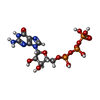 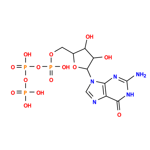 Mass: 523.180 Da / Num. of mol.: 2 / Source method: obtained synthetically / Formula: C10H16N5O14P3 / Comment: GTP, energy-carrying molecule*YM Mass: 523.180 Da / Num. of mol.: 2 / Source method: obtained synthetically / Formula: C10H16N5O14P3 / Comment: GTP, energy-carrying molecule*YM#4: Chemical | ChemComp-MG /   Mass: 24.305 Da / Num. of mol.: 4 / Source method: obtained synthetically / Formula: Mg Mass: 24.305 Da / Num. of mol.: 4 / Source method: obtained synthetically / Formula: Mg#5: Chemical |  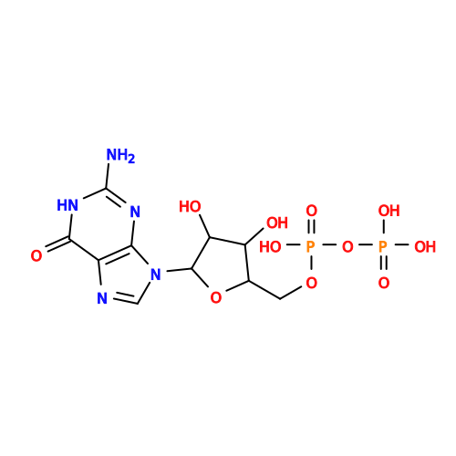 Type: RNA linking / Mass: 443.201 Da / Num. of mol.: 2 / Source method: obtained synthetically / Formula: C10H15N5O11P2 / Comment: GDP, energy-carrying molecule*YM Type: RNA linking / Mass: 443.201 Da / Num. of mol.: 2 / Source method: obtained synthetically / Formula: C10H15N5O11P2 / Comment: GDP, energy-carrying molecule*YMHas ligand of interest | N | Has protein modification | N | |
|---|
-Experimental details
-Experiment
| Experiment | Method: ELECTRON MICROSCOPY |
|---|---|
| EM experiment | Aggregation state: FILAMENT / 3D reconstruction method: helical reconstruction |
- Sample preparation
| Component | Name: Microtubule, axonal / Type: ORGANELLE OR CELLULAR COMPONENT Details: explant axon from thalamus primary mouse embryo E15.5 tissue after ghost preparation (hypotonic treatment and mechanical plasma membrane removal) Entity ID: #1-#2 / Source: NATURAL |
|---|---|
| Molecular weight | Experimental value: NO |
| Source (natural) | Organism: Mus musculus (house mouse) / Strain: CD-1 / Cellular location: Axon / Organ: Brain / Tissue: Thalamus |
| Buffer solution | pH: 7.2 / Details: Gibco Neurobasal Media (diluted 1:3 to 160 mOsm) |
| Specimen | Embedding applied: NO / Shadowing applied: NO / Staining applied: NO / Vitrification applied: YES Details: ghost neurons prepared by hypotonic and mechanical treatment |
| Specimen support | Details: coated with poly-L-lysine and laminin / Grid material: GOLD / Grid mesh size: 200 divisions/in. / Grid type: Quantifoil R2/4 |
| Vitrification | Instrument: FEI VITROBOT MARK IV / Cryogen name: ETHANE / Humidity: 100 % / Chamber temperature: 303 K / Details: blot force 4, blot time 4 s |
- Electron microscopy imaging
| Experimental equipment |  Model: Titan Krios / Image courtesy: FEI Company |
|---|---|
| Microscopy | Model: TFS KRIOS |
| Electron gun | Electron source:  FIELD EMISSION GUN / Accelerating voltage: 300 kV / Illumination mode: FLOOD BEAM FIELD EMISSION GUN / Accelerating voltage: 300 kV / Illumination mode: FLOOD BEAM |
| Electron lens | Mode: BRIGHT FIELD / Nominal magnification: 105000 X / Nominal defocus max: 3000 nm / Nominal defocus min: 800 nm / Cs: 2.7 mm / Alignment procedure: COMA FREE |
| Specimen holder | Cryogen: NITROGEN / Specimen holder model: FEI TITAN KRIOS AUTOGRID HOLDER |
| Image recording | Electron dose: 52.2 e/Å2 / Film or detector model: GATAN K3 BIOQUANTUM (6k x 4k) / Num. of grids imaged: 10 / Num. of real images: 4172 / Details: curated image number |
| EM imaging optics | Energyfilter name: BIOCONTINUUM |
| Image scans | Width: 11520 / Height: 8184 |
- Processing
| EM software |
| ||||||||||||||||||||||||||||||||||||||||||||
|---|---|---|---|---|---|---|---|---|---|---|---|---|---|---|---|---|---|---|---|---|---|---|---|---|---|---|---|---|---|---|---|---|---|---|---|---|---|---|---|---|---|---|---|---|---|
| CTF correction | Type: PHASE FLIPPING AND AMPLITUDE CORRECTION | ||||||||||||||||||||||||||||||||||||||||||||
| Helical symmerty | Angular rotation/subunit: 0 ° / Axial rise/subunit: 83.11 Å / Axial symmetry: C1 | ||||||||||||||||||||||||||||||||||||||||||||
| Particle selection | Num. of particles selected: 158108 | ||||||||||||||||||||||||||||||||||||||||||||
| 3D reconstruction | Resolution: 3.96 Å / Resolution method: FSC 0.143 CUT-OFF / Num. of particles: 30423 / Algorithm: FOURIER SPACE / Num. of class averages: 1 / Symmetry type: HELICAL | ||||||||||||||||||||||||||||||||||||||||||||
| Atomic model building | Protocol: FLEXIBLE FIT / Space: REAL Details: iterative rounds of manual building and automated real-space refinement | ||||||||||||||||||||||||||||||||||||||||||||
| Atomic model building | PDB-ID: 9PND Accession code: 9PND / Source name: PDB / Type: experimental model | ||||||||||||||||||||||||||||||||||||||||||||
| Refinement | Highest resolution: 3.96 Å Stereochemistry target values: REAL-SPACE (WEIGHTED MAP SUM AT ATOM CENTERS) | ||||||||||||||||||||||||||||||||||||||||||||
| Refine LS restraints |
|
