 Movie
Movie Controller


+ Open data

- Basic information
| Entry |  | |||||||||
|---|---|---|---|---|---|---|---|---|---|---|
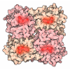
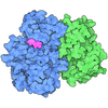
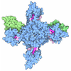
| Title | Cryo-EM structure of human nonmuscle myosin-2B, Class 3 | |||||||||
 Map data Map data | IHM | |||||||||
 Sample Sample |
| |||||||||
 Keywords Keywords | myosin / ATPase / MOTOR PROTEIN | |||||||||
| Function / homology |  Function and homology information Function and homology informationmyosin II filament / postsynaptic actin cytoskeleton organization / unconventional myosin complex / actin filament-based movement / actomyosin / myosin filament / RHO GTPases Activate ROCKs / actomyosin structure organization / RHO GTPases activate CIT / postsynaptic actin cytoskeleton ...myosin II filament / postsynaptic actin cytoskeleton organization / unconventional myosin complex / actin filament-based movement / actomyosin / myosin filament / RHO GTPases Activate ROCKs / actomyosin structure organization / RHO GTPases activate CIT / postsynaptic actin cytoskeleton / Sema4D induced cell migration and growth-cone collapse / muscle filament sliding / myosin complex / myosin II complex / skeletal muscle tissue development / EPHA-mediated growth cone collapse / structural constituent of muscle / microfilament motor activity / myosin heavy chain binding / cleavage furrow / mitotic cytokinesis / RHO GTPases activate PAKs / myofibril / cytoskeletal motor activity / brush border / Smooth Muscle Contraction / RHO GTPases activate PKNs / stress fiber / muscle contraction / positive regulation of protein secretion / neuromuscular junction / ADP binding / RNA stem-loop binding / mRNA 5'-UTR binding / spindle / Z disc / cytoplasmic side of plasma membrane / regulation of cell shape / actin filament binding / lamellipodium / growth cone / virus receptor activity / actin binding / midbody / cell cortex / vesicle / dendritic spine / calmodulin binding / cell adhesion / neuronal cell body / calcium ion binding / symbiont entry into host cell / glutamatergic synapse / cell surface / extracellular exosome / ATP binding / membrane / nucleus / cytosol / cytoplasm Similarity search - Function | |||||||||
| Biological species |  Homo sapiens (human) Homo sapiens (human) | |||||||||
| Method | single particle reconstruction / cryo EM / Resolution: 4.1 Å | |||||||||
 Authors Authors | Heissler SM / Chinthalapudi K | |||||||||
| Funding support |  United States, 1 items United States, 1 items
| |||||||||
 Citation Citation | Journal: Nat Commun / Year: 2026 Title: Structural basis of nonmuscle myosin-2 autoinhibition mechanisms. Authors: Sarah M Heissler / Giovanna Grandinetti / James R Sellers / Krishna Chinthalapudi / Abstract: Nonmuscle myosin-2 (NM2) is a fundamental actin-based mechanochemical ATPase that regulates cellular architecture, migration, adhesion, and force generation across diverse biological contexts. NM2 ...Nonmuscle myosin-2 (NM2) is a fundamental actin-based mechanochemical ATPase that regulates cellular architecture, migration, adhesion, and force generation across diverse biological contexts. NM2 function is tightly regulated by a structural transition between an autoinhibited monomeric (10S) conformation in which ATPase activity, actin binding, and filament assembly are coordinately suppressed and an enzymatically active, filamentous conformation. The autoinhibited conformation is critical for the spatial and temporal control of contractility in nonmuscle cells, yet structural insights into the 10S conformation remain largely elusive. Here, we report a ~53-nm elongated full-length structure of NM2B in the 10S conformation and four distinct cryo-EM structures representing the conformational landscape within the human NM2B 10S state. These structures reveal a tri-segmented tail fold that sequesters interfaces essential for actin binding and filament assembly. The asymmetric arrangement of myosin heavy and light chains provides a mechanistic foundation for understanding how regulatory post-translational modifications and disease-associated mutations shift NM2 conformational equilibria and may enable the development of structure-based interventions for cytoskeletal diseases including hearing loss, neurodegeneration, and cancer. | |||||||||
| History |
|
- Structure visualization
| Supplemental images |
|---|

- Downloads & links
-EMDB archive
| Map data | emd_73811.map.gz | 483.6 MB | EMDB map data format | |
|---|---|---|---|---|
| Header (meta data) | emd-73811-v30.xmlemd-73811.xml | 23.9 KB 23.9 KB | Display Display | EMDB header |
| FSC (resolution estimation) | emd_73811_fsc.xml | 16.9 KB | Display | FSC data file |
| Images |  emd_73811.png emd_73811.png | 84.6 KB | ||
| Filedesc metadata | emd-73811.cif.gz | 7.6 KB | ||
| Others | emd_73811_half_map_1.map.gzemd_73811_half_map_2.map.gz | 475.8 MB 475.8 MB | ||
| Archive directory |  http://ftp.pdbj.org/pub/emdb/structures/EMD-73811ftp://ftp.pdbj.org/pub/emdb/structures/EMD-73811 http://ftp.pdbj.org/pub/emdb/structures/EMD-73811ftp://ftp.pdbj.org/pub/emdb/structures/EMD-73811 | HTTPS FTP |
-Related structure data
| Related structure data |  9z4qMC  9z3wC  9z3zC  9z40C M: atomic model generated by this map C: citing same article ( |
|---|---|
| Similar structure data |


-Links
| EMDB pages | EMDB (EBI/PDBe) / EMDataResource |
|---|---|
| Related items in Molecule of the Month |











-Map
| File | Download / File: emd_73811.map.gz / Format: CCP4 / Size: 512 MB / Type: IMAGE STORED AS FLOATING POINT NUMBER (4 BYTES) | ||||||||||||||||||||||||||||||||||||
|---|---|---|---|---|---|---|---|---|---|---|---|---|---|---|---|---|---|---|---|---|---|---|---|---|---|---|---|---|---|---|---|---|---|---|---|---|---|
| Annotation | IHM | ||||||||||||||||||||||||||||||||||||
| Projections & slices | Image control
Images are generated by Spider. | ||||||||||||||||||||||||||||||||||||
| Voxel size | X=Y=Z: 1.06 Å | ||||||||||||||||||||||||||||||||||||
| Density |
| ||||||||||||||||||||||||||||||||||||
| Symmetry | Space group: 1 | ||||||||||||||||||||||||||||||||||||
| Details | EMDB XML:
|
 Z (Sec.)
 Y (Row.)
 X (Col.)


-Supplemental data
-Half map: #2
| File | emd_73811_half_map_1.map | ||||||||||||
|---|---|---|---|---|---|---|---|---|---|---|---|---|---|
| Projections & Slices |
| ||||||||||||
| Density Histograms |


-Half map: #1
| File | emd_73811_half_map_2.map | ||||||||||||
|---|---|---|---|---|---|---|---|---|---|---|---|---|---|
| Projections & Slices |
| ||||||||||||
| Density Histograms |


- Sample components
-Entire : Non-muscle myosin-2B
| Entire | Name: Non-muscle myosin-2B |
|---|---|
| Components |
|
-Supramolecule #1: Non-muscle myosin-2B
| Supramolecule | Name: Non-muscle myosin-2B / type: complex / ID: 1 / Parent: 0 / Macromolecule list: #1-#3 |
|---|---|
| Source (natural) | Organism: Homo sapiens (human) |
-Macromolecule #1: Myosin-10
| Macromolecule | Name: Myosin-10 / type: protein_or_peptide / ID: 1 / Number of copies: 2 / Enantiomer: LEVO |
|---|---|
| Source (natural) | Organism: Homo sapiens (human) |
| Molecular weight | Theoretical: 229.361547 KDa |
| Recombinant expression | Organism:   Spodoptera frugiperda (fall armyworm) Spodoptera frugiperda (fall armyworm) |
| Sequence | String: MAQRTGLEDP ERYLFVDRAV IYNPATQADW TAKKLVWIPS ERHGFEAASI KEERGDEVMV ELAENGKKAM VNKDDIQKMN PPKFSKVED MAELTCLNEA SVLHNLKDRY YSGLIYTYSG LFCVVINPYK NLPIYSENII EMYRGKKRHE MPPHIYAISE S AYRCMLQD ...String: MAQRTGLEDP ERYLFVDRAV IYNPATQADW TAKKLVWIPS ERHGFEAASI KEERGDEVMV ELAENGKKAM VNKDDIQKMN PPKFSKVED MAELTCLNEA SVLHNLKDRY YSGLIYTYSG LFCVVINPYK NLPIYSENII EMYRGKKRHE MPPHIYAISE S AYRCMLQD REDQSILCTG ESGAGKTENT KKVIQYLAHV ASSHKGRKDH NIPGELERQL LQANPILESF GNAKTVKNDN SS RFGKFIR INFDVTGYIV GANIETYLLE KSRAVRQAKD ERTFHIFYQL LSGAGEHLKS DLLLEGFNNY RFLSNGYIPI PGQ QDKDNF QETMEAMHIM GFSHEEILSM LKVVSSVLQF GNISFKKERN TDQASMPENT VAQKLCHLLG MNVMEFTRAI LTPR IKVGR DYVQKAQTKE QADFAVEALA KATYERLFRW LVHRINKALD RTKRQGASFI GILDIAGFEI FELNSFEQLC INYTN EKLQ QLFNHTMFIL EQEEYQREGI EWNFIDFGLD LQPCIDLIER PANPPGVLAL LDEECWFPKA TDKTFVEKLV QEQGSH SKF QKPRQLKDKA DFCIIHYAGK VDYKADEWLM KNMDPLNDNV ATLLHQSSDR FVAELWKDVD RIVGLDQVTG MTETAFG SA YKTKKGMFRT VGQLYKESLT KLMATLRNTN PNFVRCIIPN HEKRAGKLDP HLVLDQLRCN GVLEGIRICR QGFPNRIV F QEFRQRYEIL TPNAIPKGFM DGKQACERMI RALELDPNLY RIGQSKIFFR AGVLAHLEEE RDLKITDIII FFQAVCRGY LARKAFAKKQ QQLSALKVLQ RNCAAYLKLR HWQWWRVFTK VKPLLQVTRQ EEELQAKDEE LLKVKEKQTK VEGELEEMER KHQQLLEEK NILAEQLQAE TELFAEAEEM RARLAAKKQE LEEILHDLES RVEEEEERNQ ILQNEKKKMQ AHIQDLEEQL D EEEGARQK LQLEKVTAEA KIKKMEEEIL LLEDQNSKFI KEKKLMEDRI AECSSQLAEE EEKAKNLAKI RNKQEVMISD LE ERLKKEE KTRQELEKAK RKLDGETTDL QDQIAELQAQ IDELKLQLAK KEEELQGALA RGDDETLHKN NALKVVRELQ AQI AELQED FESEKASRNK AEKQKRDLSE ELEALKTELE DTLDTTAAQQ ELRTKREQEV AELKKALEEE TKNHEAQIQD MRQR HATAL EELSEQLEQA KRFKANLEKN KQGLETDNKE LACEVKVLQQ VKAESEHKRK KLDAQVQELH AKVSEGDRLR VELAE KASK LQNELDNVST LLEEAEKKGI KFAKDAASLE SQLQDTQELL QEETRQKLNL SSRIRQLEEE KNSLQEQQEE EEEARK NLE KQVLALQSQL ADTKKKVDDD LGTIESLEEA KKKLLKDAEA LSQRLEEKAL AYDKLEKTKN RLQQELDDLT VDLDHQR QV ASNLEKKQKK FDQLLAEEKS ISARYAEERD RAEAEAREKE TKALSLARAL EEALEAKEEF ERQNKQLRAD MEDLMSSK D DVGKNVHELE KSKRALEQQV EEMRTQLEEL EDELQATEDA KLRLEVNMQA MKAQFERDLQ TRDEQNEEKK RLLIKQVRE LEAELEDERK QRALAVASKK KMEIDLKDLE AQIEAANKAR DEVIKQLRKL QAQMKDYQRE LEEARASRDE IFAQSKESEK KLKSLEAEI LQLQEELASS ERARRHAEQE RDELADEITN SASGKSALLD EKRRLEARIA QLEEELEEEQ SNMELLNDRF R KTTLQVDT LNAELAAERS AAQKSDNARQ QLERQNKELK AKLQELEGAV KSKFKATISA LEAKIGQLEE QLEQEAKERA AA NKLVRRT EKKLKEIFMQ VEDERRHADQ YKEQMEKANA RMKQLKRQLE EAEEEATRAN ASRRKLQREL DDATEANEGL SRE VSTLKN RLRRGGPISF SSSRSGRRQL HLEGASLELS DDDTESKTSD VNETQPPQSE UniProtKB: Myosin-10 |
-Macromolecule #2: Myosin light polypeptide 6
| Macromolecule | Name: Myosin light polypeptide 6 / type: protein_or_peptide / ID: 2 / Details: ELC, MYL6 / Number of copies: 2 / Enantiomer: LEVO |
|---|---|
| Source (natural) | Organism: Homo sapiens (human) |
| Molecular weight | Theoretical: 16.948031 KDa |
| Recombinant expression | Organism: Spodoptera frugiperda (fall armyworm) |
| Sequence | String: MCDFTEDQTA EFKEAFQLFD RTGDGKILYS QCGDVMRALG QNPTNAEVLK VLGNPKSDEM NVKVLDFEHF LPMLQTVAKN KDQGTYEDY VEGLRVFDKE GNGTVMGAEI RHVLVTLGEK MTEEEVEMLV AGHEDSNGCI NYEAFVRHIL SG UniProtKB: Myosin light polypeptide 6 |
-Macromolecule #3: Myosin regulatory light chain 12B
| Macromolecule | Name: Myosin regulatory light chain 12B / type: protein_or_peptide / ID: 3 Details: The first methionine residue is cleaved after the expression. So the first residue number is serine-1. Number of copies: 2 / Enantiomer: LEVO |
|---|---|
| Source (natural) | Organism: Homo sapiens (human) |
| Molecular weight | Theoretical: 19.80416 KDa |
| Recombinant expression | Organism: Spodoptera frugiperda (fall armyworm) |
| Sequence | String: MSSKKAKTKT TKKRPQRATS NVFAMFDQSQ IQEFKEAFNM IDQNRDGFID KEDLHDMLAS LGKNPTDAYL DAMMNEAPGP INFTMFLTM FGEKLNGTDP EDVIRNAFAC FDEEATGTIQ EDYLRELLTT MGDRFTDEEV DELYREAPID KKGNFNYIEF T RILKHGAK DKDD UniProtKB: Myosin regulatory light chain 12B |
-Macromolecule #4: MAGNESIUM ION
| Macromolecule | Name: MAGNESIUM ION / type: ligand / ID: 4 / Number of copies: 2 / Formula: MG |
|---|---|
| Molecular weight | Theoretical: 24.305 Da |
-Macromolecule #5: ADENOSINE-5'-DIPHOSPHATE
| Macromolecule | Name: ADENOSINE-5'-DIPHOSPHATE / type: ligand / ID: 5 / Number of copies: 2 / Formula: ADP |
|---|---|
| Molecular weight | Theoretical: 427.201 Da |
| Chemical component information |  ChemComp-ADP: |
-Macromolecule #6: PHOSPHATE ION
| Macromolecule | Name: PHOSPHATE ION / type: ligand / ID: 6 / Number of copies: 2 / Formula: PO4 |
|---|---|
| Molecular weight | Theoretical: 94.971 Da |
| Chemical component information | 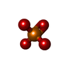 ChemComp-PO4: |
-Experimental details
-Structure determination
| Method | cryo EM |
|---|---|
 Processing Processing | single particle reconstruction |
| Aggregation state | particle |
-Sample preparation
| Buffer | pH: 7.4 |
|---|---|
| Grid | Model: C-flat-1.2/1.3 / Material: GOLD / Mesh: 300 / Pretreatment - Type: GLOW DISCHARGE |
| Vitrification | Cryogen name: ETHANE / Chamber humidity: 95 % / Instrument: LEICA EM GP |
- Electron microscopy
| Microscope | TFS KRIOS |
|---|---|
| Image recording | Film or detector model: GATAN K2 QUANTUM (4k x 4k) / Detector mode: SUPER-RESOLUTION / Average electron dose: 72.0 e/Å2 |
| Electron beam | Acceleration voltage: 300 kV / Electron source:  FIELD EMISSION GUN FIELD EMISSION GUN |
| Electron optics | Illumination mode: FLOOD BEAM / Imaging mode: BRIGHT FIELD / Nominal defocus max: 3.5 µm / Nominal defocus min: 1.0 µm |
| Experimental equipment |  Model: Titan Krios / Image courtesy: FEI Company |
+Image processing


-Atomic model buiding 1
| Refinement | Space: REAL / Protocol: FLEXIBLE FIT |
|---|---|
| Output model | PDB-9z4q: |
