[English] 日本語
Yorodumi
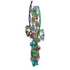
- EMDB-73030: The structure of the cardiac native crossbridge in the rigor stat... -

+
Open data


ID or keywords:

Loading...

-
Basic information

Entry
Database: EMDB / ID: EMD-73030
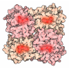
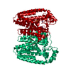
TitleThe structure of the cardiac native crossbridge in the rigor state, myosin heads bound to actin molecules 2 and 3
Map datamap
Sample
  • Complex: complex of heavy meromyosin bound to cardiac thin filament
    • Protein or peptide: Actin, alpha cardiac muscle 1
    • Protein or peptide: Tropomyosin alpha-1 chain
    • Protein or peptide: Troponin C, slow skeletal and cardiac muscles
    • Protein or peptide: Troponin I, cardiac muscle
    • Protein or peptide: Troponin T2, cardiac type
    • Protein or peptide: Myosin-7
    • Protein or peptide: Myosin light chain 3
  • Ligand: ADENOSINE-5'-DIPHOSPHATE
  • Ligand: MAGNESIUM ION
  • Ligand: CALCIUM ION
Keywordscardiac myosin / tropomyosin / thin filament / troponin / MOTOR PROTEIN
Function / homology
Function and homology information


RHOB GTPase cycle / Striated Muscle Contraction / RHOA GTPase cycle / Regulation of CDH1 Function / Formation of the dystrophin-glycoprotein complex (DGC) / regulation of striated muscle contraction / regulation of muscle filament sliding speed / troponin T binding / cardiac Troponin complex / regulation of the force of heart contraction ...RHOB GTPase cycle / Striated Muscle Contraction / RHOA GTPase cycle / Regulation of CDH1 Function / Formation of the dystrophin-glycoprotein complex (DGC) / regulation of striated muscle contraction / regulation of muscle filament sliding speed / troponin T binding / cardiac Troponin complex / regulation of the force of heart contraction / troponin complex / actin-myosin filament sliding / A band / regulation of muscle contraction / adult heart development / myosin filament / muscle filament sliding / myosin II complex / I band / cardiac muscle cell contraction / heart contraction / ventricular cardiac muscle tissue morphogenesis / myosin binding / microfilament motor activity / troponin I binding / mesenchyme migration / myofibril / cytoskeletal motor activity / actin monomer binding / cardiac muscle contraction / skeletal muscle contraction / actin filament organization / sarcomere / filopodium / actin filament / structural constituent of cytoskeleton / calcium-dependent protein binding / actin filament binding / actin cytoskeleton / lamellipodium / actin binding / cell body / calmodulin binding / protein heterodimerization activity / positive regulation of gene expression / calcium ion binding / protein homodimerization activity / ATP binding / identical protein binding / cytosol / cytoplasm
Similarity search - Function
Troponin T / Troponin I residues 1-32 / Troponin I residues 1-32 / : / Troponin / Troponin domain superfamily / Troponin / Tropomyosins signature. / Tropomyosin / Tropomyosin ...Troponin T / Troponin I residues 1-32 / Troponin I residues 1-32 / : / Troponin / Troponin domain superfamily / Troponin / Tropomyosins signature. / Tropomyosin / Tropomyosin / DNA repair protein XRCC4-like, C-terminal / Myosin tail / Myosin tail / Myosin N-terminal SH3-like domain / Myosin S1 fragment, N-terminal / EF-hand domain pair / Myosin, N-terminal, SH3-like / Myosin N-terminal SH3-like domain profile. / Myosin motor domain profile. / Myosin head, motor domain / Myosin head (motor domain) / Myosin. Large ATPases. / IQ motif profile. / Kinesin motor domain superfamily / : / Actins signature 1. / Actin, conserved site / Actins signature 2. / Actin/actin-like conserved site / Actins and actin-related proteins signature. / Actin / Actin family / Actin / EF-hand domain pair / EF-hand, calcium binding motif / EF-Hand 1, calcium-binding site / EF-hand calcium-binding domain. / ATPase, nucleotide binding domain / EF-hand calcium-binding domain profile. / EF-hand domain / EF-hand domain pair / P-loop containing nucleoside triphosphate hydrolase
Similarity search - Domain/homology
Troponin T, cardiac muscle / Troponin I, cardiac muscle / Actin, alpha cardiac muscle 1 / Myosin light chain 3 / Tropomyosin alpha-1 chain / Troponin C, slow skeletal and cardiac muscles / Myosin-7
Similarity search - Component
Biological speciesSus scrofa (pig)
Methodsingle particle reconstruction / cryo EM / Resolution: 4.8 Å
AuthorsGalkin VE / Risi CM
Funding support United States, 2 items
OrganizationGrant numberCountry
National Institutes of Health/National Heart, Lung, and Blood Institute (NIH/NHLBI)R01HL160966 United States
National Institutes of Health/National Institute of General Medical Sciences (NIH/NIGMS)R01GM120137 United States
CitationJournal: To Be Published
Title: The structure of the native cardiac cross-bridge
Authors: Galkin VE / Risi CM
History
DepositionOct 3, 2025-
Header (metadata) releaseJul 22, 2026-
Map releaseJul 22, 2026-
UpdateJul 22, 2026-
Current statusJul 22, 2026Processing site: RCSB / Status: Released

-
Structure visualization

Supplemental images

Downloads & links

-
Map

FileDownload / File: emd_73030.map.gz / Format: CCP4 / Size: 236.9 MB / Type: IMAGE STORED AS FLOATING POINT NUMBER (4 BYTES)
Annotationmap
Projections & slices

Image control

Size
Brightness
Contrast
Others
AxesZ (Sec.)Y (Row.)X (Col.)
1.36 Å/pix.
x 396 pix.
= 536.976 Å
1.36 Å/pix.
x 396 pix.
= 536.976 Å
1.36 Å/pix.
x 396 pix.
= 536.976 Å

Surface

Projections

Slices (1/3)

Slices (1/2)

Slices (2/3)

Images are generated by Spider.

Voxel sizeX=Y=Z: 1.356 Å
Density
Contour LevelBy AUTHOR: 1.4
Minimum - Maximum-5.4403925 - 11.968043
Average (Standard dev.)0.00019771286 (±0.19595133)
SymmetrySpace group: 1
Details

EMDB XML:

Map geometry
Axis orderXYZ
Origin000
Dimensions396396396
Spacing396396396
CellA=B=C: 536.97595 Å
α=β=γ: 90.0 °

-
Supplemental data

-
Half map: half map 1

Fileemd_73030_half_map_1.map
Annotationhalf map 1
Projections & Slices
AxesZYX

Projections

Slices (1/2)
Density Histograms

-
Half map: half map 2

Fileemd_73030_half_map_2.map
Annotationhalf map 2
Projections & Slices
AxesZYX

Projections

Slices (1/2)
Density Histograms

-
Sample components

+
Entire : complex of heavy meromyosin bound to cardiac thin filament

EntireName: complex of heavy meromyosin bound to cardiac thin filament
Components
  • Complex: complex of heavy meromyosin bound to cardiac thin filament
    • Protein or peptide: Actin, alpha cardiac muscle 1
    • Protein or peptide: Tropomyosin alpha-1 chain
    • Protein or peptide: Troponin C, slow skeletal and cardiac muscles
    • Protein or peptide: Troponin I, cardiac muscle
    • Protein or peptide: Troponin T2, cardiac type
    • Protein or peptide: Myosin-7
    • Protein or peptide: Myosin light chain 3
  • Ligand: ADENOSINE-5'-DIPHOSPHATE
  • Ligand: MAGNESIUM ION
  • Ligand: CALCIUM ION

+
Supramolecule #1: complex of heavy meromyosin bound to cardiac thin filament

SupramoleculeName: complex of heavy meromyosin bound to cardiac thin filament
type: complex / ID: 1 / Parent: 0 / Macromolecule list: #1-#7
Source (natural)Organism: Sus scrofa (pig)

+
Macromolecule #1: Actin, alpha cardiac muscle 1

MacromoleculeName: Actin, alpha cardiac muscle 1 / type: protein_or_peptide / ID: 1 / Number of copies: 4 / Enantiomer: LEVO
Source (natural)Organism: Sus scrofa (pig)
Molecular weightTheoretical: 42.064891 KDa
SequenceString: MCDDEETTAL VCDNGSGLVK AGFAGDDAPR AVFPSIVGRP RHQGVMVGMG QKDSYVGDEA QSKRGILTLK YPIEHGIITN WDDMEKIWH HTFYNELRVA PEEHPTLLTE APLNPKANRE KMTQIMFETF NVPAMYVAIQ AVLSLYASGR TTGIVLDSGD G VTHNVPIY ...String:
MCDDEETTAL VCDNGSGLVK AGFAGDDAPR AVFPSIVGRP RHQGVMVGMG QKDSYVGDEA QSKRGILTLK YPIEHGIITN WDDMEKIWH HTFYNELRVA PEEHPTLLTE APLNPKANRE KMTQIMFETF NVPAMYVAIQ AVLSLYASGR TTGIVLDSGD G VTHNVPIY EGYALPHAIM RLDLAGRDLT DYLMKILTER GYSFVTTAER EIVRDIKEKL CYVALDFENE MATAASSSSL EK SYELPDG QVITIGNERF RCPETLFQPS FIGMESAGIH ETTYNSIMKC DIDIRKDLYA NNVLSGGTTM YPGIADRMQK EIT ALAPST MKIKIIAPPE RKYSVWIGGS ILASLSTFQQ MWISKQEYDE AGPSIVHRKC F

UniProtKB: Actin, alpha cardiac muscle 1

+
Macromolecule #2: Tropomyosin alpha-1 chain

MacromoleculeName: Tropomyosin alpha-1 chain / type: protein_or_peptide / ID: 2 / Number of copies: 2 / Enantiomer: LEVO
Source (natural)Organism: Sus scrofa (pig)
Molecular weightTheoretical: 32.762656 KDa
SequenceString: MDAIKKKMQM LKLDKENALD RAEQAEADKK AAEDRSKRLE DELVSLQKKL KATEDELDKY SEAPKDAQEK LELAEKKATD AEADVASLN RRIQLVEEEL DRAQERLATA LQKLEEAEKA ADESERGMKV IESRAQKDEE KMEIQEIQLK EAKHIAEDAD R KYEEVARK ...String:
MDAIKKKMQM LKLDKENALD RAEQAEADKK AAEDRSKRLE DELVSLQKKL KATEDELDKY SEAPKDAQEK LELAEKKATD AEADVASLN RRIQLVEEEL DRAQERLATA LQKLEEAEKA ADESERGMKV IESRAQKDEE KMEIQEIQLK EAKHIAEDAD R KYEEVARK LVIIESDLER AEERAELSEG KCAELEEELK TVTNNLKSLE AQAEKYSQKE DKYEEEIKVL SDKLKEAETR AE FAERSVT KLEKSIDDLE DELYAQKLKY KAISEELDHA LNDMTSI

UniProtKB: Tropomyosin alpha-1 chain

+
Macromolecule #3: Troponin C, slow skeletal and cardiac muscles

MacromoleculeName: Troponin C, slow skeletal and cardiac muscles / type: protein_or_peptide / ID: 3 / Number of copies: 1 / Enantiomer: LEVO
Source (natural)Organism: Sus scrofa (pig)
Molecular weightTheoretical: 18.433508 KDa
SequenceString:
MDDIYKAAVE QLTEEQKNEF KAAFDIFVLG AEDGCISTKE LGKVMRMLGQ NPTPEELQEM IDEVDEDGSG TVDFDEFLVM MVRCMKDDS KGKSEEELSD LFRMFDKNAD GYIDLEELKI MLQATGETIT EDDIEELMKD GDKNNDGRID YDEFLEFMKG V E

UniProtKB: Troponin C, slow skeletal and cardiac muscles

+
Macromolecule #4: Troponin I, cardiac muscle

MacromoleculeName: Troponin I, cardiac muscle / type: protein_or_peptide / ID: 4 / Number of copies: 1 / Enantiomer: LEVO
Source (natural)Organism: Sus scrofa (pig)
Molecular weightTheoretical: 24.120732 KDa
SequenceString: MADRSGDAAG DSRPAPAPVR RRSSANYRAY ATEPHAKKKS KISASRKLQL KTLMLQIAKQ ELEREAEERR GEKGRALSTR CQPLELAGL SFAELQDLCR QLHARVDKVD EERYDVEVKV TKNITEIADL NQKIFDLRGK FKRPTLRRVR ISADAMMQAL L GARAKETL ...String:
MADRSGDAAG DSRPAPAPVR RRSSANYRAY ATEPHAKKKS KISASRKLQL KTLMLQIAKQ ELEREAEERR GEKGRALSTR CQPLELAGL SFAELQDLCR QLHARVDKVD EERYDVEVKV TKNITEIADL NQKIFDLRGK FKRPTLRRVR ISADAMMQAL L GARAKETL DLRAHLKQVK KEDTEKENRE VGDWRKNIDA LSGMEGRKKK FEG

UniProtKB: Troponin I, cardiac muscle

+
Macromolecule #5: Troponin T2, cardiac type

MacromoleculeName: Troponin T2, cardiac type / type: protein_or_peptide / ID: 5 / Number of copies: 1 / Enantiomer: LEVO
Source (natural)Organism: Sus scrofa (pig)
Molecular weightTheoretical: 33.941738 KDa
SequenceString: MSDVEETVDE YEEEQEEGAA EEQEEAVEEE AGGEAEAEEA NAEEAGQEED GREAEDGPME ESKPKPRLFM PNLVPPKIPD GERVDFDDI HRKRMKDLNE LQTLIEAHFE NRKKEEELVS LKDRIEKRRA ERAEQQRIRT EREKERQTRL AEERARREEE E NRRKAEDE ...String:
MSDVEETVDE YEEEQEEGAA EEQEEAVEEE AGGEAEAEEA NAEEAGQEED GREAEDGPME ESKPKPRLFM PNLVPPKIPD GERVDFDDI HRKRMKDLNE LQTLIEAHFE NRKKEEELVS LKDRIEKRRA ERAEQQRIRT EREKERQTRL AEERARREEE E NRRKAEDE ARKKKALSNM MHFGGYIQKQ AQTERKSGKR QTEREKKKKI LAERRKVLAI DHLNEDQLRE KAKELWQSIY NL EAEKFDL QEKFKQQKYE INVLRNRIND NQKVSKTRGK AKVTGRWK

UniProtKB: Troponin T, cardiac muscle

+
Macromolecule #6: Myosin-7

MacromoleculeName: Myosin-7 / type: protein_or_peptide / ID: 6 / Number of copies: 2 / Enantiomer: LEVO
Source (natural)Organism: Sus scrofa (pig)
Molecular weightTheoretical: 223.649094 KDa
SequenceString: MVDAEMAAFG EAAPYLRKSE KERLEAQTRP FDLKKDVYVP DDKEEFVKAK ILSREGGKVT AETEHGKTVT VKEDQVLQQN PPKFDKIED MAMLTFLHEP AVLYNLKERY ASWMIYTYSG LFCVTINPYK WLPVYNAEVV AAYRGKKRSE APPHIFSISD N AYQYMLTD ...String:
MVDAEMAAFG EAAPYLRKSE KERLEAQTRP FDLKKDVYVP DDKEEFVKAK ILSREGGKVT AETEHGKTVT VKEDQVLQQN PPKFDKIED MAMLTFLHEP AVLYNLKERY ASWMIYTYSG LFCVTINPYK WLPVYNAEVV AAYRGKKRSE APPHIFSISD N AYQYMLTD RENQSILITG ESGAGKTVNT KRVIQYFAVI AAIGDRSKKE QTPGKGTLED QIIQANPALE AFGNAKTVRN DN SSRFGKF IRIHFGATGK LASADIETYL LEKSRVIFQL KAERDYHIFY QILSNKKPEL LDMLLITNNP YDYAFISQGE TTV ASIDDA EELMATDNAF DVLGFTSEEK NSMYKLTGAI MHFGNMKFKL KQREEQAEPD GTEEADKSAY LMGLNSADLL KGLC HPRVK VGNEYVTKGQ NVQQVMYATG ALAKAVYEKM FNWMVTRINT TLETKQPRQY FIGVLDIAGF EIFDFNSFEQ LCINF TNEK LQQFFNHHMF VLEQEEYKKE GIEWEFIDFG MDLQACIDLI EKPMGIMSIL EEECMFPKAT DMTFKAKLYD NHLGKS NNF QKPRNIKGRP EAHFALIHYA GTVDYNIIGW LQKNKDPLNE TVVDLYKKSS LKLLSNLFAN YAGADTPVEK GKGKAKK GS SFQTVSALHR ENLNKLMTNL RSTHPHFVRC IIPNETKSPG VIDNPLVMHQ LRCNGVLEGI RICRKGFPNR ILYGDFRQ R YRILNPAAIP EGQFIDSRKG AEKLLGSLDI DHNQYKFGHT KVFFKAGLLG LLEEMRDERL SRIITRIQAQ SRGVLSRME FKKLLERRDS LLIIQWNIRA FMSVKNWPWM KLYFKIKPLL KSAETEKEMA TMKEEFGRLK EALEKSEARR KELEEKMVSL LQEKNDLQL QVQAEQDNLA DAEERCDQLI KNKIQLEAKV KEMTERLEDE EEMNAELTAK KRKLEDECSE LKRDIDDLEL T LAKVEKEK HATENKVKNL TEEMAGLDEI IAKLTKEKKA LQEAHQQALD DLQAEEDKVN TLTKAKVKLE QHVDDLEGSL EQ EKKVRMD LERAKRKLEG DLKLTQESIM DLENDKQQLD ERLKKKDFEL NALNARIEDE QALGSQLQKK LKELQARIEE LEE ELEAER TARAKVEKLR SDLSRELEEI SERLEEAGGA TSVQIEMNKK REAEFQKMRR DLEEATLQHE ATAAALRKKH ADSV AELGE QIDNLQRVKQ KLEKEKSEFK LELDDVTSNM EQIIKAKANL EKMCRTLEDQ MNEHRSKAEE TQRSVNDLTS QRAKL QTEN GELSRQLDEK EALISQLTRG KLTYTQQLED LKRQLEEEVK AKNALAHALQ SARHDCDLLR EQYEEETEAK AELQRV LSK ANSEVAQWRT KYETDAIQRT EELEEAKKKL AQRLQDAEEA VEAVNAKCSS LEKTKHRLQN EIEDLMVDVE RSNAAAA AL DKKQRNFDKI LAEWKQKYEE SQSELESSQK EARSLSTELF KLKNAYEESL EHLETFKREN KNLQEEISDL TEQLGSSG K TIHELEKVRK QLEAEKLELQ SALEEAEASL EHEEGKILRA QLEFNQIKAE MERKLAEKDE EMEQAKRNHL RVVDSLQTS LDAETRSRNE ALRVKKKMEG DLNEMEIQLS HANRMAAEAQ KQVKSLQSLL KDTQIQLDDA VRANDDLKEN IAIVERRNNL LQAELEELR AVVEQTERSR KLAEQELIET SERVQLLHSQ NTSLINQKKK MEADLSQLQT EVEEAVQECR NAEEKAKKAI T DAAMMAEE LKKEQDTSAH LERMKKNMEQ TIKDLQHRLD EAEQIALKGG KKQLQKLEAR VRELENELEA EQKRNAESVK GM RKSERRI KELTYQTEED RKNLLRLQDL VDKLQLKVKA YKRQAEEAEE QANTNLSKFR KVQHELDEAE ERADIAESQV NKL RAKSRD IGTKGLNEE

UniProtKB: Myosin-7

+
Macromolecule #7: Myosin light chain 3

MacromoleculeName: Myosin light chain 3 / type: protein_or_peptide / ID: 7 / Number of copies: 2 / Enantiomer: LEVO
Source (natural)Organism: Sus scrofa (pig)
Molecular weightTheoretical: 21.788875 KDa
SequenceString: MAPKKPEPKK DDAKAAAKAA PAPAPAPAPA PEPKEPEFDA SKIKIEFTPE QIEEFKEAFM LFDRTPKCEM KITYGQCGDV LRALGQNPT QAEVLRVLGK PKQEELNSKM MDFDTFLPML QHISKNKDTG TYEDFVEGLR VFDKEGNGTV MGAELRHVLA T LGERLTED ...String:
MAPKKPEPKK DDAKAAAKAA PAPAPAPAPA PEPKEPEFDA SKIKIEFTPE QIEEFKEAFM LFDRTPKCEM KITYGQCGDV LRALGQNPT QAEVLRVLGK PKQEELNSKM MDFDTFLPML QHISKNKDTG TYEDFVEGLR VFDKEGNGTV MGAELRHVLA T LGERLTED EVEKLMAGQE DSNGCINYEA FVKHIMAS

UniProtKB: Myosin light chain 3

+
Macromolecule #8: ADENOSINE-5'-DIPHOSPHATE

MacromoleculeName: ADENOSINE-5'-DIPHOSPHATE / type: ligand / ID: 8 / Number of copies: 4 / Formula: ADP
Molecular weightTheoretical: 427.201 Da
Chemical component information

ChemComp-ADP:
ADENOSINE-5'-DIPHOSPHATE / ADP, energy-carrying molecule*YM

+
Macromolecule #9: MAGNESIUM ION

MacromoleculeName: MAGNESIUM ION / type: ligand / ID: 9 / Number of copies: 4 / Formula: MG
Molecular weightTheoretical: 24.305 Da

+
Macromolecule #10: CALCIUM ION

MacromoleculeName: CALCIUM ION / type: ligand / ID: 10 / Number of copies: 3 / Formula: CA
Molecular weightTheoretical: 40.078 Da

-
Experimental details

-
Structure determination

Methodcryo EM
Processingsingle particle reconstruction
Aggregation statefilament

-
Sample preparation

BufferpH: 7.5
GridModel: EMS Lacey Carbon / Material: COPPER / Mesh: 300 / Support film - Material: CARBON / Support film - topology: LACEY / Pretreatment - Type: GLOW DISCHARGE / Pretreatment - Time: 45 sec.
VitrificationCryogen name: ETHANE / Chamber humidity: 100 % / Chamber temperature: 278 K / Instrument: FEI VITROBOT MARK IV

-
Electron microscopy

MicroscopeTFS KRIOS
Image recordingFilm or detector model: GATAN K3 (6k x 4k) / Number grids imaged: 10 / Average electron dose: 34.0 e/Å2
Details: Images were collected in a movie mode, 40 frames with the dose of 0.85 e/A2 per frame
Electron beamAcceleration voltage: 300 kV / Electron source: FIELD EMISSION GUN
Electron opticsIllumination mode: FLOOD BEAM / Imaging mode: BRIGHT FIELD / Nominal defocus max: 3.5 µm / Nominal defocus min: 0.5 µm
Sample stageSpecimen holder model: FEI TITAN KRIOS AUTOGRID HOLDER / Cooling holder cryogen: NITROGEN
Experimental equipment
Model: Titan Krios / Image courtesy: FEI Company

+
Image processing

Particle selectionNumber selected: 5235506
CTF correctionSoftware - Name: CTFFIND (ver. 4) / Type: PHASE FLIPPING AND AMPLITUDE CORRECTION
Startup modelType of model: OTHER / Details: model created from 5H53 and 8UZX
Final reconstructionNumber classes used: 1 / Algorithm: BACK PROJECTION / Resolution.type: BY AUTHOR / Resolution: 4.8 Å / Resolution method: FSC 0.143 CUT-OFF / Software - Name: RELION (ver. 3.1) / Number images used: 31180
Initial angle assignmentType: MAXIMUM LIKELIHOOD / Software - Name: RELION (ver. 3.1)
Final angle assignmentType: MAXIMUM LIKELIHOOD / Software - Name: RELION (ver. 3.1)
Final 3D classificationNumber classes: 40 / Software - Name: RELION (ver. 3.1)
FSC plot (resolution estimation)

-
Atomic model buiding 1

Initial model
PDB IDChain

source_name: PDB, initial_model_type: experimental model

source_name: PDB, initial_model_type: experimental model

source_name: PDB, initial_model_type: experimental model

source_name: PDB, initial_model_type: experimental model

source_name: PDB, initial_model_type: experimental model
RefinementSpace: REAL / Protocol: FLEXIBLE FIT
Output model

PDB-9yjp:
The structure of the cardiac native crossbridge in the rigor state, myosin heads bound to actin molecules 2 and 3

+
About Yorodumi

-
News

-
Feb 9, 2022. New format data for meta-information of EMDB entries

New format data for meta-information of EMDB entries

  • Version 3 of the EMDB header file is now the official format.
  • The previous official version 1.9 will be removed from the archive.

Related info.:EMDB header

External links:wwPDB to switch to version 3 of the EMDB data model

-
Aug 12, 2020. Covid-19 info

Covid-19 info

URL: https://pdbj.org/emnavi/covid19.php

New page: Covid-19 featured information page in EM Navigator.

Related info.:Covid-19 info / Mar 5, 2020. Novel coronavirus structure data

+
Mar 5, 2020. Novel coronavirus structure data

Novel coronavirus structure data

Related info.:Yorodumi Speices / Aug 12, 2020. Covid-19 info

External links:COVID-19 featured content - PDBj / Molecule of the Month (242):Coronavirus Proteases

+
Jan 31, 2019. EMDB accession codes are about to change! (news from PDBe EMDB page)

EMDB accession codes are about to change! (news from PDBe EMDB page)

  • The allocation of 4 digits for EMDB accession codes will soon come to an end. Whilst these codes will remain in use, new EMDB accession codes will include an additional digit and will expand incrementally as the available range of codes is exhausted. The current 4-digit format prefixed with “EMD-” (i.e. EMD-XXXX) will advance to a 5-digit format (i.e. EMD-XXXXX), and so on. It is currently estimated that the 4-digit codes will be depleted around Spring 2019, at which point the 5-digit format will come into force.
  • The EM Navigator/Yorodumi systems omit the EMD- prefix.

Related info.:Q: What is EMD? / ID/Accession-code notation in Yorodumi/EM Navigator

External links:EMDB Accession Codes are Changing Soon! / Contact to PDBj

+
Jul 12, 2017. Major update of PDB

Major update of PDB

  • wwPDB released updated PDB data conforming to the new PDBx/mmCIF dictionary.
  • This is a major update changing the version number from 4 to 5, and with Remediation, in which all the entries are updated.
  • In this update, many items about electron microscopy experimental information are reorganized (e.g. em_software).
  • Now, EM Navigator and Yorodumi are based on the updated data.

External links:wwPDB Remediation / Enriched Model Files Conforming to OneDep Data Standards Now Available in the PDB FTP Archive

-
Yorodumi

Thousand views of thousand structures

  • Yorodumi is a browser for structure data from EMDB, PDB, SASBDB, etc.
  • This page is also the successor to EM Navigator detail page, and also detail information page/front-end page for Omokage search.
  • The word "yorodu" (or yorozu) is an old Japanese word meaning "ten thousand". "mi" (miru) is to see.

Related info.:EMDB / PDB / SASBDB / Comparison of 3 databanks / Yorodumi Search / Aug 31, 2016. New EM Navigator & Yorodumi / Yorodumi Papers / Jmol/JSmol / Function and homology information / Changes in new EM Navigator and Yorodumi

Read more