Movie
Movie Controller

[English] 日本語
 Yorodumi
- PDB-9yjp: The structure of the cardiac native crossbridge in the rigor stat... -
+ Open data

- Basic information
| Entry | Database: PDB / ID: 9yjp | ||||||||||||||||||||||||
|---|---|---|---|---|---|---|---|---|---|---|---|---|---|---|---|---|---|---|---|---|---|---|---|---|---|
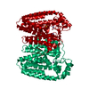
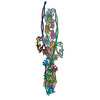
| Title | The structure of the cardiac native crossbridge in the rigor state, myosin heads bound to actin molecules 2 and 3 | ||||||||||||||||||||||||
 Components Components |
| ||||||||||||||||||||||||
 Keywords Keywords | MOTOR PROTEIN / cardiac myosin / tropomyosin / thin filament / troponin | ||||||||||||||||||||||||
| Function / homology |  Function and homology information Function and homology informationRHOB GTPase cycle / Striated Muscle Contraction / RHOA GTPase cycle / Regulation of CDH1 Function / Formation of the dystrophin-glycoprotein complex (DGC) / Activation of STAT3 by cadherin engagement / regulation of striated muscle contraction / regulation of muscle filament sliding speed / troponin T binding / cardiac Troponin complex ...RHOB GTPase cycle / Striated Muscle Contraction / RHOA GTPase cycle / Regulation of CDH1 Function / Formation of the dystrophin-glycoprotein complex (DGC) / Activation of STAT3 by cadherin engagement / regulation of striated muscle contraction / regulation of muscle filament sliding speed / troponin T binding / cardiac Troponin complex / regulation of the force of heart contraction / troponin complex / actin-myosin filament sliding / regulation of muscle contraction / adult heart development / myosin filament / muscle filament sliding / myosin II complex / I band / cardiac muscle cell contraction / heart contraction / microfilament motor activity / ventricular cardiac muscle tissue morphogenesis / myosin binding / A band / troponin I binding / mesenchyme migration / myofibril / cytoskeletal motor activity / actin monomer binding / cardiac muscle contraction / skeletal muscle contraction / actin filament organization / sarcomere / filopodium / actin filament / structural constituent of cytoskeleton / calcium-dependent protein binding / actin filament binding / actin cytoskeleton / lamellipodium / cell body / calmodulin binding / protein heterodimerization activity / positive regulation of gene expression / calcium ion binding / protein homodimerization activity / ATP binding / identical protein binding / cytosol / cytoplasm Similarity search - Function | ||||||||||||||||||||||||
| Biological species |   Sus scrofa (pig) Sus scrofa (pig) | ||||||||||||||||||||||||
| Method | ELECTRON MICROSCOPY / single particle reconstruction / cryo EM / Resolution: 4.8 Å | ||||||||||||||||||||||||
 Authors Authors | Galkin, V.E. / Risi, C.M. | ||||||||||||||||||||||||
| Funding support |  United States, 2items United States, 2items
| ||||||||||||||||||||||||
 Citation Citation | Journal: Sci Adv / Year: 2026 Title: The structure of the native cardiac crossbridge in the rigor state. Authors: Cristina M Risi / Tyler Nguyen / Betty Belknap / Howard D White / Jose R Pinto / Prescott B Chase / Vitold E Galkin / Abstract: Cardiac contraction is driven by double-headed myosin cycling on cardiac thin filaments, where troponin-tropomyosin regulates myosin access to actin. Prior high-resolution structural studies used a ...Cardiac contraction is driven by double-headed myosin cycling on cardiac thin filaments, where troponin-tropomyosin regulates myosin access to actin. Prior high-resolution structural studies used a single-headed myosin bound to bare actin, thereby limiting insight into coordination between myosin heads and the influence of troponin-tropomyosin on actomyosin interactions. Here, we report a high-resolution structure of the native cardiac rigor cross-bridge formed by heavy meromyosin bound to the thin filament. We show that direct communication between the two bound heads, uneven interactions between the heads and tropomyosin, and spatial constraints imposed by troponin govern myosin placement along the thin filament. Additionally, the two heads display non-equivalent motor-light chain interactions, yielding distinct lever-arm conformations indicative of asymmetric intramolecular strain. Together, these findings provide a structural framework for how the two myosin heads coordinate and how the components of the thin filament are integrated into force generation by active cross-bridges. | ||||||||||||||||||||||||
| History |
|
- Structure visualization
| Structure viewer | Molecule: MolmilJmol/JSmol |
|---|
- Downloads & links
-Download
| PDBx/mmCIF format | 9yjp.cif.gz | 760.6 KB | Display | PDBx/mmCIF format |
|---|---|---|---|---|
| PDB format | pdb9yjp.ent.gz | 592.7 KB | Display | PDB format |
| PDBx/mmJSON format | 9yjp.json.gz | Tree view | PDBx/mmJSON format | |
| Others |  Other downloads Other downloads |
-Validation report
| Arichive directory | https://data.pdbj.org/pub/pdb/validation_reports/yj/9yjpftp://data.pdbj.org/pub/pdb/validation_reports/yj/9yjp | HTTPS FTP |
|---|
-Related structure data
| Related structure data |  73030MC  9ya8C  9yaqC  9yk9C  9yknC M: map data used to model this data C: citing same article ( |
|---|---|
| Similar structure data |


-Links
 PDBj
PDBj




- Assembly
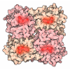
| Deposited unit | 
|
|---|---|
| 1 |
|
-Components
-Protein , 7 types, 13 molecules ABCDEFGHIJKLM
| #1: Protein | Mass: 42064.891 Da / Num. of mol.: 4 / Source method: isolated from a natural source / Source: (natural) Sus scrofa (pig) / References: UniProt: B6VNT8#2: Protein | Mass: 32762.656 Da / Num. of mol.: 2 / Source method: isolated from a natural source / Source: (natural) Sus scrofa (pig) / References: UniProt: P42639#3: Protein | | Mass: 18433.508 Da / Num. of mol.: 1 / Source method: isolated from a natural source / Source: (natural) Sus scrofa (pig) / References: UniProt: P63317#4: Protein | | Mass: 24120.732 Da / Num. of mol.: 1 / Source method: isolated from a natural source / Source: (natural) Sus scrofa (pig) / References: UniProt: A5X5T5#5: Protein | | Mass: 33941.738 Da / Num. of mol.: 1 / Source method: isolated from a natural source / Source: (natural) Sus scrofa (pig) / References: UniProt: A0A5G2Q8N0#6: Protein | Mass: 223649.094 Da / Num. of mol.: 2 / Source method: isolated from a natural source / Source: (natural) Sus scrofa (pig) / References: UniProt: P79293#7: Protein | Mass: 21788.875 Da / Num. of mol.: 2 / Source method: isolated from a natural source / Source: (natural) Sus scrofa (pig) / References: UniProt: F1SNW4 |
|---|
-Non-polymers , 3 types, 11 molecules 


| #8: Chemical | ChemComp-ADP / 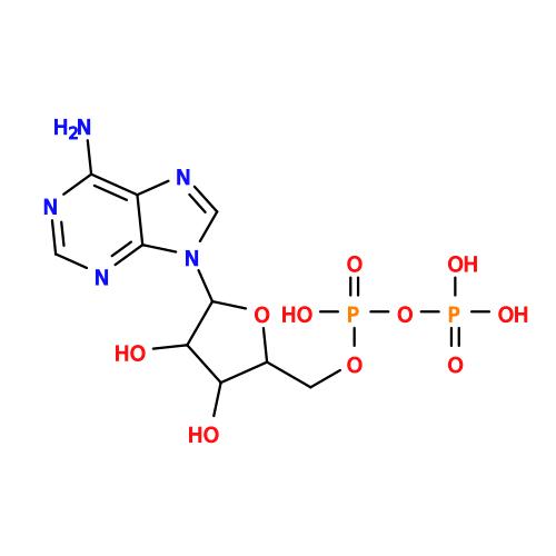 Mass: 427.201 Da / Num. of mol.: 4 / Source method: obtained synthetically / Formula: C10H15N5O10P2 / Comment: ADP, energy-carrying molecule*YM Mass: 427.201 Da / Num. of mol.: 4 / Source method: obtained synthetically / Formula: C10H15N5O10P2 / Comment: ADP, energy-carrying molecule*YM#9: Chemical | ChemComp-MG /  Mass: 24.305 Da / Num. of mol.: 4 / Source method: obtained synthetically / Formula: Mg Mass: 24.305 Da / Num. of mol.: 4 / Source method: obtained synthetically / Formula: Mg#10: Chemical |  Mass: 40.078 Da / Num. of mol.: 3 / Source method: obtained synthetically / Formula: Ca Mass: 40.078 Da / Num. of mol.: 3 / Source method: obtained synthetically / Formula: Ca |
|---|
-Details
| Has ligand of interest | N |
|---|---|
| Has protein modification | N |
-Experimental details
-Experiment
| Experiment | Method: ELECTRON MICROSCOPY |
|---|---|
| EM experiment | Aggregation state: FILAMENT / 3D reconstruction method: single particle reconstruction |
- Sample preparation
| Component | Name: complex of heavy meromyosin bound to cardiac thin filament Type: COMPLEX / Entity ID: #1-#7 / Source: NATURAL |
|---|---|
| Molecular weight | Experimental value: NO |
| Source (natural) | Organism: Sus scrofa (pig) |
| Buffer solution | pH: 7.5 |
| Specimen | Embedding applied: NO / Shadowing applied: NO / Staining applied: NO / Vitrification applied: YES |
| Specimen support | Grid material: COPPER / Grid mesh size: 300 divisions/in. / Grid type: EMS Lacey Carbon |
| Vitrification | Instrument: FEI VITROBOT MARK IV / Cryogen name: ETHANE / Humidity: 100 % / Chamber temperature: 278 K |
- Electron microscopy imaging
| Experimental equipment |  Model: Titan Krios / Image courtesy: FEI Company |
|---|---|
| Microscopy | Model: TFS KRIOS |
| Electron gun | Electron source:  FIELD EMISSION GUN / Accelerating voltage: 300 kV / Illumination mode: FLOOD BEAM FIELD EMISSION GUN / Accelerating voltage: 300 kV / Illumination mode: FLOOD BEAM |
| Electron lens | Mode: BRIGHT FIELD / Nominal defocus max: 3500 nm / Nominal defocus min: 500 nm |
| Specimen holder | Cryogen: NITROGEN / Specimen holder model: FEI TITAN KRIOS AUTOGRID HOLDER |
| Image recording | Electron dose: 34 e/Å2 / Film or detector model: GATAN K3 (6k x 4k) / Num. of grids imaged: 10 Details: Images were collected in a movie mode, 40 frames with the dose of 0.85 e/A2 per frame |
- Processing
| EM software |
| ||||||||||||||||||||||||||||||||||||||||||||||||||||
|---|---|---|---|---|---|---|---|---|---|---|---|---|---|---|---|---|---|---|---|---|---|---|---|---|---|---|---|---|---|---|---|---|---|---|---|---|---|---|---|---|---|---|---|---|---|---|---|---|---|---|---|---|---|
| CTF correction | Type: PHASE FLIPPING AND AMPLITUDE CORRECTION | ||||||||||||||||||||||||||||||||||||||||||||||||||||
| Particle selection | Num. of particles selected: 5235506 | ||||||||||||||||||||||||||||||||||||||||||||||||||||
| 3D reconstruction | Resolution: 4.8 Å / Resolution method: FSC 0.143 CUT-OFF / Num. of particles: 31180 / Algorithm: BACK PROJECTION / Num. of class averages: 1 / Symmetry type: POINT | ||||||||||||||||||||||||||||||||||||||||||||||||||||
| Atomic model building | Protocol: FLEXIBLE FIT / Space: REAL | ||||||||||||||||||||||||||||||||||||||||||||||||||||
| Atomic model building |
|