ムービー
ムービー コントローラー


+ データを開く

- 基本情報
基本情報
| 登録情報 | データベース: EMDB / ID: EMD-6258 | |||||||||
|---|---|---|---|---|---|---|---|---|---|---|
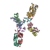
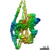
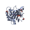
| タイトル | Electron cryo-microscopy of the G protein effector, PDE6 | |||||||||
 マップデータ マップデータ | 3D reconstruction of the complex of the phosphodiesterase of rod photoreceptor cells (PDE6) with Ros-1 Fab (PDE6-Fab). C2 symmetry was imposed during the reconstruction. | |||||||||
 試料 試料 |
| |||||||||
 キーワード キーワード | phosphodiesterase / photoreceptor. | |||||||||
| 機能・相同性 |  機能・相同性情報 機能・相同性情報cGMP effects / Smooth Muscle Contraction / RHOBTB1 GTPase cycle / cyclic-nucleotide phosphodiesterase activity / GMP catabolic process / cellular response to macrophage colony-stimulating factor stimulus / 3',5'-cyclic-GMP phosphodiesterase / cellular response to cGMP / positive regulation of G protein-coupled receptor signaling pathway / Inactivation, recovery and regulation of the phototransduction cascade ...cGMP effects / Smooth Muscle Contraction / RHOBTB1 GTPase cycle / cyclic-nucleotide phosphodiesterase activity / GMP catabolic process / cellular response to macrophage colony-stimulating factor stimulus / 3',5'-cyclic-GMP phosphodiesterase / cellular response to cGMP / positive regulation of G protein-coupled receptor signaling pathway / Inactivation, recovery and regulation of the phototransduction cascade / Activation of the phototransduction cascade / positive regulation of vascular permeability / ion binding / cellular response to granulocyte macrophage colony-stimulating factor stimulus / response to stimulus / negative regulation of vascular permeability / establishment of endothelial barrier / negative regulation of cAMP-mediated signaling / regulation of mitochondrion organization / Ca2+ pathway / positive regulation of epidermal growth factor receptor signaling pathway / photoreceptor outer segment membrane / cGMP-stimulated cyclic-nucleotide phosphodiesterase activity / 3',5'-cyclic-nucleotide phosphodiesterase / negative regulation of cGMP-mediated signaling / cGMP-mediated signaling / cGMP catabolic process / cGMP binding / 3',5'-cyclic-GMP phosphodiesterase activity / 3',5'-cyclic-AMP phosphodiesterase activity / regulation of cAMP-mediated signaling / visual perception / cAMP-mediated signaling / synaptic membrane / photoreceptor disc membrane / positive regulation of inflammatory response / cellular response to mechanical stimulus / presynaptic membrane / mitochondrial outer membrane / mitochondrial inner membrane / molecular adaptor activity / mitochondrial matrix / positive regulation of gene expression / perinuclear region of cytoplasm / Golgi apparatus / negative regulation of transcription by RNA polymerase II / endoplasmic reticulum / signal transduction / protein homodimerization activity / zinc ion binding / nucleus / metal ion binding / plasma membrane / cytoplasm / cytosol 類似検索 - 分子機能 | |||||||||
| 生物種 |  | |||||||||
| 手法 | 単粒子再構成法 / クライオ電子顕微鏡法 / 解像度: 11.0 Å | |||||||||
 データ登録者 データ登録者 | Zhang Z / He F / Constantine R / Baker ML / Baehr W / Schmid MF / Wensel TG / Agosto MA | |||||||||
 引用 引用 | ジャーナル: J Biol Chem / 年: 2015 タイトル: Domain organization and conformational plasticity of the G protein effector, PDE6. 著者: Zhixian Zhang / Feng He / Ryan Constantine / Matthew L Baker / Wolfgang Baehr / Michael F Schmid / Theodore G Wensel / Melina A Agosto /  要旨: The cGMP phosphodiesterase of rod photoreceptor cells, PDE6, is the key effector enzyme in phototransduction. Two large catalytic subunits, PDE6α and -β, each contain one catalytic domain and two ...The cGMP phosphodiesterase of rod photoreceptor cells, PDE6, is the key effector enzyme in phototransduction. Two large catalytic subunits, PDE6α and -β, each contain one catalytic domain and two non-catalytic GAF domains, whereas two small inhibitory PDE6γ subunits allow tight regulation by the G protein transducin. The structure of holo-PDE6 in complex with the ROS-1 antibody Fab fragment was determined by cryo-electron microscopy. The ∼11 Å map revealed previously unseen features of PDE6, and each domain was readily fit with high resolution structures. A structure of PDE6 in complex with prenyl-binding protein (PrBP/δ) indicated the location of the PDE6 C-terminal prenylations. Reconstructions of complexes with Fab fragments bound to N or C termini of PDE6γ revealed that PDE6γ stretches from the catalytic domain at one end of the holoenzyme to the GAF-A domain at the other. Removal of PDE6γ caused dramatic structural rearrangements, which were reversed upon its restoration. | |||||||||
| 履歴 |
|
- 構造の表示
構造の表示
| ムービー |
ムービービューア |
|---|---|
| 構造ビューア | EMマップ: SurfViewMolmilJmol/JSmol |
| 添付画像 |

- ダウンロードとリンク
-EMDBアーカイブ
| マップデータ | emd_6258.map.gz | 47.2 MB | EMDBマップデータ形式 | |
|---|---|---|---|---|
| ヘッダ (付随情報) | emd-6258-v30.xmlemd-6258.xml | 15.7 KB 15.7 KB | 表示 表示 | EMDBヘッダ |
| 画像 | 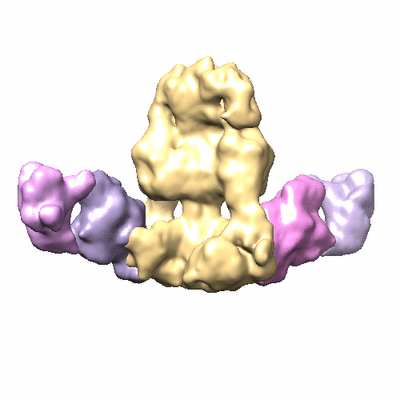 400_6258.gif 400_6258.gif 80_6258.gif 80_6258.gif | 38 KB 3.2 KB | ||
| アーカイブディレクトリ |  http://ftp.pdbj.org/pub/emdb/structures/EMD-6258ftp://ftp.pdbj.org/pub/emdb/structures/EMD-6258 http://ftp.pdbj.org/pub/emdb/structures/EMD-6258ftp://ftp.pdbj.org/pub/emdb/structures/EMD-6258 | HTTPS FTP |
-検証レポート
| 文書・要旨 | emd_6258_validation.pdf.gz | 287.9 KB | 表示 | EMDB検証レポート |
|---|---|---|---|---|
| 文書・詳細版 | emd_6258_full_validation.pdf.gz | 287.5 KB | 表示 | |
| XML形式データ | emd_6258_validation.xml.gz | 6.3 KB | 表示 | |
| アーカイブディレクトリ | https://ftp.pdbj.org/pub/emdb/validation_reports/EMD-6258ftp://ftp.pdbj.org/pub/emdb/validation_reports/EMD-6258 | HTTPS FTP |
-関連構造データ

-リンク
| EMDBのページ | EMDB (EBI/PDBe) / EMDataResource |
|---|---|
| 「今月の分子」の関連する項目 |


-マップ
| ファイル | ダウンロード / ファイル: emd_6258.map.gz / 形式: CCP4 / 大きさ: 51.5 MB / タイプ: IMAGE STORED AS FLOATING POINT NUMBER (4 BYTES) | ||||||||||||||||||||||||||||||||||||||||||||||||||||||||||||
|---|---|---|---|---|---|---|---|---|---|---|---|---|---|---|---|---|---|---|---|---|---|---|---|---|---|---|---|---|---|---|---|---|---|---|---|---|---|---|---|---|---|---|---|---|---|---|---|---|---|---|---|---|---|---|---|---|---|---|---|---|---|
| 注釈 | 3D reconstruction of the complex of the phosphodiesterase of rod photoreceptor cells (PDE6) with Ros-1 Fab (PDE6-Fab). C2 symmetry was imposed during the reconstruction. | ||||||||||||||||||||||||||||||||||||||||||||||||||||||||||||
| ボクセルのサイズ | X=Y=Z: 1.81 Å | ||||||||||||||||||||||||||||||||||||||||||||||||||||||||||||
| 密度 |
| ||||||||||||||||||||||||||||||||||||||||||||||||||||||||||||
| 対称性 | 空間群: 1 | ||||||||||||||||||||||||||||||||||||||||||||||||||||||||||||
| 詳細 | EMDB XML:
CCP4マップ ヘッダ情報:
| ||||||||||||||||||||||||||||||||||||||||||||||||||||||||||||
-添付データ
- 試料の構成要素
-全体 : Bovine rod PDE6 holoenzyme in complex with the Fab fragment from ...
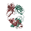
| 全体 | 名称: Bovine rod PDE6 holoenzyme in complex with the Fab fragment from the ROS-1 monoclonal antibody |
|---|---|
| 要素 |
|
-超分子 #1000: Bovine rod PDE6 holoenzyme in complex with the Fab fragment from ...
| 超分子 | 名称: Bovine rod PDE6 holoenzyme in complex with the Fab fragment from the ROS-1 monoclonal antibody タイプ: sample / ID: 1000 / 集合状態: Two Fab molecules bind to PDE6 / Number unique components: 2 |
|---|---|
| 分子量 | 理論値: 320 KDa |
-分子 #1: Rod cGMP-specific 3',5'-cyclic phosphodiesterase
| 分子 | 名称: Rod cGMP-specific 3',5'-cyclic phosphodiesterase / タイプ: protein_or_peptide / ID: 1 / Name.synonym: PDE6 詳細: PDE6 holoenzyme contains PDE6a (UniProt P11541), PDE6b (UniProt P23439), and PDE6g (UniProt P04972). コピー数: 1 / 集合状態: dimer / 組換発現: No / データベース: NCBI |
|---|---|
| 由来(天然) | 生物種: |
| 分子量 | 理論値: 200 KDa |
-分子 #2: monoclonal antibody Fab fragment of ROS-1
| 分子 | 名称: monoclonal antibody Fab fragment of ROS-1 / タイプ: protein_or_peptide / ID: 2 詳細: Fab fragment was purified using immobilized protein L. コピー数: 2 / 組換発現: No / データベース: NCBI |
|---|---|
| 由来(天然) | 生物種: |
| 分子量 | 理論値: 50 KDa |
-実験情報
-構造解析
| 手法 | クライオ電子顕微鏡法 |
|---|---|
 解析 解析 | 単粒子再構成法 |
| 試料の集合状態 | particle |
-試料調製
| 濃度 | 0.5 mg/mL |
|---|---|
| 緩衝液 | pH: 7.5 / 詳細: 20 mM sodium phosphate, 150 mM sodium chloride |
| グリッド | 詳細: 400 mesh glow-discharged Quantifoil grids with 2.0 A holes |
| 凍結 | 凍結剤: ETHANE / チャンバー内湿度: 95 % / チャンバー内温度: 93 K / 装置: FEI VITROBOT MARK III 手法: Applied 3 uL sample per grid and blotted for 1 second before plunging. |
- 電子顕微鏡法
| 顕微鏡 | JEOL 2010F |
|---|---|
| 温度 | 最高: 94 K |
| アライメント法 | Legacy - 非点収差: Objective lens astigmatism was corrected at 100,000 times magnification. |
| 特殊光学系 | エネルギーフィルター - 名称: FEI |
| 詳細 | Parallel beam illumination |
| 日付 | 2011年8月8日 |
| 撮影 | カテゴリ: CCD フィルム・検出器のモデル: GATAN ULTRASCAN 4000 (4k x 4k) 平均電子線量: 15 e/Å2 |
| 電子線 | 加速電圧: 200 kV / 電子線源:  FIELD EMISSION GUN FIELD EMISSION GUN |
| 電子光学系 | 照射モード: FLOOD BEAM / 撮影モード: BRIGHT FIELD / Cs: 2.0 mm / 倍率(公称値): 60000 |
| 試料ステージ | 試料ホルダーモデル: GATAN LIQUID NITROGEN |
-画像解析
| 詳細 | Image processing was performed using EMAN. 21,100 particles were manually boxed from ice images and CTF-corrected using Ctfit. Initial models were generated from reference-free class averages and a cylindrical starting model. The two refined models were essentially similar. Three noise-seeded models were generated and used as initial models in the Multirefine procedure. A model (with two Ros-1 Fab bound) with a population of ~15,000 particles emerged and was subjected to further refinement using standard iterative projection matching, class averaging, and Fourier reconstruction. |
|---|---|
| CTF補正 | 詳細: citfit (EMAN) for each particle |
| 最終 再構成 | アルゴリズム: OTHER / 解像度のタイプ: BY AUTHOR / 解像度: 11.0 Å / 解像度の算出法: OTHER / ソフトウェア - 名称: EMAN 詳細: A total of 21,100 particles were picked from ice images and CTF-corrected using Ctfit. After an initial 3D model was generated as described for PDE6, three noise-seeded models were generated ...詳細: A total of 21,100 particles were picked from ice images and CTF-corrected using Ctfit. After an initial 3D model was generated as described for PDE6, three noise-seeded models were generated and used as initial models in the Multirefine procedure. A model (with two Ros-1 Fab bound) with a population of ~15,000 particles emerged and was subjected to further refinement using standard iterative projection matching, class averaging, and Fourier reconstruction. The final 3D maps with C2 symmetry were generated from 12,373 particles. 使用した粒子像数: 12373 |
| 最終 2次元分類 | クラス数: 20 |
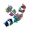
-原子モデル構築 1
| 初期モデル | PDB ID: Chain - Chain ID: B |
|---|---|
| ソフトウェア | 名称: Chimera |
| 詳細 | The domains were separately fitted by manual docking using Chimera and refined using "Fit in Map" within Chimera. The fitting of GFA(A,B) was confirmed using Folderhunter. |
| 精密化 | 空間: REAL / プロトコル: RIGID BODY FIT |
| 得られたモデル |  PDB-3jab:  PDB-3jbq: |

-原子モデル構築 2
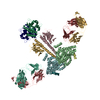
| 初期モデル | PDB ID: Chain - #0 - Chain ID: B / Chain - #1 - Chain ID: D |
|---|---|
| ソフトウェア | 名称: Chimera |
| 詳細 | The domains were fitted by manual docking using Chimera. |
| 精密化 | 空間: REAL / プロトコル: RIGID BODY FIT |
| 得られたモデル | PDB-3jab: PDB-3jbq: |
-原子モデル構築 3
| 初期モデル | PDB ID: Chain - Chain ID: A |
|---|---|
| ソフトウェア | 名称: Chimera |
| 詳細 | The domains were separately fitted by manual docking using Chimera and refined using "Fit in Map" within Chimera. The fitting of GFA(A,B) were confirmed using Folderhunter. |
| 精密化 | 空間: REAL / プロトコル: RIGID BODY FIT |
| 得られたモデル | PDB-3jab: PDB-3jbq: |
-原子モデル構築 4
| 初期モデル | PDB ID: Chain - #0 - Chain ID: H / Chain - #1 - Chain ID: L |
|---|---|
| ソフトウェア | 名称: Chimera |
| 詳細 | The domains were fitted by manual docking using Chimera. |
| 精密化 | 空間: REAL / プロトコル: RIGID BODY FIT |
| 得られたモデル | PDB-3jab: PDB-3jbq: |