ムービー
ムービー コントローラー


+ データを開く

- 基本情報
基本情報
| 登録情報 | データベース: EMDB / ID: EMD-6124 | |||||||||
|---|---|---|---|---|---|---|---|---|---|---|
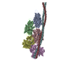
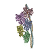
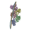
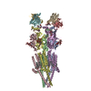
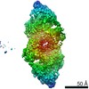
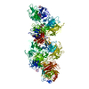
| タイトル | Structure of the F-actin-tropomyosin complex | |||||||||
 マップデータ マップデータ | Reconstruction of F-actin tropomyosin complex | |||||||||
 試料 試料 |
| |||||||||
 キーワード キーワード | F-actin / tropomyosin / filament / protein polymers / muscle / thin filament / cytoskeleton | |||||||||
| 機能・相同性 |  機能・相同性情報 機能・相同性情報positive regulation of heart rate by epinephrine / muscle thin filament tropomyosin / Striated Muscle Contraction / Smooth Muscle Contraction / muscle filament sliding / actin filament capping / ruffle organization / ventricular cardiac muscle tissue morphogenesis / cytoskeletal motor activator activity / myofibril ...positive regulation of heart rate by epinephrine / muscle thin filament tropomyosin / Striated Muscle Contraction / Smooth Muscle Contraction / muscle filament sliding / actin filament capping / ruffle organization / ventricular cardiac muscle tissue morphogenesis / cytoskeletal motor activator activity / myofibril / tropomyosin binding / mesenchyme migration / troponin I binding / myosin heavy chain binding / filamentous actin / actin filament bundle / skeletal muscle thin filament assembly / striated muscle thin filament / actin filament bundle assembly / skeletal muscle myofibril / positive regulation of cell adhesion / actin monomer binding / skeletal muscle fiber development / cardiac muscle contraction / stress fiber / titin binding / positive regulation of stress fiber assembly / actin filament polymerization / negative regulation of cell migration / filopodium / actin filament organization / actin filament / 加水分解酵素; 酸無水物に作用; 酸無水物に作用・細胞または細胞小器官の運動に関与 / wound healing / structural constituent of cytoskeleton / calcium-dependent protein binding / disordered domain specific binding / actin filament binding / actin cytoskeleton / lamellipodium / actin binding / cell body / in utero embryonic development / hydrolase activity / protein heterodimerization activity / protein domain specific binding / calcium ion binding / positive regulation of gene expression / magnesium ion binding / protein homodimerization activity / protein-containing complex / ATP binding / identical protein binding / cytoplasm 類似検索 - 分子機能 | |||||||||
| 生物種 |  | |||||||||
| 手法 | らせん対称体再構成法 / クライオ電子顕微鏡法 / 解像度: 3.7 Å | |||||||||
 データ登録者 データ登録者 | von der Ecken J / Mueller M / Lehman W / Manstein DJ / Penczek PA / Raunser S | |||||||||
 引用 引用 | ジャーナル: Nature / 年: 2015 タイトル: Structure of the F-actin-tropomyosin complex. 著者: Julian von der Ecken / Mirco Müller / William Lehman / Dietmar J Manstein / Pawel A Penczek / Stefan Raunser /   要旨: Filamentous actin (F-actin) is the major protein of muscle thin filaments, and actin microfilaments are the main component of the eukaryotic cytoskeleton. Mutations in different actin isoforms lead ...Filamentous actin (F-actin) is the major protein of muscle thin filaments, and actin microfilaments are the main component of the eukaryotic cytoskeleton. Mutations in different actin isoforms lead to early-onset autosomal dominant non-syndromic hearing loss, familial thoracic aortic aneurysms and dissections, and multiple variations of myopathies. In striated muscle fibres, the binding of myosin motors to actin filaments is mainly regulated by tropomyosin and troponin. Tropomyosin also binds to F-actin in smooth muscle and in non-muscle cells and stabilizes and regulates the filaments there in the absence of troponin. Although crystal structures for monomeric actin (G-actin) are available, a high-resolution structure of F-actin is still missing, hampering our understanding of how disease-causing mutations affect the function of thin muscle filaments and microfilaments. Here we report the three-dimensional structure of F-actin at a resolution of 3.7 Å in complex with tropomyosin at a resolution of 6.5 Å, determined by electron cryomicroscopy. The structure reveals that the D-loop is ordered and acts as a central region for hydrophobic and electrostatic interactions that stabilize the F-actin filament. We clearly identify map density corresponding to ADP and Mg(2+) and explain the possible effect of prominent disease-causing mutants. A comparison of F-actin with G-actin reveals the conformational changes during filament formation and identifies the D-loop as their key mediator. We also confirm that negatively charged tropomyosin interacts with a positively charged groove on F-actin. Comparison of the position of tropomyosin in F-actin-tropomyosin with its position in our previously determined F-actin-tropomyosin-myosin structure reveals a myosin-induced transition of tropomyosin. Our results allow us to understand the role of individual mutations in the genesis of actin- and tropomyosin-related diseases and will serve as a strong foundation for the targeted development of drugs. | |||||||||
| 履歴 |
|
- 構造の表示
構造の表示
| ムービー |
ムービービューア |
|---|---|
| 構造ビューア | EMマップ: SurfViewMolmilJmol/JSmol |
| 添付画像 |

- ダウンロードとリンク
-EMDBアーカイブ
| マップデータ | emd_6124.map.gz | 5.5 MB | EMDBマップデータ形式 | |
|---|---|---|---|---|
| ヘッダ (付随情報) | emd-6124-v30.xmlemd-6124.xml | 11.7 KB 11.7 KB | 表示 表示 | EMDBヘッダ |
| 画像 | 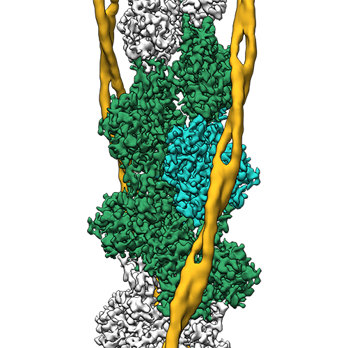 emd_6124.jpg emd_6124.jpg | 210.6 KB | ||
| アーカイブディレクトリ |  http://ftp.pdbj.org/pub/emdb/structures/EMD-6124ftp://ftp.pdbj.org/pub/emdb/structures/EMD-6124 http://ftp.pdbj.org/pub/emdb/structures/EMD-6124ftp://ftp.pdbj.org/pub/emdb/structures/EMD-6124 | HTTPS FTP |
-関連構造データ
-リンク
| EMDBのページ | EMDB (EBI/PDBe) / EMDataResource |
|---|---|
| 「今月の分子」の関連する項目 |


-マップ
| ファイル | ダウンロード / ファイル: emd_6124.map.gz / 形式: CCP4 / 大きさ: 6.4 MB / タイプ: IMAGE STORED AS FLOATING POINT NUMBER (4 BYTES) | ||||||||||||||||||||||||||||||||||||||||||||||||||||||||||||||||||||
|---|---|---|---|---|---|---|---|---|---|---|---|---|---|---|---|---|---|---|---|---|---|---|---|---|---|---|---|---|---|---|---|---|---|---|---|---|---|---|---|---|---|---|---|---|---|---|---|---|---|---|---|---|---|---|---|---|---|---|---|---|---|---|---|---|---|---|---|---|---|
| 注釈 | Reconstruction of F-actin tropomyosin complex | ||||||||||||||||||||||||||||||||||||||||||||||||||||||||||||||||||||
| ボクセルのサイズ | X=Y=Z: 1.12 Å | ||||||||||||||||||||||||||||||||||||||||||||||||||||||||||||||||||||
| 密度 |
| ||||||||||||||||||||||||||||||||||||||||||||||||||||||||||||||||||||
| 対称性 | 空間群: 1 | ||||||||||||||||||||||||||||||||||||||||||||||||||||||||||||||||||||
| 詳細 | EMDB XML:
CCP4マップ ヘッダ情報:
| ||||||||||||||||||||||||||||||||||||||||||||||||||||||||||||||||||||
-添付データ
- 試料の構成要素
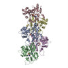
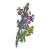
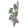
-全体 : F-actin-tropomyosin complex
| 全体 | 名称: F-actin-tropomyosin complex |
|---|---|
| 要素 |
|
-超分子 #1000: F-actin-tropomyosin complex
| 超分子 | 名称: F-actin-tropomyosin complex / タイプ: sample / ID: 1000 / 集合状態: filament / Number unique components: 2 |
|---|
-分子 #1: F-actin (alpha)
| 分子 | 名称: F-actin (alpha) / タイプ: protein_or_peptide / ID: 1 / コピー数: 5 / 集合状態: filament / 組換発現: No / データベース: NCBI |
|---|---|
| 由来(天然) | 生物種: |
| 配列 | UniProtKB: Actin, alpha skeletal muscle |
-分子 #2: tropomyosin (alpha)
| 分子 | 名称: tropomyosin (alpha) / タイプ: protein_or_peptide / ID: 2 / 集合状態: filament (dimer) / 組換発現: Yes |
|---|---|
| 由来(天然) | 生物種: |
| 組換発現 | 生物種:  |
| 配列 | UniProtKB: Tropomyosin alpha-1 chain |
-実験情報
-構造解析
| 手法 | クライオ電子顕微鏡法 |
|---|---|
 解析 解析 | らせん対称体再構成法 |
| 試料の集合状態 | filament |
-試料調製
| 緩衝液 | pH: 7.5 / 詳細: 5 mM Tris, 1 mM DTT, 100 mM KCL, 2 mM MgCl2 |
|---|---|
| グリッド | 詳細: C-flats 2/1 copper 300 mesh, Protochips, glow-discharged |
| 凍結 | 凍結剤: ETHANE / チャンバー内湿度: 90 % / チャンバー内温度: 106 K / 装置: GATAN CRYOPLUNGE 3 手法: Sample was applied to grid, incubated for 10 seconds, and manually blotted for 3 seconds from the backside with filter paper. |
- 電子顕微鏡法
| 顕微鏡 | FEI TITAN KRIOS |
|---|---|
| Cs | 0 |
| 詳細 | Cs-corrected microscope |
| 日付 | 2013年10月17日 |
| 撮影 | カテゴリ: CCD フィルム・検出器のモデル: FEI FALCON II (4k x 4k) 実像数: 1311 / 平均電子線量: 14.6 e/Å2 詳細: Every image is the average of seven frames recorded by the direct electron detector after beam-induced motion correction. Only 689 images were used for the refinement. |
| 電子線 | 加速電圧: 300 kV / 電子線源:  FIELD EMISSION GUN FIELD EMISSION GUN |
| 電子光学系 | 照射モード: FLOOD BEAM / 撮影モード: BRIGHT FIELD / 最大 デフォーカス(公称値): 2.6 µm / 最小 デフォーカス(公称値): 0.8 µm / 倍率(公称値): 59000 / Cs: mm |
| 試料ステージ | 試料ホルダーモデル: FEI TITAN KRIOS AUTOGRID HOLDER |
| 実験機器 |  モデル: Titan Krios / 画像提供: FEI Company |
-画像解析
| 最終 再構成 | 想定した対称性 - らせんパラメータ - Δz: 27.5 Å 想定した対称性 - らせんパラメータ - ΔΦ: 166.4 ° 想定した対称性 - らせんパラメータ - 軸対称性: C1 (非対称) アルゴリズム: OTHER / 解像度のタイプ: BY AUTHOR / 解像度: 3.7 Å / 解像度の算出法: OTHER / ソフトウェア - 名称: SPARX 詳細: The tropomyosin map filtered to 6.5 Angstrom was merged with the final F-actin map (3.7 Angstrom) to obtain a map of the entire F-actin tropomyosin complex. |
|---|---|
| CTF補正 | 詳細: each micrograph |
-原子モデル構築 1
| 初期モデル | PDB ID: Chain - #0 - Chain ID: A / Chain - #1 - Chain ID: B / Chain - #2 - Chain ID: C / Chain - #3 - Chain ID: D / Chain - #4 - Chain ID: E |
|---|---|
| ソフトウェア | 名称: SPARX, PHENIX, COOT |
| 精密化 | 空間: RECIPROCAL / プロトコル: FLEXIBLE FIT / 温度因子: 55.4 / 当てはまり具合の基準: R-factor |
| 得られたモデル |  PDB-3j8a: |