+ Open data
- Basic information
| Entry |  | |||||||||||||||
|---|---|---|---|---|---|---|---|---|---|---|---|---|---|---|---|---|
| Title | Structure of the bacteriophage T5 tail tip complex | |||||||||||||||
 Map data Map data | ||||||||||||||||
 Sample Sample |
| |||||||||||||||
 Keywords Keywords | Complex / VIRAL PROTEIN | |||||||||||||||
| Function / homology |  Function and homology information Function and homology information | |||||||||||||||
| Biological species |  Escherichia phage T5 (virus) Escherichia phage T5 (virus) | |||||||||||||||
| Method | single particle reconstruction / cryo EM / Resolution: 3.9 Å | |||||||||||||||
 Authors Authors | Peng YN / Liu HR | |||||||||||||||
| Funding support |  China, 4 items China, 4 items
| |||||||||||||||
 Citation Citation |  Journal: Int J Mol Sci / Year: 2024 Journal: Int J Mol Sci / Year: 2024Title: Structures of Mature and Urea-Treated Empty Bacteriophage T5: Insights into Siphophage Infection and DNA Ejection. Authors: Yuning Peng / Huanrong Tang / Hao Xiao / Wenyuan Chen / Jingdong Song / Jing Zheng / Hongrong Liu /  Abstract: T5 is a siphophage that has been extensively studied by structural and biochemical methods. However, the complete in situ structures of T5 before and after DNA ejection remain unknown. In this study, ...T5 is a siphophage that has been extensively studied by structural and biochemical methods. However, the complete in situ structures of T5 before and after DNA ejection remain unknown. In this study, we used cryo-electron microscopy (cryo-EM) to determine the structures of mature T5 (a laboratory-adapted, fiberless T5 mutant) and urea-treated empty T5 (lacking the tip complex) at near-atomic resolutions. Atomic models of the head, connector complex, tail tube, and tail tip were built for mature T5, and atomic models of the connector complex, comprising the portal protein pb7, adaptor protein p144, and tail terminator protein p142, were built for urea-treated empty T5. Our findings revealed that the aforementioned proteins did not undergo global conformational changes before and after DNA ejection, indicating that these structural features were conserved among most myophages and siphophages. The present study elucidates the underlying mechanisms of siphophage infection and DNA ejection. | |||||||||||||||
| History |
|
- Structure visualization
| Supplemental images |
|---|
- Downloads & links
-EMDB archive
| Map data |  emd_60750.map.gz emd_60750.map.gz | 28.5 MB |  EMDB map data format EMDB map data format | |
|---|---|---|---|---|
| Header (meta data) |  emd-60750-v30.xml emd-60750-v30.xml emd-60750.xml emd-60750.xml | 18.9 KB 18.9 KB | Display Display |  EMDB header EMDB header |
| Images |  emd_60750.png emd_60750.png | 64 KB | ||
| Filedesc metadata |  emd-60750.cif.gz emd-60750.cif.gz | 6.2 KB | ||
| Others |  emd_60750_half_map_1.map.gz emd_60750_half_map_1.map.gz emd_60750_half_map_2.map.gz emd_60750_half_map_2.map.gz | 23.5 MB 23.5 MB | ||
| Archive directory |  http://ftp.pdbj.org/pub/emdb/structures/EMD-60750 http://ftp.pdbj.org/pub/emdb/structures/EMD-60750 ftp://ftp.pdbj.org/pub/emdb/structures/EMD-60750 ftp://ftp.pdbj.org/pub/emdb/structures/EMD-60750 | HTTPS FTP |
-Related structure data
| Related structure data | 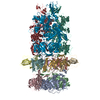 9iozMC 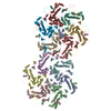 8zviC 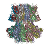 9ilpC 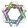 9ilvC 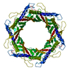 9imhC 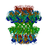 9imvC 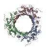 9inyC M: atomic model generated by this map C: citing same article ( |
|---|---|
| Similar structure data | Similarity search - Function & homology  F&H Search F&H Search |
- Links
Links
| EMDB pages |  EMDB (EBI/PDBe) / EMDB (EBI/PDBe) /  EMDataResource EMDataResource |
|---|
- Map
Map
| File |  Download / File: emd_60750.map.gz / Format: CCP4 / Size: 30.5 MB / Type: IMAGE STORED AS FLOATING POINT NUMBER (4 BYTES) Download / File: emd_60750.map.gz / Format: CCP4 / Size: 30.5 MB / Type: IMAGE STORED AS FLOATING POINT NUMBER (4 BYTES) | ||||||||||||||||||||||||||||||||||||
|---|---|---|---|---|---|---|---|---|---|---|---|---|---|---|---|---|---|---|---|---|---|---|---|---|---|---|---|---|---|---|---|---|---|---|---|---|---|
| Projections & slices | Image control
Images are generated by Spider. | ||||||||||||||||||||||||||||||||||||
| Voxel size | X=Y=Z: 1.3 Å | ||||||||||||||||||||||||||||||||||||
| Density |
| ||||||||||||||||||||||||||||||||||||
| Symmetry | Space group: 1 | ||||||||||||||||||||||||||||||||||||
| Details | EMDB XML:
|
-Supplemental data
-Half map: #2
| File | emd_60750_half_map_1.map | ||||||||||||
|---|---|---|---|---|---|---|---|---|---|---|---|---|---|
| Projections & Slices |
| ||||||||||||
| Density Histograms |
-Half map: #1
| File | emd_60750_half_map_2.map | ||||||||||||
|---|---|---|---|---|---|---|---|---|---|---|---|---|---|
| Projections & Slices |
| ||||||||||||
| Density Histograms |
- Sample components
-Entire : Escherichia phage T5
| Entire | Name:  Escherichia phage T5 (virus) Escherichia phage T5 (virus) |
|---|---|
| Components |
|
-Supramolecule #1: Escherichia phage T5
| Supramolecule | Name: Escherichia phage T5 / type: virus / ID: 1 / Parent: 0 / Macromolecule list: all / NCBI-ID: 2695836 / Sci species name: Escherichia phage T5 / Virus type: VIRION / Virus isolate: SPECIES / Virus enveloped: No / Virus empty: No |
|---|
-Macromolecule #1: Baseplate tube protein p140
| Macromolecule | Name: Baseplate tube protein p140 / type: protein_or_peptide / ID: 1 / Number of copies: 3 / Enantiomer: LEVO |
|---|---|
| Source (natural) | Organism:  Escherichia phage T5 (virus) Escherichia phage T5 (virus) |
| Molecular weight | Theoretical: 34.367605 KDa |
| Sequence | String: MFYSLMRESK IVIEYDGRGY HFDALSNYDA STSFQEFKTL RRTIHNRTNY ADSIINAQDP SSISLAINFS TTLIESNFFD WMGFTREGN SLFLPRNTPN IEPIMFNMYI INHNNSCIYF ENCYVSTVDF SLDKSIPILN VGIESGKFSE VSTFRDGYTI T QGEVLPYS ...String: MFYSLMRESK IVIEYDGRGY HFDALSNYDA STSFQEFKTL RRTIHNRTNY ADSIINAQDP SSISLAINFS TTLIESNFFD WMGFTREGN SLFLPRNTPN IEPIMFNMYI INHNNSCIYF ENCYVSTVDF SLDKSIPILN VGIESGKFSE VSTFRDGYTI T QGEVLPYS APAVYTNSSP LPALISASMS FQQQCSWRED RNIFDINKIY TNKRAYVNEM NASATLAFYY VKRLVGDKFL NL DPETRTP LIIKNKYVSI TFPLARISKR LNFSDLYQVE YDVIPTADSD PVEINFFGER K UniProtKB: Baseplate tube protein p140 |
-Macromolecule #2: Distal tail protein pb9
| Macromolecule | Name: Distal tail protein pb9 / type: protein_or_peptide / ID: 2 / Number of copies: 6 / Enantiomer: LEVO |
|---|---|
| Source (natural) | Organism:  Escherichia phage T5 (virus) Escherichia phage T5 (virus) |
| Molecular weight | Theoretical: 22.798641 KDa |
| Sequence | String: MRLPDPYTNP EYPGLGFESV NLVDNDPMIR DELPNGKVKE VKISAQYWGI NISYPELFPD EYAFLDSRLL EYKRTGDYLD VLLPQYEAF RVRGDTKSVT IPAGQKGSQI ILNTNGTLTG QPKAGDLFKL STHPKVYKIT NFSSSGNVWN ISLYPDLFIT T TGSEKPVF ...String: MRLPDPYTNP EYPGLGFESV NLVDNDPMIR DELPNGKVKE VKISAQYWGI NISYPELFPD EYAFLDSRLL EYKRTGDYLD VLLPQYEAF RVRGDTKSVT IPAGQKGSQI ILNTNGTLTG QPKAGDLFKL STHPKVYKIT NFSSSGNVWN ISLYPDLFIT T TGSEKPVF NGILFRTKLM NGDSFGSTLN NNGTYSGISL SLRESL UniProtKB: Distal tail protein pb9 |
-Macromolecule #3: Baseplate hub protein pb3
| Macromolecule | Name: Baseplate hub protein pb3 / type: protein_or_peptide / ID: 3 / Number of copies: 3 / Enantiomer: LEVO |
|---|---|
| Source (natural) | Organism:  Escherichia phage T5 (virus) Escherichia phage T5 (virus) |
| Molecular weight | Theoretical: 107.279766 KDa |
| Sequence | String: MKKILDSAKN YLNTHDKLKT ACLIALELPS SSGSAATYIY LTDYFRDVTY NGILYRSGKV KSISSHKQNR QLSIGSLSFT ITGTAEDEV LKLVQNGVSF LDRGITIHQA IINEEGNILP VDPDTDGPLL FFRGRITGGG IKDNVNTSGI GTSVITWNCS N QFYDFDRV ...String: MKKILDSAKN YLNTHDKLKT ACLIALELPS SSGSAATYIY LTDYFRDVTY NGILYRSGKV KSISSHKQNR QLSIGSLSFT ITGTAEDEV LKLVQNGVSF LDRGITIHQA IINEEGNILP VDPDTDGPLL FFRGRITGGG IKDNVNTSGI GTSVITWNCS N QFYDFDRV NGRYTDDASH RGLEVVNGTL QPSNGAKRPE YQEDYGFFHS NKSTTILAKY QVKEERYKLQ SKKKLFGLSR SY SLKKYYE TVTKEVDLDF NLAAKFIPVV YGVQKIPGIP IFADTELNNP NIVYVVYAFA EGEIDGFLDF YIGDSPMICF DET DSDTRT CFGRKKIVGD TMHRLAAGTS TSQPSVHGQE YKYNDGNGDI RIWTFHGKPD QTAAQVLVDI AKKKGFYLQN QNGN GPEYW DSRYKLLDTA YAIVRFTINE NRTEIPEISA EVQGKKVKVY NSDGTIKADK TSLNGIWQLM DYLTSDRYGA DITLD QFPL QKVISEAKIL DIIDESYQTS WQPYWRYVGW NDPLSENRQI VQLNTILDTS ESVFKNVQGI LESFGGAINN LSGEYR ITV EKYSTNPLRI NFLDTYGDLD LSDTTGRNKF NSVQASLVDP ALSWKTNSIT FYNSKFKEQD KGLDKKLQLS FANITNY YT ARSYADRELK KSRYSRTLSF SVPYKFIGIE PNDPIAFTYE RYGWKDKFFL VDEVENTRDG KINLVLQEYG EDVFINSE Q VDNSGNDIPD ISNNVLPPRD FKYTPTPGGV VGAIGKNGEL SWLPSLTNNV VYYSIAHSGH VNPYIVQQLE NNPNERMIQ EIIGEPAGLA IFELRAVDIN GRRSSPVTLS VDLNSAKNLS VVSNFRVVNT ASGDVTEFVG PDVKLAWDKI PEEEIIPEIY YTLEIYDSQ DRMLRSVRIE DVYTYDYLLT YNKADFALLN SGALGINRKL RFRIRAEGEN GEQSVGWATI UniProtKB: Baseplate hub protein pb3 |
-Experimental details
-Structure determination
| Method | cryo EM |
|---|---|
 Processing Processing | single particle reconstruction |
| Aggregation state | particle |
- Sample preparation
| Buffer | pH: 7.4 |
|---|---|
| Vitrification | Cryogen name: ETHANE |
- Electron microscopy
| Microscope | FEI TITAN KRIOS |
|---|---|
| Image recording | Film or detector model: GATAN K3 (6k x 4k) / Average electron dose: 32.0 e/Å2 |
| Electron beam | Acceleration voltage: 300 kV / Electron source:  FIELD EMISSION GUN FIELD EMISSION GUN |
| Electron optics | Illumination mode: FLOOD BEAM / Imaging mode: BRIGHT FIELD / Nominal defocus max: 2.2 µm / Nominal defocus min: 1.6 µm |
| Experimental equipment |  Model: Titan Krios / Image courtesy: FEI Company |
 Movie
Movie Controller




 Z (Sec.)
 Y (Row.)
 X (Col.)


