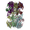
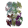
MCM core complex / Assembly of the pre-replicative complex / Switching of origins to a post-replicative state / nuclear DNA replication / MCM complex binding / premeiotic DNA replication / pre-replicative complex assembly involved in nuclear cell cycle DNA replication / Activation of the pre-replicative complex / CMG complex / nuclear pre-replicative complex ...MCM core complex / Assembly of the pre-replicative complex / Switching of origins to a post-replicative state / nuclear DNA replication / MCM complex binding / premeiotic DNA replication / pre-replicative complex assembly involved in nuclear cell cycle DNA replication / Activation of the pre-replicative complex / CMG complex / nuclear pre-replicative complex / Activation of ATR in response to replication stress / DNA replication preinitiation complex / single-stranded DNA helicase activity / MCM complex / mitotic DNA replication initiation / mitotic DNA replication / silent mating-type cassette heterochromatin formation / double-strand break repair via break-induced replication / DNA strand elongation involved in DNA replication / nuclear replication fork / DNA replication origin binding / subtelomeric heterochromatin formation / DNA replication initiation / regulation of DNA-templated DNA replication initiation / helicase activity / transcription elongation by RNA polymerase II / peroxisome / single-stranded DNA binding / heterochromatin formation / DNA helicase / DNA replication / DNA helicase activity / chromosome, telomeric region / chromatin binding / DNA damage response / ATP hydrolysis activity / nucleoplasm / ATP binding / nucleus / cytoplasm Similarity search - Function
Biotechnology and Biological Sciences Research Council (BBSRC)
BB/T005378/1
United Kingdom
Cancer Research UK
DRCNPG-May21/100006
United Kingdom
Citation
Journal: Nat Commun / Year: 2026 Title: Structural insights into Sld3-Sld7-dependent Cdc45 loading during replication initiation. Authors: Yasunori Noguchi / Almutasem Saleh / Sarah Schneider / Marina E Ivanova / Zhuo Angel Chen / Lepakshi Ranjha / Ricardo Aramayo / Silvia Tognetti / Sarah V Faull / Juri Rappsilber / Christian Speck / Abstract: Regulated helicase activation by DDK kinase is central for genome stability. However, how DDK phosphorylation primes the MCM2-7 double hexamer (DH) for Sld3-Sld7 binding and Cdc45 loading remained ...Regulated helicase activation by DDK kinase is central for genome stability. However, how DDK phosphorylation primes the MCM2-7 double hexamer (DH) for Sld3-Sld7 binding and Cdc45 loading remained unclear. We define this mechanism through cryo-EM structures of MCM2-7 DH-Sld3-Sld7 (MS) and MCM2-7 DH-Sld3-Sld7-Cdc45 (MSC). We reveal that the autoinhibitory Mcm4 tail engages not only Mcm4 but also Mcm6. Upon DDK-dependent phosphorylation, both of these sites become accessible. In the context of the MS structure, we identify that two short Sld3 motifs that contact Mcm4 and Mcm6 read out the DH phosphorylation state, while the Sld3 Treslin domain (STD) binds to Mcm2. In the MSC structure, Cdc45 dislodges the Sld3 STD from Mcm2, allowing Sld3 to position Cdc45 at the Mcm2/Mcm5 interface. Mutagenesis of the Sld3 STD-Cdc45 interface disrupts Cdc45 loading, validating this interaction. Together, our data reveal a phosphorylation-encoded mechanism coupling DDK-activated Mcm4/Mcm6 surfaces to distal Cdc45 placement, explaining how firing factors choreograph the DH-to-CMG transition.
In the structure databanks used in Yorodumi, some data are registered as the other names, "COVID-19 virus" and "2019-nCoV". Here are the details of the virus and the list of structure data.
Jan 31, 2019. EMDB accession codes are about to change! (news from PDBe EMDB page)
EMDB accession codes are about to change! (news from PDBe EMDB page)
The allocation of 4 digits for EMDB accession codes will soon come to an end. Whilst these codes will remain in use, new EMDB accession codes will include an additional digit and will expand incrementally as the available range of codes is exhausted. The current 4-digit format prefixed with “EMD-” (i.e. EMD-XXXX) will advance to a 5-digit format (i.e. EMD-XXXXX), and so on. It is currently estimated that the 4-digit codes will be depleted around Spring 2019, at which point the 5-digit format will come into force.
The EM Navigator/Yorodumi systems omit the EMD- prefix.
Related info.:Q: What is EMD? / ID/Accession-code notation in Yorodumi/EM Navigator
Yorodumi is a browser for structure data from EMDB, PDB, SASBDB, etc.
This page is also the successor to EM Navigator detail page, and also detail information page/front-end page for Omokage search.
The word "yorodu" (or yorozu) is an old Japanese word meaning "ten thousand". "mi" (miru) is to see.
Related info.:EMDB / PDB / SASBDB / Comparison of 3 databanks / Yorodumi Search / Aug 31, 2016. New EM Navigator & Yorodumi / Yorodumi Papers / Jmol/JSmol / Function and homology information / Changes in new EM Navigator and Yorodumi
 Movie
Movie Controller


 Open data
 Basic information
 Map data
 Sample
 Keywords
 Function and homology information
 Saccharomyces cerevisiae (strain ATCC 204508 / S288c) (yeast) /
  Authors
 United Kingdom, 3 items
  Citation

 Structure visualization
 Downloads & links
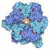
 emd_55897.png
 https://data.pdbj.org/pub/emdb/structures/EMD-55897


 X (Sec.)
 Y (Row.)
 Z (Col.)


 Sample components

 Processing
 Electron microscopy
 FIELD EMISSION GUN