Movie
Movie Controller

[English] 日本語
 Yorodumi
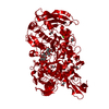
- EMDB-53103: Structure of the GH13 and MucBP domains of Ruminococcus bromii Amy12 -
+ Open data

- Basic information
| Entry |  | |||||||||
|---|---|---|---|---|---|---|---|---|---|---|
| Title | Structure of the GH13 and MucBP domains of Ruminococcus bromii Amy12 | |||||||||
 Map data Map data | ||||||||||
 Sample Sample |
| |||||||||
 Keywords Keywords | GH13 / Ruminococcus bromii / Amy12 / MucBP / HYDROLASE | |||||||||
| Biological species |  Ruminococcus bromii L2-63 (bacteria) Ruminococcus bromii L2-63 (bacteria) | |||||||||
| Method | single particle reconstruction / cryo EM / Resolution: 3.1 Å | |||||||||
 Authors Authors | Wimmer BH / Medalia O | |||||||||
| Funding support |  Switzerland, 1 items Switzerland, 1 items
| |||||||||
 Citation Citation | Journal: Nat Commun / Year: 2025 Title: Spatial constraints drive amylosome-mediated resistant starch degradation by Ruminococcus bromii in the human colon. Authors: Benedikt H Wimmer / Sarah Moraïs / Itai Amit / Omar Tovar-Herrera / Meltem Tatli / Anke Trautwein-Schult / Barbara Pfister / Ran Zalk / Paloma Tödtli / Sebastian Simoni / Matteo Lisibach / ...Authors: Benedikt H Wimmer / Sarah Moraïs / Itai Amit / Omar Tovar-Herrera / Meltem Tatli / Anke Trautwein-Schult / Barbara Pfister / Ran Zalk / Paloma Tödtli / Sebastian Simoni / Matteo Lisibach / Liron Levin / Dörte Becher / Edward A Bayer / Ohad Medalia / Itzhak Mizrahi /   Abstract: Degradation of complex dietary fiber by gut microbes is essential for colonic fermentation, short-chain fatty acid production, and microbiome function. Ruminococcus bromii is the primary resistant ...Degradation of complex dietary fiber by gut microbes is essential for colonic fermentation, short-chain fatty acid production, and microbiome function. Ruminococcus bromii is the primary resistant starch (RS) degrader in humans, which relies on the amylosome, a specialized cell-bound enzymatic complex. To unravel its architecture, function, and the interplay among its components, we applied a holistic multilayered approach: Cryo-electron tomography reveals that the amylosome comprises a constitutive extracellular layer extending toward the RS substrate. Proteomics demonstrates remodeling of its contents across different growth conditions, with Amy4 and Amy16 comprising 60% of the amylosome in response to RS. Structural and biochemical analyses reveal complementarity and synergistic RS degradation by these enzymes. We demonstrate that amylosome composition and RS degradation are regulated at two levels: structural constraints and expression-driven shifts in enzyme proportions enforce enzyme proximity, which allows R. bromii to fine-tune its adaptation to dietary fiber and shape colonic metabolism. | |||||||||
| History |
|
- Structure visualization
| Supplemental images |
|---|

- Downloads & links
-EMDB archive
| Map data | emd_53103.map.gz | 229.9 MB |  EMDB map data format EMDB map data format | |
|---|---|---|---|---|
| Header (meta data) | emd-53103-v30.xmlemd-53103.xml | 22.8 KB 22.8 KB | Display Display | EMDB header |
| FSC (resolution estimation) | emd_53103_fsc.xml | 15 KB | Display | FSC data file |
| Images |  emd_53103.png emd_53103.png | 41 KB | ||
| Masks | emd_53103_msk_1.map | 244.1 MB | Mask map | |
| Filedesc metadata | emd-53103.cif.gz | 7.2 KB | ||
| Others | emd_53103_half_map_1.map.gzemd_53103_half_map_2.map.gz | 226.8 MB 226.8 MB | ||
| Archive directory |  http://ftp.pdbj.org/pub/emdb/structures/EMD-53103ftp://ftp.pdbj.org/pub/emdb/structures/EMD-53103 http://ftp.pdbj.org/pub/emdb/structures/EMD-53103ftp://ftp.pdbj.org/pub/emdb/structures/EMD-53103 | HTTPS FTP |
-Related structure data
| Related structure data |  9qfaMC  9qf3C  9qf8C  9qf9C M: atomic model generated by this map C: citing same article ( |
|---|


-Links
| EMDB pages | EMDB (EBI/PDBe) / EMDataResource |
|---|
-Map
| File | Download / File: emd_53103.map.gz / Format: CCP4 / Size: 244.1 MB / Type: IMAGE STORED AS FLOATING POINT NUMBER (4 BYTES) | ||||||||||||||||||||||||||||||||||||
|---|---|---|---|---|---|---|---|---|---|---|---|---|---|---|---|---|---|---|---|---|---|---|---|---|---|---|---|---|---|---|---|---|---|---|---|---|---|
| Projections & slices | Image control
Images are generated by Spider. | ||||||||||||||||||||||||||||||||||||
| Voxel size | X=Y=Z: 0.651 Å | ||||||||||||||||||||||||||||||||||||
| Density |
| ||||||||||||||||||||||||||||||||||||
| Symmetry | Space group: 1 | ||||||||||||||||||||||||||||||||||||
| Details | EMDB XML:
|
 Z (Sec.)
 Y (Row.)
 X (Col.)


-Supplemental data
-Mask #1
| File | emd_53103_msk_1.map | ||||||||||||
|---|---|---|---|---|---|---|---|---|---|---|---|---|---|
| Projections & Slices |
| ||||||||||||
| Density Histograms |


-Half map: #2
| File | emd_53103_half_map_1.map | ||||||||||||
|---|---|---|---|---|---|---|---|---|---|---|---|---|---|
| Projections & Slices |
| ||||||||||||
| Density Histograms |


-Half map: #1
| File | emd_53103_half_map_2.map | ||||||||||||
|---|---|---|---|---|---|---|---|---|---|---|---|---|---|
| Projections & Slices |
| ||||||||||||
| Density Histograms |


- Sample components
-Entire : Ruminococcus bromii Amy12
| Entire | Name: Ruminococcus bromii Amy12 |
|---|---|
| Components |
|
-Supramolecule #1: Ruminococcus bromii Amy12
| Supramolecule | Name: Ruminococcus bromii Amy12 / type: complex / ID: 1 / Parent: 0 / Macromolecule list: all |
|---|---|
| Source (natural) | Organism: Ruminococcus bromii L2-63 (bacteria) |
| Molecular weight | Theoretical: 114.5 KDa |
-Macromolecule #1: Ruminococcus bromii Amy12
| Macromolecule | Name: Ruminococcus bromii Amy12 / type: protein_or_peptide / ID: 1 Details: removal of signalling peptide for secretion, addition of a N-terminal 6x His-Tag Number of copies: 1 / Enantiomer: LEVO / EC number: pullulanase |
|---|---|
| Source (natural) | Organism: Ruminococcus bromii L2-63 (bacteria) |
| Molecular weight | Theoretical: 114.603805 KDa |
| Recombinant expression | Organism: |
| Sequence | String: MAHHHHHHAT ADDSSAVSSD YARDNSYTKA AEDIDAQYAY SGNDLGVTYT KDATTFKVWS PTATGVKLNI FTKGSDDEQG ASKVASYTL EKMLVDGEWN GVWTITLVGE WKDYYYTYSV TTTDTTHIGS DATKTYETQD VYSTATGVNG KRSMIVDLDE T DPEGWSND ...String: MAHHHHHHAT ADDSSAVSSD YARDNSYTKA AEDIDAQYAY SGNDLGVTYT KDATTFKVWS PTATGVKLNI FTKGSDDEQG ASKVASYTL EKMLVDGEWN GVWTITLVGE WKDYYYTYSV TTTDTTHIGS DATKTYETQD VYSTATGVNG KRSMIVDLDE T DPEGWSND SHVLLDKSTK SSVWELHIKD FSYDKASGVS DANRGKYLAF TENGTTLNGE GKVSTCIDYL KELGVTTVQL NP FYDFQSV NEAGDDSQFN WGYDPVNYNV PEGSYSSNPY DGKVRIKECK EMIKALHDAG ISVVMDVVYN HTYSTDSCFQ YTV PNYYYR MKTTGAFSDG SGCGNEGATE RAMYRQYVID SLKYWVNEYH VDGFRFDLMG LMDVETMNMA REALDQIDPR ITMW GEGWA GGDSYHPTNT CSGTKFYPAT QANASRLSDR IAIFNDGIRD GIKGSAMDIS DVGFIQGSKS SAKGVSYGVR ANSSG TYKW KAQAPSQCVT YDACHDNATL YDQIIASTGL ADYGERNSEA VKMNRLASAI IYTSQGISFT LAGEEMARSK DGDTNS YKS AANLNMIKWQ NVVDYADVVS YYKGMMQIKS AFSPLTAMDN SYADKYTFTK KVSASTNQIS FTIQNDVEGE WNKMAVI YN NATTAADVTL SDTSVTDWVV IANGETAGLD SLGEVTGSTF TVPARSAIVA VDKAGYESAG IHSSKGKVKV NYVYEATG E KLEDSVILQG SVGSGYVTVP SAVIPDTYIV SRIGGNAEGK YTSDMQEVTY YYTDYIPESL KNADFNGDGE INVIDATLL QKYIAKLETP TVDESTLDLN YDGTFNIEDS TMIMKYVARI PVSSGKVTVN YYYTDADGKQ QKLTDSIVFA GRAGSTYKST AFKVVGYAV DPDRMPENQS GLIPYGEAEV NYYYIASSLD IKLHVKHNGS LTWTPYLWIW GSDLKGKDSG NFMSEWPGDP M TEGENGWF DYSFTYKGAG TYNVIVSDNA TNQTIDYKGF VDNEMWIVID DSAVMGSTYL TFYTDNPDTN PNAPIAEQVT LG |
-Experimental details
-Structure determination
| Method | cryo EM |
|---|---|
 Processing Processing | single particle reconstruction |
| Aggregation state | particle |
-Sample preparation
| Concentration | 0.08 mg/mL | ||||||||||||||||||
|---|---|---|---|---|---|---|---|---|---|---|---|---|---|---|---|---|---|---|---|
| Buffer | pH: 7.4 Component:
| ||||||||||||||||||
| Grid | Model: Quantifoil R1.2/1.3 / Material: GOLD / Mesh: 200 / Support film - Material: CARBON / Support film - topology: HOLEY / Pretreatment - Type: GLOW DISCHARGE / Pretreatment - Time: 45 sec. | ||||||||||||||||||
| Vitrification | Cryogen name: ETHANE / Chamber humidity: 100 % / Chamber temperature: 277 K / Instrument: FEI VITROBOT MARK IV |
- Electron microscopy
| Microscope | TFS KRIOS |
|---|---|
| Image recording | Film or detector model: GATAN K3 (6k x 4k) / Number grids imaged: 2 / Number real images: 27350 / Average electron dose: 65.0 e/Å2 |
| Electron beam | Acceleration voltage: 300 kV / Electron source:  FIELD EMISSION GUN FIELD EMISSION GUN |
| Electron optics | C2 aperture diameter: 50.0 µm / Illumination mode: FLOOD BEAM / Imaging mode: BRIGHT FIELD / Cs: 2.7 mm / Nominal defocus max: 2.4 µm / Nominal defocus min: 0.8 µm / Nominal magnification: 130000 |
| Sample stage | Specimen holder model: FEI TITAN KRIOS AUTOGRID HOLDER / Cooling holder cryogen: NITROGEN |
| Experimental equipment |  Model: Titan Krios / Image courtesy: FEI Company |