 ムービー
ムービー コントローラー


+ データを開く

- 基本情報
基本情報
| 登録情報 | データベース: EMDB / ID: EMD-5235 | |||||||||
|---|---|---|---|---|---|---|---|---|---|---|
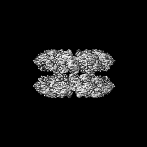
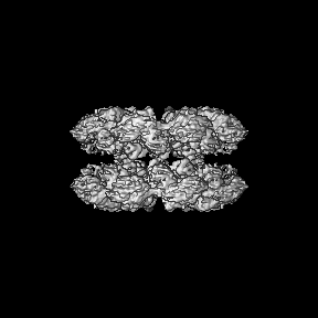
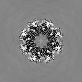
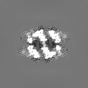
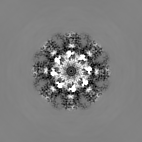
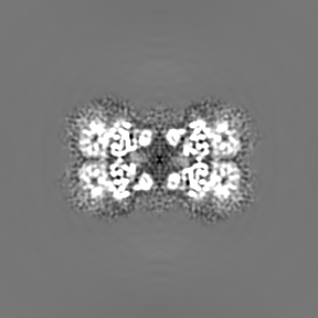
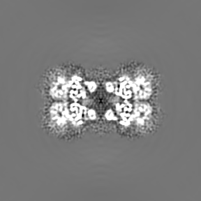
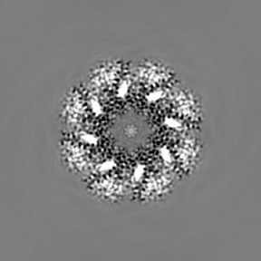
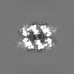
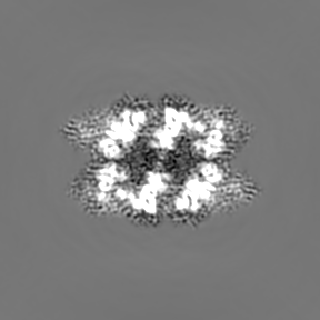
| タイトル | Structure of the Drosophila apoptosome | |||||||||
 マップデータ マップデータ | map of the Drosophila apoptosome | |||||||||
 試料 試料 |
| |||||||||
 キーワード キーワード | Drosophila apoptosome / apoptosis / programmed cell death | |||||||||
| 機能・相同性 |  機能・相同性情報 機能・相同性情報negative regulation of humoral immune response / positive regulation of glial cell apoptotic process / Formation of apoptosome / salivary gland histolysis / melanization defense response / : / Activation of caspases through apoptosome-mediated cleavage / Regulation of the apoptosome activity / compound eye retinal cell programmed cell death / central nervous system formation ...negative regulation of humoral immune response / positive regulation of glial cell apoptotic process / Formation of apoptosome / salivary gland histolysis / melanization defense response / : / Activation of caspases through apoptosome-mediated cleavage / Regulation of the apoptosome activity / compound eye retinal cell programmed cell death / central nervous system formation / positive regulation of apoptotic process involved in development / sperm individualization / apoptosome / autophagic cell death / chaeta development / Neutrophil degranulation / L-methionine cycle / CARD domain binding / programmed cell death / triglyceride homeostasis / dendrite morphogenesis / response to starvation / response to gamma radiation / neuron cellular homeostasis / ADP binding / apoptotic process / structural molecule activity / ATP binding / identical protein binding 類似検索 - 分子機能 | |||||||||
| 生物種 |  | |||||||||
| 手法 | 単粒子再構成法 / クライオ電子顕微鏡法 / 解像度: 6.9 Å | |||||||||
 データ登録者 データ登録者 | Yuan S / Yu X / Topf M / Ludtke SJ / Akey CW | |||||||||
 引用 引用 | ジャーナル: Structure / 年: 2011 タイトル: Structure of the Drosophila apoptosome at 6.9 å resolution. 著者: Shujun Yuan / Xinchao Yu / Maya Topf / Loretta Dorstyn / Sharad Kumar / Steven J Ludtke / Christopher W Akey /  要旨: The Drosophila Apaf-1 related killer forms an apoptosome in the intrinsic cell death pathway. In this study we show that Dark forms a single ring when initiator procaspases are bound. This Dark-Dronc ...The Drosophila Apaf-1 related killer forms an apoptosome in the intrinsic cell death pathway. In this study we show that Dark forms a single ring when initiator procaspases are bound. This Dark-Dronc complex cleaves DrICE efficiently; hence, a single ring represents the Drosophila apoptosome. We then determined the 3D structure of a double ring at ∼6.9 Å resolution and created a model of the apoptosome. Subunit interactions in the Dark complex are similar to those in Apaf-1 and CED-4 apoptosomes, but there are significant differences. In particular, Dark has "lost" a loop in the nucleotide-binding pocket, which opens a path for possible dATP exchange in the apoptosome. In addition, caspase recruitment domains (CARDs) form a crown on the central hub of the Dark apoptosome. This CARD geometry suggests that conformational changes will be required to form active Dark-Dronc complexes. When taken together, these data provide insights into apoptosome structure, function, and evolution. | |||||||||
| 履歴 |
|
- 構造の表示
構造の表示
| ムービー |
ムービービューア |
|---|---|
| 構造ビューア | EMマップ: SurfViewMolmilJmol/JSmol |
| 添付画像 |

- ダウンロードとリンク
-EMDBアーカイブ
| マップデータ | emd_5235.map.gz | 83.8 MB | EMDBマップデータ形式 | |
|---|---|---|---|---|
| ヘッダ (付随情報) | emd-5235-v30.xmlemd-5235.xml | 11.2 KB 11.2 KB | 表示 表示 | EMDBヘッダ |
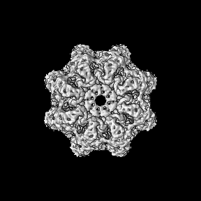
| 画像 | 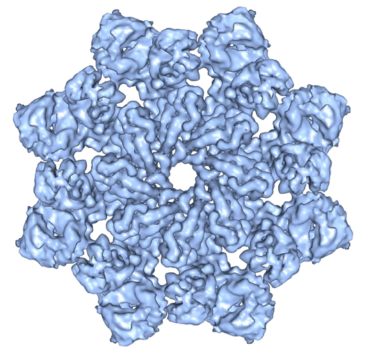 emd_5235_1.jpg emd_5235_1.jpg | 208.3 KB | ||
| アーカイブディレクトリ |  http://ftp.pdbj.org/pub/emdb/structures/EMD-5235ftp://ftp.pdbj.org/pub/emdb/structures/EMD-5235 http://ftp.pdbj.org/pub/emdb/structures/EMD-5235ftp://ftp.pdbj.org/pub/emdb/structures/EMD-5235 | HTTPS FTP |
-関連構造データ


-リンク
| EMDBのページ | EMDB (EBI/PDBe) / EMDataResource |
|---|---|
| 「今月の分子」の関連する項目 |


-マップ
| ファイル | ダウンロード / ファイル: emd_5235.map.gz / 形式: CCP4 / 大きさ: 89 MB / タイプ: IMAGE STORED AS FLOATING POINT NUMBER (4 BYTES) | ||||||||||||||||||||||||||||||||||||||||||||||||||||||||||||||||||||
|---|---|---|---|---|---|---|---|---|---|---|---|---|---|---|---|---|---|---|---|---|---|---|---|---|---|---|---|---|---|---|---|---|---|---|---|---|---|---|---|---|---|---|---|---|---|---|---|---|---|---|---|---|---|---|---|---|---|---|---|---|---|---|---|---|---|---|---|---|---|
| 注釈 | map of the Drosophila apoptosome | ||||||||||||||||||||||||||||||||||||||||||||||||||||||||||||||||||||
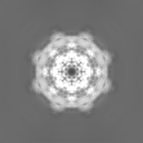
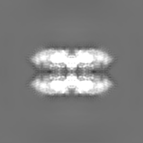
| 投影像・断面図 | 画像のコントロール
画像は Spider により作成 | ||||||||||||||||||||||||||||||||||||||||||||||||||||||||||||||||||||
| ボクセルのサイズ | X=Y=Z: 1.72 Å | ||||||||||||||||||||||||||||||||||||||||||||||||||||||||||||||||||||
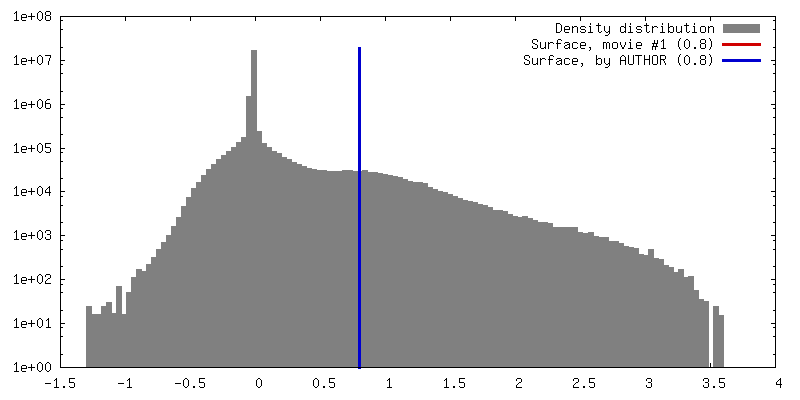
| 密度 |
| ||||||||||||||||||||||||||||||||||||||||||||||||||||||||||||||||||||
| 対称性 | 空間群: 1 | ||||||||||||||||||||||||||||||||||||||||||||||||||||||||||||||||||||
| 詳細 | EMDB XML:
CCP4マップ ヘッダ情報:
| ||||||||||||||||||||||||||||||||||||||||||||||||||||||||||||||||||||
 Z (Sec.)
 Y (Row.)
 X (Col.)


-添付データ
- 試料の構成要素
-全体 : Drosophila apoptosome (double ring)
| 全体 | 名称: Drosophila apoptosome (double ring) |
|---|---|
| 要素 |
|
-超分子 #1000: Drosophila apoptosome (double ring)
| 超分子 | 名称: Drosophila apoptosome (double ring) / タイプ: sample / ID: 1000 詳細: Sample assembled in low salt buffer (20 mM HEPES pH 7.5, 10 mM KCl, 1.5 mM MgCl2, 1 mM EDTA, 1 mM EGTA, 1 mM DTT) at about 0.5 mg per ml with dATP 集合状態: hexadecamer of Dark molecules / Number unique components: 1 |
|---|---|
| 分子量 | 理論値: 2.5 MDa |
-分子 #1: Drosophila Apaf-1 like protein
| 分子 | 名称: Drosophila Apaf-1 like protein / タイプ: protein_or_peptide / ID: 1 / Name.synonym: Dark / コピー数: 16 / 集合状態: hexadecamer / 組換発現: Yes |
|---|---|
| 由来(天然) | 生物種: 別称: fruit fly / 細胞中の位置: cytosol |
| 分子量 | 理論値: 160 KDa |
| 組換発現 | 生物種: sf21 insect cells (unknown) / 組換プラスミド: pFastBac |
-実験情報
-構造解析
| 手法 | クライオ電子顕微鏡法 |
|---|---|
 解析 解析 | 単粒子再構成法 |
| 試料の集合状態 | particle |
-試料調製
| 濃度 | 2 mg/mL |
|---|---|
| 緩衝液 | pH: 7.5 詳細: 20mM HEPES, 10mM KCl, 1.5mM MgCl2, 1mM EDTA, 1mM EGTA, 1mM DTT |
| グリッド | 詳細: thin carbon film covered holey grid |
| 凍結 | 凍結剤: ETHANE / チャンバー内湿度: 100 % / チャンバー内温度: 77 K / 装置: OTHER 詳細: Vitrification instrument: FEI Vitrobot. blotting at room temperature with sample at room temperature 手法: Blot for 2-2.5s before plunging |
- 電子顕微鏡法
| 顕微鏡 | FEI TECNAI F20 |
|---|---|
| 温度 | 最低: 93 K / 最高: 100 K / 平均: 93 K |
| アライメント法 | Legacy - 非点収差: objective lens astigmatism was corrected at 200,000 times magnification |
| 詳細 | actual magnification at the ccd 87000, camera pixel size 15um, 1.72 angstrom per pixel, data collected semi-automatically with EMTools |
| 日付 | 2009年9月15日 |
| 撮影 | カテゴリ: CCD フィルム・検出器のモデル: GENERIC TVIPS (4k x 4k) 実像数: 1100 / 平均電子線量: 20 e/Å2 詳細: the frames were 4096 x 4096 tiffs collected with the TVIPS CCD |
| 電子線 | 加速電圧: 160 kV / 電子線源:  FIELD EMISSION GUN FIELD EMISSION GUN |
| 電子光学系 | 倍率(補正後): 50000 / 照射モード: FLOOD BEAM / 撮影モード: BRIGHT FIELD / Cs: 2.0 mm / 最大 デフォーカス(公称値): 3.0 µm / 最小 デフォーカス(公称値): 1.5 µm / 倍率(公称値): 50000 |
| 試料ステージ | 試料ホルダー: Side entry liquid nitrogen-cooled cryo specimen holder 試料ホルダーモデル: GATAN LIQUID NITROGEN |
| 実験機器 |  モデル: Tecnai F20 / 画像提供: FEI Company |
-画像解析
| 詳細 | actual class number 1353 |
|---|---|
| CTF補正 | 詳細: each image |
| 最終 再構成 | アルゴリズム: OTHER / 解像度のタイプ: BY AUTHOR / 解像度: 6.9 Å / 解像度の算出法: FSC 0.5 CUT-OFF / ソフトウェア - 名称: EMAN2 詳細: Projection matching was done with Fourier ring correlation, model-based masking and SSNR weighting over an 80-6 angstrom resolution range. The final refinement steps used an angular step of 2. ...詳細: Projection matching was done with Fourier ring correlation, model-based masking and SSNR weighting over an 80-6 angstrom resolution range. The final refinement steps used an angular step of 2.5 degrees and each of the 48,000 particles was matched to the best two projection classes (1353). In total, 45,000 particles were used in the final reconstruction and the 3D map was amplitude corrected then Gaussian low-pass filtered with a Fourier half-width of 0.12. 使用した粒子像数: 48271 |
| 最終 2次元分類 | クラス数: 1000 |
