 Movie
Movie Controller

[English] 日本語
 Yorodumi
- EMDB-50803: Cryo-EM structure of single-layered nucleoprotein-RNA helical ass... -
+ Open data

- Basic information
| Entry |  | |||||||||
|---|---|---|---|---|---|---|---|---|---|---|
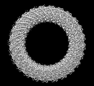
| Title | Cryo-EM structure of single-layered nucleoprotein-RNA helical assembly from Marburg virus, trimeric repeat unit | |||||||||
 Map data Map data | ||||||||||
 Sample Sample |
| |||||||||
 Keywords Keywords | RNA-binding protein / VIRAL PROTEIN | |||||||||
| Function / homology |  Function and homology information Function and homology informationviral RNA genome packaging / helical viral capsid / viral budding via host ESCRT complex / viral nucleocapsid / host cell cytoplasm / ribonucleoprotein complex / RNA binding Similarity search - Function | |||||||||
| Biological species |  Marburg virus - Musoke, Kenya, 1980 Marburg virus - Musoke, Kenya, 1980 | |||||||||
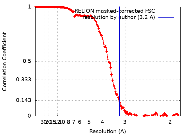
| Method | helical reconstruction / cryo EM / Resolution: 3.2 Å | |||||||||
 Authors Authors | Zinzula L / Beck F / Camasta M / Bohn S / Liu C / Morado D / Bracher A / Plitzko JM / Baumeister W | |||||||||
| Funding support | 1 items
| |||||||||
 Citation Citation | Journal: Nat Commun / Year: 2024 Title: Cryo-EM structure of single-layered nucleoprotein-RNA complex from Marburg virus. Authors: Luca Zinzula / Florian Beck / Marianna Camasta / Stefan Bohn / Chuan Liu / Dustin Morado / Andreas Bracher / Juergen M Plitzko / Wolfgang Baumeister /    Abstract: Marburg virus (MARV) causes lethal hemorrhagic fever in humans, posing a threat to global health. We determined by cryogenic electron microscopy (cryo-EM) the MARV helical ribonucleoprotein (RNP) ...Marburg virus (MARV) causes lethal hemorrhagic fever in humans, posing a threat to global health. We determined by cryogenic electron microscopy (cryo-EM) the MARV helical ribonucleoprotein (RNP) complex structure in single-layered conformation, which differs from the previously reported structure of a double-layered helix. Our findings illuminate novel RNP interactions and expand knowledge on MARV genome packaging and nucleocapsid assembly, both processes representing attractive targets for the development of antiviral therapeutics against MARV disease. | |||||||||
| History |
|
- Structure visualization
| Supplemental images |
|---|
- Downloads & links
-EMDB archive
| Map data | emd_50803.map.gz | 411.8 MB | EMDB map data format | |
|---|---|---|---|---|
| Header (meta data) | emd-50803-v30.xmlemd-50803.xml | 17.4 KB 17.4 KB | Display Display | EMDB header |
| FSC (resolution estimation) | emd_50803_fsc.xml | 18.1 KB | Display | FSC data file |
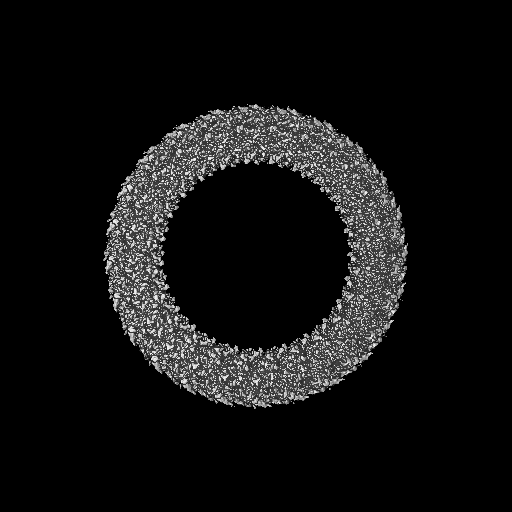
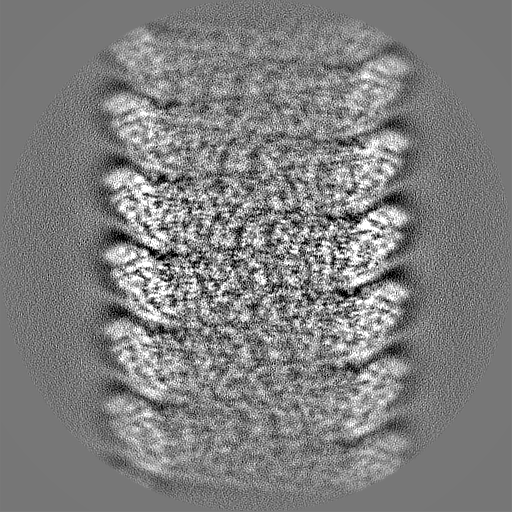
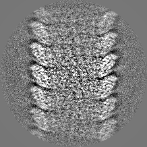
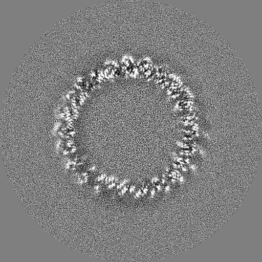
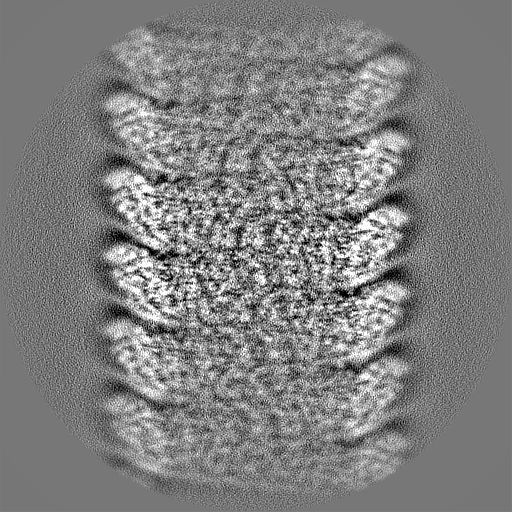
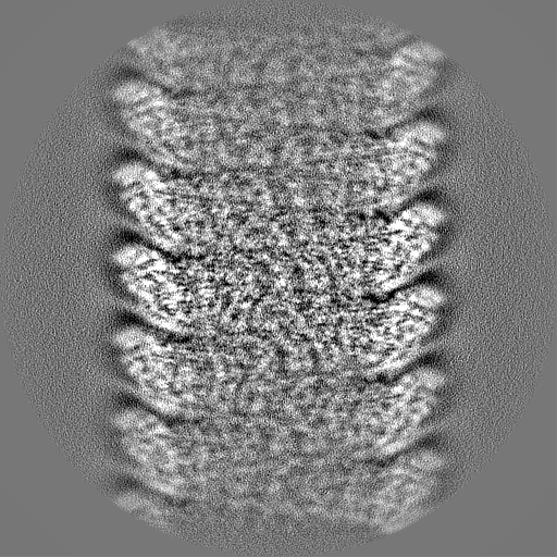
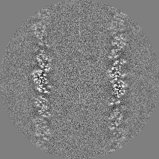
| Images |  emd_50803.png emd_50803.png | 128 KB | ||
| Masks | emd_50803_msk_1.map | 512 MB | Mask map | |
| Filedesc metadata | emd-50803.cif.gz | 6.2 KB | ||
| Others | emd_50803_half_map_1.map.gzemd_50803_half_map_2.map.gz | 411.7 MB 411.7 MB | ||
| Archive directory |  http://ftp.pdbj.org/pub/emdb/structures/EMD-50803ftp://ftp.pdbj.org/pub/emdb/structures/EMD-50803 http://ftp.pdbj.org/pub/emdb/structures/EMD-50803ftp://ftp.pdbj.org/pub/emdb/structures/EMD-50803 | HTTPS FTP |
-Related structure data
| Related structure data |  9fvdMC M: atomic model generated by this map C: citing same article ( |
|---|---|
| Similar structure data |
-Links
| EMDB pages | EMDB (EBI/PDBe) / EMDataResource |
|---|---|
| Related items in Molecule of the Month |

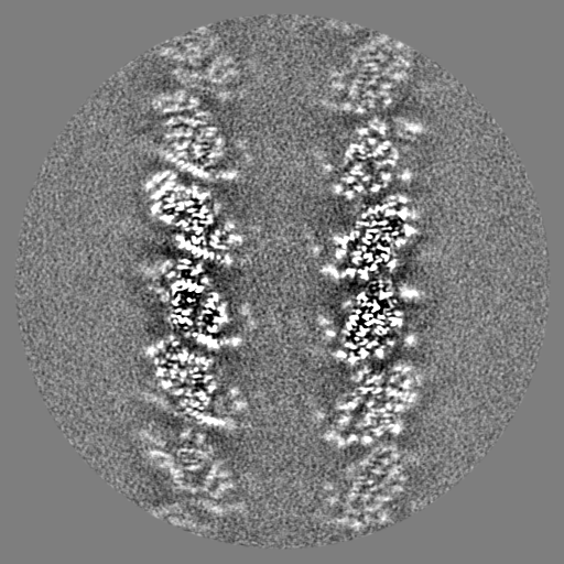
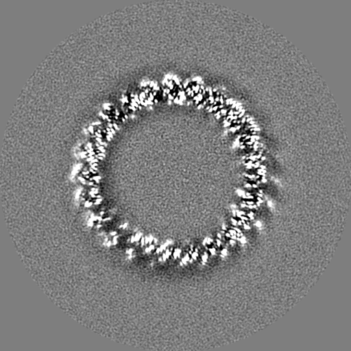
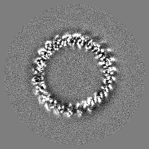
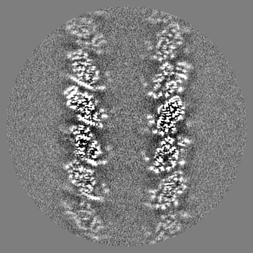
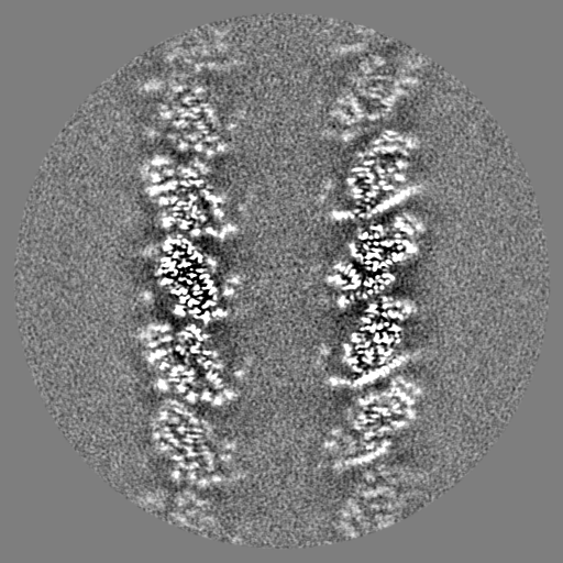
-Map
| File | Download / File: emd_50803.map.gz / Format: CCP4 / Size: 512 MB / Type: IMAGE STORED AS FLOATING POINT NUMBER (4 BYTES) | ||||||||||||||||||||||||||||||||||||
|---|---|---|---|---|---|---|---|---|---|---|---|---|---|---|---|---|---|---|---|---|---|---|---|---|---|---|---|---|---|---|---|---|---|---|---|---|---|
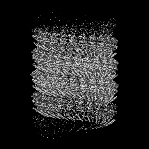
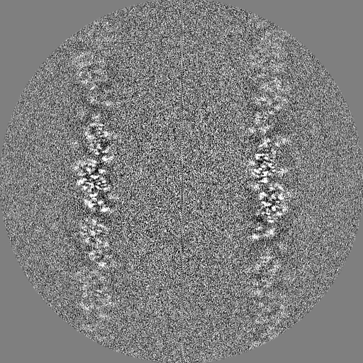
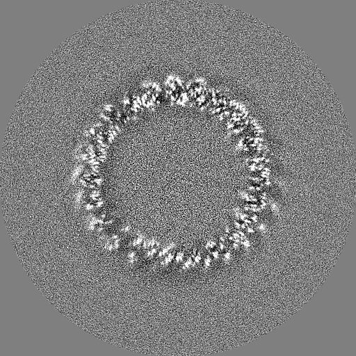
| Projections & slices | Image control
Images are generated by Spider. | ||||||||||||||||||||||||||||||||||||
| Voxel size | X=Y=Z: 0.93 Å | ||||||||||||||||||||||||||||||||||||
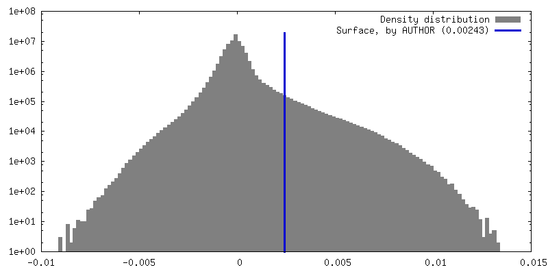
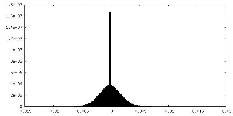
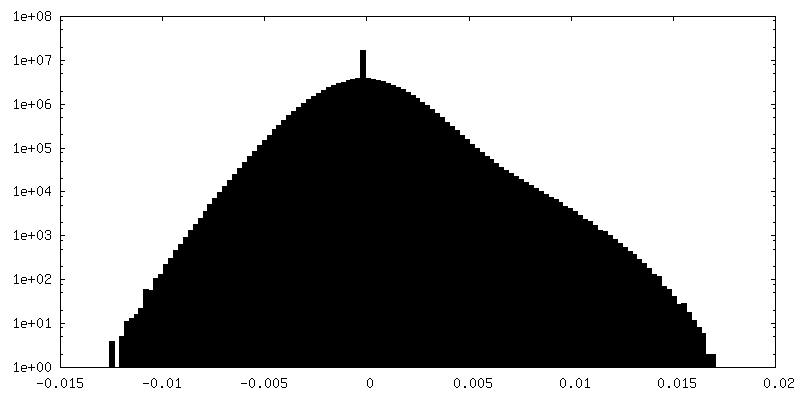
| Density |
| ||||||||||||||||||||||||||||||||||||
| Symmetry | Space group: 1 | ||||||||||||||||||||||||||||||||||||
| Details | EMDB XML:
|
 Z (Sec.)
 Y (Row.)
 X (Col.)


-Supplemental data
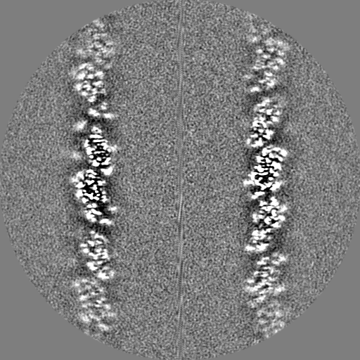
-Mask #1
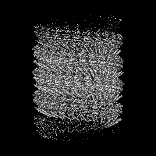
| File | emd_50803_msk_1.map | ||||||||||||
|---|---|---|---|---|---|---|---|---|---|---|---|---|---|
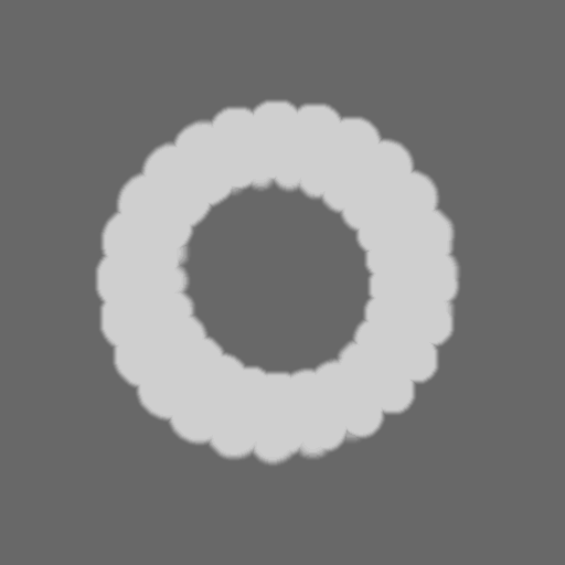
| Projections & Slices |
| ||||||||||||
| Density Histograms |


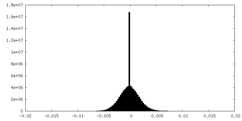
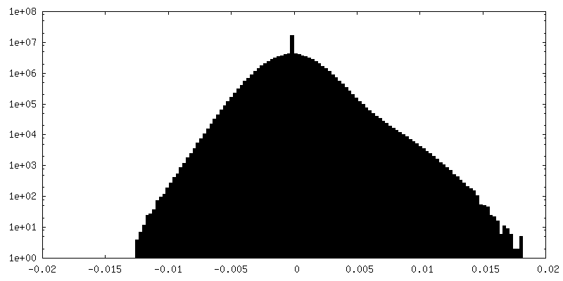
-Half map: #1
| File | emd_50803_half_map_1.map | ||||||||||||
|---|---|---|---|---|---|---|---|---|---|---|---|---|---|
| Projections & Slices |
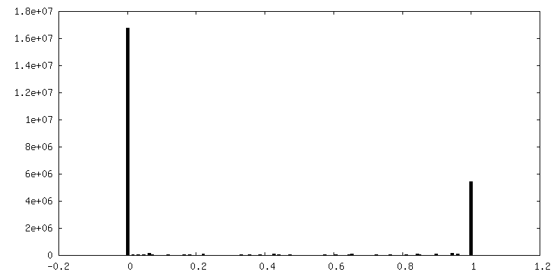
| ||||||||||||
| Density Histograms |

-Half map: #2
| File | emd_50803_half_map_2.map | ||||||||||||
|---|---|---|---|---|---|---|---|---|---|---|---|---|---|
| Projections & Slices |
| ||||||||||||
| Density Histograms |
- Sample components
-Entire : Helical assembly of nucleoprotein-RNA complex from Marburg virus
| Entire | Name: Helical assembly of nucleoprotein-RNA complex from Marburg virus |
|---|---|
| Components |
|
-Supramolecule #1: Helical assembly of nucleoprotein-RNA complex from Marburg virus
| Supramolecule | Name: Helical assembly of nucleoprotein-RNA complex from Marburg virus type: complex / ID: 1 / Parent: 0 / Macromolecule list: all |
|---|---|
| Source (natural) | Organism: Marburg virus - Musoke, Kenya, 1980 |
-Macromolecule #1: Nucleoprotein
| Macromolecule | Name: Nucleoprotein / type: protein_or_peptide / ID: 1 / Number of copies: 3 / Enantiomer: LEVO |
|---|---|
| Source (natural) | Organism: Marburg virus - Musoke, Kenya, 1980 |
| Molecular weight | Theoretical: 49.495105 KDa |
| Recombinant expression | Organism:  |
| Sequence | String: GMDLHSLLEL GTKPTAPHVR NKKVILFDTN HQVSICNQII DAINSGIDLG DLLEGGLLTL CVEHYYNSDK DKFNTSPIAK YLRDAGYEF DVIKNADATR FLDVIPNEPH YSPLILALKT LESTESQRGR IGLFLSFCSL FLPKLVVGDR ASIEKALRQV T VHQEQGIV ...String: GMDLHSLLEL GTKPTAPHVR NKKVILFDTN HQVSICNQII DAINSGIDLG DLLEGGLLTL CVEHYYNSDK DKFNTSPIAK YLRDAGYEF DVIKNADATR FLDVIPNEPH YSPLILALKT LESTESQRGR IGLFLSFCSL FLPKLVVGDR ASIEKALRQV T VHQEQGIV TYPNHWLTTG HMKVIFGILR SSFILKFVLI HQGVNLVTGH DAYDSIISNS VGQTRFSGLL IVKTVLEFIL QK TDSGVTL HPLVRTSKVK NEVASFKQAL SNLARHGEYA PFARVLNLSG INNLEHGLYP QLSAIALGVA TAHGSTLAGV NVG EQYQQL REAAHDAEVK LQRRHEHQEI QAIAEDDEER KILEQFHLQK TEITHSQTLA VLSQKREKLA RLAAEIENNI VEDQ GFKQS QNRVSQSFLN DPTPVEVTVQ ARPMNRVEHH HHHHHH UniProtKB: Nucleoprotein |
-Macromolecule #2: RNA (5'-R(P*AP*GP*AP*CP*AP*CP*AP*CP*AP*AP*AP*AP*AP*CP*AP*AP*GP*A)-3')
| Macromolecule | Name: RNA (5'-R(P*AP*GP*AP*CP*AP*CP*AP*CP*AP*AP*AP*AP*AP*CP*AP*AP*GP*A)-3') type: rna / ID: 2 / Number of copies: 5 |
|---|---|
| Source (natural) | Organism: Marburg virus - Musoke, Kenya, 1980 |
| Molecular weight | Theoretical: 5.816651 KDa |
| Sequence | String: AGACACACAA AAACAAGA |
-Experimental details
-Structure determination
| Method | cryo EM |
|---|---|
 Processing Processing | helical reconstruction |
| Aggregation state | helical array |
-Sample preparation
| Concentration | 1.25 mg/mL | |||||||||
|---|---|---|---|---|---|---|---|---|---|---|
| Buffer | pH: 8 Component:
| |||||||||
| Grid | Model: Quantifoil R1.2/1.3 / Material: COPPER / Mesh: 400 | |||||||||
| Vitrification | Cryogen name: ETHANE-PROPANE / Chamber humidity: 95 % / Chamber temperature: 277.15 K / Instrument: FEI VITROBOT MARK IV |
- Electron microscopy
| Microscope | FEI TITAN KRIOS |
|---|---|
| Image recording | Film or detector model: FEI FALCON IV (4k x 4k) / Number real images: 5518 / Average electron dose: 30.0 e/Å2 |
| Electron beam | Acceleration voltage: 300 kV / Electron source:  FIELD EMISSION GUN FIELD EMISSION GUN |
| Electron optics | Illumination mode: FLOOD BEAM / Imaging mode: BRIGHT FIELD / Nominal defocus max: 5.0 µm / Nominal defocus min: 0.5 µm |
| Sample stage | Specimen holder model: FEI TITAN KRIOS AUTOGRID HOLDER |
| Experimental equipment |  Model: Titan Krios / Image courtesy: FEI Company |
+Image processing
-Atomic model buiding 1
| Initial model | Chain - Source name: AlphaFold / Chain - Initial model type: in silico model |
|---|---|
| Refinement | Space: REAL / Protocol: AB INITIO MODEL |
| Output model | PDB-9fvd: |
