Septum site-determining protein MinD / : / Septum site-determining protein MinD / Probable septum site-determining protein MinC Similarity search - Component
Biological species
Pseudomonas aeruginosa (bacteria)
Method
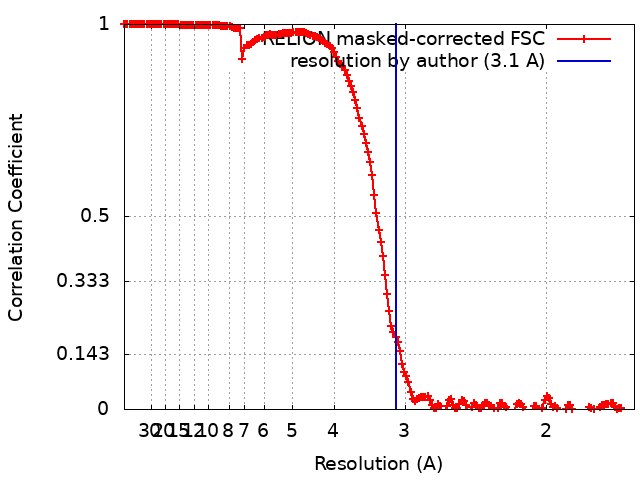
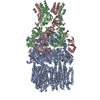
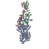
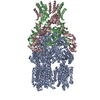
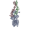
helical reconstruction / cryo EM / Resolution: 3.1 Å
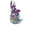
Journal: FEBS Lett / Year: 2019 Title: Cryo-EM structure of the MinCD copolymeric filament from Pseudomonas aeruginosa at 3.1 Å resolution. Authors: Andrzej Szewczak-Harris / James Wagstaff / Jan Löwe / Abstract: Positioning of the division site in many bacterial species relies on the MinCDE system, which prevents the cytokinetic Z-ring from assembling anywhere but the mid-cell, through an oscillatory ...Positioning of the division site in many bacterial species relies on the MinCDE system, which prevents the cytokinetic Z-ring from assembling anywhere but the mid-cell, through an oscillatory diffusion-reaction mechanism. MinD dimers bind to membranes and, via their partner MinC, inhibit the polymerization of cell division protein FtsZ into the Z-ring. MinC and MinD form polymeric assemblies in solution and on cell membranes. Here, we report the high-resolution cryo-EM structure of the copolymeric filaments of Pseudomonas aeruginosa MinCD. The filaments consist of three protofilaments made of alternating MinC and MinD dimers. The MinCD protofilaments are almost completely straight and assemble as single protofilaments on lipid membranes, which we also visualized by cryo-EM.
History
Deposition
Apr 24, 2019
-
Header (metadata) release
May 1, 2019
-
Map release
Jun 19, 2019
-
Update
Oct 1, 2025
-
Current status
Oct 1, 2025
Processing site: PDBe / Status: Released
-
Structure visualization
Movie
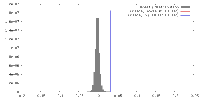
Surface view with section colored by density value
In the structure databanks used in Yorodumi, some data are registered as the other names, "COVID-19 virus" and "2019-nCoV". Here are the details of the virus and the list of structure data.
Jan 31, 2019. EMDB accession codes are about to change! (news from PDBe EMDB page)
EMDB accession codes are about to change! (news from PDBe EMDB page)
The allocation of 4 digits for EMDB accession codes will soon come to an end. Whilst these codes will remain in use, new EMDB accession codes will include an additional digit and will expand incrementally as the available range of codes is exhausted. The current 4-digit format prefixed with “EMD-” (i.e. EMD-XXXX) will advance to a 5-digit format (i.e. EMD-XXXXX), and so on. It is currently estimated that the 4-digit codes will be depleted around Spring 2019, at which point the 5-digit format will come into force.
The EM Navigator/Yorodumi systems omit the EMD- prefix.
Related info.:Q: What is EMD? / ID/Accession-code notation in Yorodumi/EM Navigator
Yorodumi is a browser for structure data from EMDB, PDB, SASBDB, etc.
This page is also the successor to EM Navigator detail page, and also detail information page/front-end page for Omokage search.
The word "yorodu" (or yorozu) is an old Japanese word meaning "ten thousand". "mi" (miru) is to see.
Related info.:EMDB / PDB / SASBDB / Comparison of 3 databanks / Yorodumi Search / Aug 31, 2016. New EM Navigator & Yorodumi / Yorodumi Papers / Jmol/JSmol / Function and homology information / Changes in new EM Navigator and Yorodumi
 Movie
Movie Controller


 Open data
 Basic information
 Map data
 Sample
 Keywords
 Function and homology information
 Pseudomonas aeruginosa (bacteria)
 Authors
 United Kingdom, 2 items
  Citation
 Structure visualization
 Downloads & links
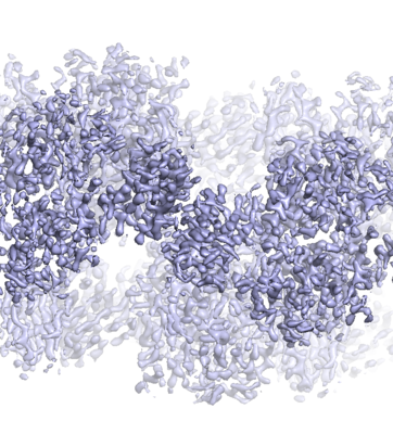 emd_4897.png
 http://ftp.pdbj.org/pub/emdb/structures/EMD-4897
 Z (Sec.)
 Y (Row.)
 X (Col.)


 Sample components
 Processing
 Electron microscopy
 FIELD EMISSION GUN