 Movie
Movie Controller


+ Open data

- Basic information
| Entry | Database: PDB / ID: 6riq | ||||||||||||||||||||||||||||||
|---|---|---|---|---|---|---|---|---|---|---|---|---|---|---|---|---|---|---|---|---|---|---|---|---|---|---|---|---|---|---|---|
| Title | MinCD filament from Pseudomonas aeruginosa | ||||||||||||||||||||||||||||||
 Components Components |
| ||||||||||||||||||||||||||||||
 Keywords Keywords | PROTEIN FIBRIL / Bacterial cell division | ||||||||||||||||||||||||||||||
| Function / homology |  Function and homology information Function and homology informationregulation of cell septum assembly / negative regulation of cell division / division septum assembly / regulation of cell division / cell morphogenesis / cytoplasmic side of plasma membrane / ATP hydrolysis activity / ATP binding / cytosol Similarity search - Function | ||||||||||||||||||||||||||||||
| Biological species |   Pseudomonas aeruginosa (bacteria) Pseudomonas aeruginosa (bacteria) | ||||||||||||||||||||||||||||||
| Method | ELECTRON MICROSCOPY / helical reconstruction / cryo EM / Resolution: 3.1 Å | ||||||||||||||||||||||||||||||
 Authors Authors | Szewczak-Harris, A. / Wagstaff, J. / Lowe, J. | ||||||||||||||||||||||||||||||
| Funding support |  United Kingdom, 2items United Kingdom, 2items
| ||||||||||||||||||||||||||||||
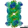
 Citation Citation | Journal: FEBS Lett / Year: 2019 Title: Cryo-EM structure of the MinCD copolymeric filament from Pseudomonas aeruginosa at 3.1 Å resolution. Authors: Andrzej Szewczak-Harris / James Wagstaff / Jan Löwe / Abstract: Positioning of the division site in many bacterial species relies on the MinCDE system, which prevents the cytokinetic Z-ring from assembling anywhere but the mid-cell, through an oscillatory ...Positioning of the division site in many bacterial species relies on the MinCDE system, which prevents the cytokinetic Z-ring from assembling anywhere but the mid-cell, through an oscillatory diffusion-reaction mechanism. MinD dimers bind to membranes and, via their partner MinC, inhibit the polymerization of cell division protein FtsZ into the Z-ring. MinC and MinD form polymeric assemblies in solution and on cell membranes. Here, we report the high-resolution cryo-EM structure of the copolymeric filaments of Pseudomonas aeruginosa MinCD. The filaments consist of three protofilaments made of alternating MinC and MinD dimers. The MinCD protofilaments are almost completely straight and assemble as single protofilaments on lipid membranes, which we also visualized by cryo-EM. | ||||||||||||||||||||||||||||||
| History |
|
- Structure visualization
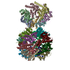
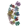
| Movie |
Movie viewer |
|---|---|
| Structure viewer | Molecule: MolmilJmol/JSmol |
- Downloads & links
-Download
| PDBx/mmCIF format | 6riq.cif.gz | 745.2 KB | Display | PDBx/mmCIF format |
|---|---|---|---|---|
| PDB format | pdb6riq.ent.gz | 615.8 KB | Display | PDB format |
| PDBx/mmJSON format | 6riq.json.gz | Tree view | PDBx/mmJSON format | |
| Others |  Other downloads Other downloads |
-Validation report
| Arichive directory | https://data.pdbj.org/pub/pdb/validation_reports/ri/6riqftp://data.pdbj.org/pub/pdb/validation_reports/ri/6riq | HTTPS FTP |
|---|
-Related structure data
| Related structure data | 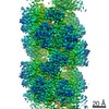 4897MC M: map data used to model this data C: citing same article ( |
|---|---|
| Similar structure data |


-Links
 PDBj
PDBj- Assembly
| Deposited unit | 
|
|---|---|
| 1 |
|
-Components
| #1: Protein | Mass: 15107.273 Da / Num. of mol.: 11 Source method: isolated from a genetically manipulated source Source: (gene. exp.) Pseudomonas aeruginosa (bacteria) / Production host: #2: Protein | Mass: 29705.143 Da / Num. of mol.: 11 Source method: isolated from a genetically manipulated source Source: (gene. exp.) Pseudomonas aeruginosa (bacteria)Gene: minD, minD_2, ALP65_02808, C0044_24365, C8257_09550, CAZ10_22480, CGU42_07990, DZ940_07140, DZ962_16115, EFK68_22340, EGV95_09885, EGY23_16190, IPC1135_03845, NCTC13719_01733, PAERUG_E15_London_ ...Gene: minD, minD_2, ALP65_02808, C0044_24365, C8257_09550, CAZ10_22480, CGU42_07990, DZ940_07140, DZ962_16115, EFK68_22340, EGV95_09885, EGY23_16190, IPC1135_03845, NCTC13719_01733, PAERUG_E15_London_28_01_14_02879, RW109_RW109_02577 Production host: #3: Chemical | ChemComp-MG /   Mass: 24.305 Da / Num. of mol.: 11 / Source method: obtained synthetically / Formula: Mg Mass: 24.305 Da / Num. of mol.: 11 / Source method: obtained synthetically / Formula: Mg#4: Chemical | ChemComp-ATP /   Mass: 507.181 Da / Num. of mol.: 11 / Source method: obtained synthetically / Formula: C10H16N5O13P3 / Comment: ATP, energy-carrying molecule*YM Mass: 507.181 Da / Num. of mol.: 11 / Source method: obtained synthetically / Formula: C10H16N5O13P3 / Comment: ATP, energy-carrying molecule*YMHas protein modification | N | |
|---|
-Experimental details
-Experiment
| Experiment | Method: ELECTRON MICROSCOPY |
|---|---|
| EM experiment | Aggregation state: HELICAL ARRAY / 3D reconstruction method: helical reconstruction |
- Sample preparation
| Component | Name: MinCD Filament / Type: COMPLEX / Entity ID: #1-#2 / Source: RECOMBINANT |
|---|---|
| Source (natural) | Organism: Pseudomonas aeruginosa (bacteria) |
| Source (recombinant) | Organism: |
| Buffer solution | pH: 7 |
| Specimen | Embedding applied: NO / Shadowing applied: NO / Staining applied: NO / Vitrification applied: YES |
| Vitrification | Instrument: FEI VITROBOT MARK IV / Cryogen name: ETHANE |
- Electron microscopy imaging
| Experimental equipment |  Model: Tecnai Polara / Image courtesy: FEI Company |
|---|---|
| Microscopy | Model: FEI POLARA 300 |
| Electron gun | Electron source:  FIELD EMISSION GUN / Accelerating voltage: 300 kV / Illumination mode: FLOOD BEAM FIELD EMISSION GUN / Accelerating voltage: 300 kV / Illumination mode: FLOOD BEAM |
| Electron lens | Mode: BRIGHT FIELD |
| Specimen holder | Cryogen: NITROGEN |
| Image recording | Electron dose: 38 e/Å2 / Detector mode: INTEGRATING / Film or detector model: FEI FALCON III (4k x 4k) / Num. of real images: 3050 |
- Processing
| CTF correction | Type: PHASE FLIPPING AND AMPLITUDE CORRECTION |
|---|---|
| Helical symmerty | Angular rotation/subunit: 116.265 ° / Axial rise/subunit: 24.998 Å / Axial symmetry: C1 |
| 3D reconstruction | Resolution: 3.1 Å / Resolution method: FSC 0.143 CUT-OFF / Num. of particles: 118659 / Symmetry type: HELICAL |
| Atomic model building | Protocol: AB INITIO MODEL / Space: RECIPROCAL |
