[English] 日本語
Yorodumi
- EMDB-47478: Cryo-EM structure of PRMT5/WDR77 in complex with 6S complex (GRG ... -

+
Open data


ID or keywords:

Loading...

-
Basic information

Entry
Database: EMDB / ID: EMD-47478
TitleCryo-EM structure of PRMT5/WDR77 in complex with 6S complex (GRG local refine)
Map dataMap of locally refined PRMT5/GRG active site
Sample
  • Complex: PRMT5/WDR77 in complex with 6S (GRG active site local refine)
    • Complex: PRMT5/WDR77
      • Protein or peptide: Protein arginine N-methyltransferase 5
    • Complex: 6S complex
      • Complex: Methylosome subunit pICln
      • Complex: Small nuclear ribonucleoprotein Sm D1
        • Protein or peptide: Small nuclear ribonucleoprotein Sm D1
      • Complex: Small nuclear ribonucleoproteins E, F, G
  • Ligand: SINEFUNGIN
KeywordsPRMT5 / Methyl transferase / WDR77 / arginine / TRANSFERASE
Function / homology
Function and homology information


positive regulation of adenylate cyclase-inhibiting dopamine receptor signaling pathway / peptidyl-arginine N-methylation / type II protein arginine methyltransferase / peptidyl-arginine methylation / protein-arginine omega-N symmetric methyltransferase activity / Golgi ribbon formation / histone arginine N-methyltransferase activity / histone H3R17 methyltransferase activity / histone H3R2 methyltransferase activity / histone H3R8 methyltransferase activity ...positive regulation of adenylate cyclase-inhibiting dopamine receptor signaling pathway / peptidyl-arginine N-methylation / type II protein arginine methyltransferase / peptidyl-arginine methylation / protein-arginine omega-N symmetric methyltransferase activity / Golgi ribbon formation / histone arginine N-methyltransferase activity / histone H3R17 methyltransferase activity / histone H3R2 methyltransferase activity / histone H3R8 methyltransferase activity / histone H3R26 methyltransferase activity / histone H4R3 methyltransferase activity / histone H3K37 methyltransferase activity / histone H4K12 methyltransferase activity / U12-type spliceosomal complex / histone H3K56 methyltransferase activity / 7-methylguanosine cap hypermethylation / protein-arginine N-methyltransferase activity / methylosome / U1 snRNP binding / pICln-Sm protein complex / positive regulation of mRNA splicing, via spliceosome / small nuclear ribonucleoprotein complex / methyl-CpG binding / SMN-Sm protein complex / spliceosomal tri-snRNP complex / U2-type precatalytic spliceosome / histone H2A Q104 methyltransferase activity / U2-type spliceosomal complex / commitment complex / U2-type prespliceosome assembly / U2-type catalytic step 2 spliceosome / U4 snRNP / endothelial cell activation / U2 snRNP / U1 snRNP / histone H3 methyltransferase activity / regulation of mitotic nuclear division / histone methyltransferase complex / precatalytic spliceosome / negative regulation of gene expression via chromosomal CpG island methylation / histone methyltransferase activity / spliceosomal complex assembly / E-box binding / mRNA Splicing - Minor Pathway / positive regulation of oligodendrocyte differentiation / negative regulation of cell differentiation / U5 snRNP / regulation of ERK1 and ERK2 cascade / spliceosomal snRNP assembly / ribonucleoprotein complex binding / U4/U6 x U5 tri-snRNP complex / catalytic step 2 spliceosome / mRNA Splicing - Major Pathway / RNA splicing / liver regeneration / regulation of signal transduction by p53 class mediator / methyltransferase activity / spliceosomal complex / circadian regulation of gene expression / DNA-templated transcription termination / mRNA splicing, via spliceosome / Regulation of TP53 Activity through Methylation / RMTs methylate histone arginines / transcription corepressor activity / p53 binding / snRNP Assembly / SARS-CoV-2 modulates host translation machinery / chromatin remodeling / protein heterodimerization activity / regulation of DNA-templated transcription / chromatin / Golgi apparatus / RNA binding / nucleoplasm / identical protein binding / nucleus / cytosol / cytoplasm
Similarity search - Function
Protein arginine N-methyltransferase PRMT5 / PRMT5, TIM barrel domain / PRMT5, oligomerisation domain / PRMT5 TIM barrel domain / PRMT5 oligomerisation domain / PRMT5 arginine-N-methyltransferase / PRMT5 arginine-N-methyltransferase / Small nuclear ribonucleoprotein D1 / Protein arginine N-methyltransferase / SAM-dependent methyltransferase PRMT-type domain profile. ...Protein arginine N-methyltransferase PRMT5 / PRMT5, TIM barrel domain / PRMT5, oligomerisation domain / PRMT5 TIM barrel domain / PRMT5 oligomerisation domain / PRMT5 arginine-N-methyltransferase / PRMT5 arginine-N-methyltransferase / Small nuclear ribonucleoprotein D1 / Protein arginine N-methyltransferase / SAM-dependent methyltransferase PRMT-type domain profile. / Like-Sm (LSM) domain containing protein, LSm4/SmD1/SmD3 / LSM domain / LSM domain, eukaryotic/archaea-type / snRNP Sm proteins / : / Sm domain profile. / LSM domain superfamily / S-adenosyl-L-methionine-dependent methyltransferase superfamily
Similarity search - Domain/homology
Protein arginine N-methyltransferase 5 / Small nuclear ribonucleoprotein Sm D1
Similarity search - Component
Biological speciesHomo sapiens (human)
Methodsingle particle reconstruction / cryo EM / Resolution: 3.19 Å
AuthorsJin CY / Hunkeler M / Fischer ES
Funding support United States, 1 items
OrganizationGrant numberCountry
National Institutes of Health/National Cancer Institute (NIH/NCI)R01CA262188 United States
CitationJournal: J Biol Chem / Year: 2025
Title: Substrate adaptors are flexible tethering modules that enhance substrate methylation by the arginine methyltransferase PRMT5.
Authors: Cyrus Y Jin / Moritz Hunkeler / Kathleen M Mulvaney / William R Sellers / Eric S Fischer /
Abstract: Protein arginine methyltransferase (PRMT) 5 is an essential arginine methyltransferase responsible for the majority of cellular symmetric dimethyl-arginine marks. PRMT5 uses substrate adaptors such ...Protein arginine methyltransferase (PRMT) 5 is an essential arginine methyltransferase responsible for the majority of cellular symmetric dimethyl-arginine marks. PRMT5 uses substrate adaptors such as pICln, RIOK1, and COPR5 to recruit and methylate a wide range of substrates. Although the substrate adaptors play important roles in substrate recognition, how they direct PRMT5 activity towards specific substrates remains incompletely understood. Using biochemistry and cryogenic electron microscopy, we show that these adaptors compete for the same binding site on PRMT5. We find that substrate adaptor and substrate complexes are bound to PRMT5 through two peptide motifs, enabling these adaptors to act as flexible tethering modules to enhance substrate methylation. Taken together, our results shed structural and mechanistic light on the PRMT5 substrate adaptor function and the biochemical nature of PRMT5 interactors.
History
DepositionOct 23, 2024-
Header (metadata) releaseFeb 19, 2025-
Map releaseFeb 19, 2025-
UpdateFeb 19, 2025-
Current statusFeb 19, 2025Processing site: RCSB / Status: Released

-
Structure visualization

Supplemental images

Downloads & links

-
Map

FileDownload / File: emd_47478.map.gz / Format: CCP4 / Size: 216 MB / Type: IMAGE STORED AS FLOATING POINT NUMBER (4 BYTES)
AnnotationMap of locally refined PRMT5/GRG active site
Projections & slices

Image control

Size
Brightness
Contrast
Others
AxesZ (Sec.)Y (Row.)X (Col.)
1.1 Å/pix.
x 384 pix.
= 422.4 Å
1.1 Å/pix.
x 384 pix.
= 422.4 Å
1.1 Å/pix.
x 384 pix.
= 422.4 Å

Surface

Projections

Slices (1/3)

Slices (1/2)

Slices (2/3)

Images are generated by Spider.

Voxel sizeX=Y=Z: 1.1 Å
Density
Contour LevelBy AUTHOR: 0.2
Minimum - Maximum-0.48362976 - 1.0668201
Average (Standard dev.)-0.0006222892 (±0.018056624)
SymmetrySpace group: 1
Details

EMDB XML:

Map geometry
Axis orderXYZ
Origin000
Dimensions384384384
Spacing384384384
CellA=B=C: 422.40002 Å
α=β=γ: 90.0 °

-
Supplemental data

-
Mask #1

Fileemd_47478_msk_1.map
Projections & Slices
AxesZYX

Projections

Slices (1/2)
Density Histograms

-
Half map: Half map of local refinement of PRMT5/GRG active site

Fileemd_47478_half_map_1.map
AnnotationHalf map of local refinement of PRMT5/GRG active site
Projections & Slices
AxesZYX

Projections

Slices (1/2)
Density Histograms

-
Half map: Half map of local refinement of PRMT5/GRG active site

Fileemd_47478_half_map_2.map
AnnotationHalf map of local refinement of PRMT5/GRG active site
Projections & Slices
AxesZYX

Projections

Slices (1/2)
Density Histograms

-
Sample components

-
Entire : PRMT5/WDR77 in complex with 6S (GRG active site local refine)

EntireName: PRMT5/WDR77 in complex with 6S (GRG active site local refine)
Components
  • Complex: PRMT5/WDR77 in complex with 6S (GRG active site local refine)
    • Complex: PRMT5/WDR77
      • Protein or peptide: Protein arginine N-methyltransferase 5
    • Complex: 6S complex
      • Complex: Methylosome subunit pICln
      • Complex: Small nuclear ribonucleoprotein Sm D1
        • Protein or peptide: Small nuclear ribonucleoprotein Sm D1
      • Complex: Small nuclear ribonucleoproteins E, F, G
  • Ligand: SINEFUNGIN

-
Supramolecule #1: PRMT5/WDR77 in complex with 6S (GRG active site local refine)

SupramoleculeName: PRMT5/WDR77 in complex with 6S (GRG active site local refine)
type: complex / ID: 1 / Parent: 0 / Macromolecule list: #1-#2
Molecular weightTheoretical: 770 KDa

-
Supramolecule #2: PRMT5/WDR77

SupramoleculeName: PRMT5/WDR77 / type: complex / ID: 2 / Parent: 1 / Macromolecule list: #1
Source (natural)Organism: Homo sapiens (human)

-
Supramolecule #3: 6S complex

SupramoleculeName: 6S complex / type: complex / ID: 3 / Parent: 1 / Macromolecule list: #2
Source (natural)Organism: Homo sapiens (human)

-
Supramolecule #4: Methylosome subunit pICln

SupramoleculeName: Methylosome subunit pICln / type: complex / ID: 4 / Parent: 3

-
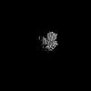
Supramolecule #5: Small nuclear ribonucleoprotein Sm D1

SupramoleculeName: Small nuclear ribonucleoprotein Sm D1 / type: complex / ID: 5 / Parent: 3 / Macromolecule list: #2

-
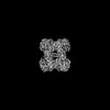
Supramolecule #6: Small nuclear ribonucleoproteins E, F, G

SupramoleculeName: Small nuclear ribonucleoproteins E, F, G / type: complex / ID: 6 / Parent: 3

-
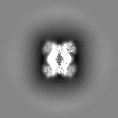
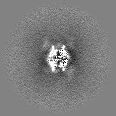
Macromolecule #1: Protein arginine N-methyltransferase 5

MacromoleculeName: Protein arginine N-methyltransferase 5 / type: protein_or_peptide / ID: 1 / Number of copies: 1 / Enantiomer: LEVO / EC number: type II protein arginine methyltransferase
Source (natural)Organism: Homo sapiens (human)
Molecular weightTheoretical: 72.766664 KDa
Recombinant expressionOrganism: Trichoplusia ni (cabbage looper)
SequenceString: MAAMAVGGAG GSRVSSGRDL NCVPEIADTL GAVAKQGFDF LCMPVFHPRF KREFIQEPAK NRPGPQTRSD LLLSGRDWNT LIVGKLSPW IRPDSKVEKI RRNSEAAMLQ ELNFGAYLGL PAFLLPLNQE DNTNLARVLT NHIHTGHHSS MFWMRVPLVA P EDLRDDII ...String:
MAAMAVGGAG GSRVSSGRDL NCVPEIADTL GAVAKQGFDF LCMPVFHPRF KREFIQEPAK NRPGPQTRSD LLLSGRDWNT LIVGKLSPW IRPDSKVEKI RRNSEAAMLQ ELNFGAYLGL PAFLLPLNQE DNTNLARVLT NHIHTGHHSS MFWMRVPLVA P EDLRDDII ENAPTTHTEE YSGEEKTWMW WHNFRTLCDY SKRIAVALEI GADLPSNHVI DRWLGEPIKA AILPTSIFLT NK KGFPVLS KMHQRLIFRL LKLEVQFIIT GTNHHSEKEF CSYLQYLEYL SQNRPPPNAY ELFAKGYEDY LQSPLQPLMD NLE SQTYEV FEKDPIKYSQ YQQAIYKCLL DRVPEEEKDT NVQVLMVLGA GRGPLVNASL RAAKQADRRI KLYAVEKNPN AVVT LENWQ FEEWGSQVTV VSSDMREWVA PEKADIIVSE LLGSFADNEL SPECLDGAQH FLKDDGVSIP GEYTSFLAPI SSSKL YNEV RACREKDRDP EAQFEMPYVV RLHNFHQLSA PQPCFTFSHP NRDPMIDNNR YCTLEFPVEV NTVLHGFAGY FETVLY QDI TLSIRPETHS PGMFSWFPIL FPIKQPITVR EGQTICVRFW RCSNSKKVWY EWAVTAPVCS AIHNPTGRSY TIGL

UniProtKB: Protein arginine N-methyltransferase 5

-
Macromolecule #2: Small nuclear ribonucleoprotein Sm D1

MacromoleculeName: Small nuclear ribonucleoprotein Sm D1 / type: protein_or_peptide / ID: 2 / Number of copies: 1 / Enantiomer: LEVO
Source (natural)Organism: Homo sapiens (human)
Molecular weightTheoretical: 13.954288 KDa
Recombinant expressionOrganism: Escherichia coli (E. coli)
SequenceString:
GGGSGGGGRM KLVRFLMKLS HETVTIELKN GTQVHGTITG VDVSMNTHLK AVKMTLKNRE PVQLETLSIR GNNIRYFILP DSLPLDTLL VDVEPKVKSK KREAVAGRGR GRGRGRGRGR GRGRGGPRR

UniProtKB: Small nuclear ribonucleoprotein Sm D1

-
Macromolecule #3: SINEFUNGIN

MacromoleculeName: SINEFUNGIN / type: ligand / ID: 3 / Number of copies: 1 / Formula: SFG
Molecular weightTheoretical: 381.387 Da
Chemical component information

ChemComp-SFG:
SINEFUNGIN

-
Experimental details

-
Structure determination

Methodcryo EM
Processingsingle particle reconstruction
Aggregation stateparticle

-
Sample preparation

Concentration1 mg/mL
BufferpH: 7.4
Component:
ConcentrationFormulaName
30.0 mMC8H18N2O4SHEPES
150.0 mMNaClsodium chloride
3.0 mMC9H15O6PTCEP
3.33 mMC15H23N7O5sinefungin

Details: 30 mM HEPES pH 7.4, 150 mM NaCl, 3 mM TCEP, 3.33 mM sinefungin
GridModel: Quantifoil R1.2/1.3 / Material: COPPER / Mesh: 300 / Support film - Material: CARBON / Support film - topology: HOLEY ARRAY
VitrificationCryogen name: ETHANE / Chamber humidity: 90 % / Chamber temperature: 283 K / Instrument: LEICA EM GP

-
Electron microscopy

MicroscopeTFS TALOS
Image recordingFilm or detector model: GATAN K3 (6k x 4k) / Number grids imaged: 1 / Number real images: 3615 / Average exposure time: 5.0 sec. / Average electron dose: 53.112 e/Å2
Electron beamAcceleration voltage: 200 kV / Electron source: FIELD EMISSION GUN
Electron opticsC2 aperture diameter: 50.0 µm / Illumination mode: FLOOD BEAM / Imaging mode: BRIGHT FIELD / Cs: 2.7 mm / Nominal defocus max: 2.2 µm / Nominal defocus min: 0.8 µm / Nominal magnification: 36000
Sample stageSpecimen holder model: FEI TITAN KRIOS AUTOGRID HOLDER / Cooling holder cryogen: NITROGEN

+
Image processing

Detailsmotioncorrected on the fly by cryosparc live
Particle selectionNumber selected: 2100293 / Details: topaz
Startup modelType of model: OTHER / Details: ab initio
Final reconstructionResolution.type: BY AUTHOR / Resolution: 3.19 Å / Resolution method: FSC 0.143 CUT-OFF / Software - Name: cryoSPARC (ver. 4.4.1) / Number images used: 530317
Initial angle assignmentType: MAXIMUM LIKELIHOOD
Final angle assignmentType: MAXIMUM LIKELIHOOD
Final 3D classificationNumber classes: 8 / Software - Name: cryoSPARC (ver. 4.41)
FSC plot (resolution estimation)

-
Atomic model buiding 1

Initial modelPDB ID:

Chain - Chain ID: A / Chain - Source name: PDB / Chain - Initial model type: experimental model
RefinementSpace: REAL / Protocol: OTHER / Overall B value: 110.3 / Target criteria: real space correlation
Output model

PDB-9e3c:
Cryo-EM structure of PRMT5/WDR77 in complex with 6S complex (GRG local refine)

+
About Yorodumi

-
News

-
Feb 9, 2022. New format data for meta-information of EMDB entries

New format data for meta-information of EMDB entries

  • Version 3 of the EMDB header file is now the official format.
  • The previous official version 1.9 will be removed from the archive.

Related info.:EMDB header

External links:wwPDB to switch to version 3 of the EMDB data model

-
Aug 12, 2020. Covid-19 info

Covid-19 info

URL: https://pdbj.org/emnavi/covid19.php

New page: Covid-19 featured information page in EM Navigator.

Related info.:Covid-19 info / Mar 5, 2020. Novel coronavirus structure data

+
Mar 5, 2020. Novel coronavirus structure data

Novel coronavirus structure data

Related info.:Yorodumi Speices / Aug 12, 2020. Covid-19 info

External links:COVID-19 featured content - PDBj / Molecule of the Month (242):Coronavirus Proteases

+
Jan 31, 2019. EMDB accession codes are about to change! (news from PDBe EMDB page)

EMDB accession codes are about to change! (news from PDBe EMDB page)

  • The allocation of 4 digits for EMDB accession codes will soon come to an end. Whilst these codes will remain in use, new EMDB accession codes will include an additional digit and will expand incrementally as the available range of codes is exhausted. The current 4-digit format prefixed with “EMD-” (i.e. EMD-XXXX) will advance to a 5-digit format (i.e. EMD-XXXXX), and so on. It is currently estimated that the 4-digit codes will be depleted around Spring 2019, at which point the 5-digit format will come into force.
  • The EM Navigator/Yorodumi systems omit the EMD- prefix.

Related info.:Q: What is EMD? / ID/Accession-code notation in Yorodumi/EM Navigator

External links:EMDB Accession Codes are Changing Soon! / Contact to PDBj

+
Jul 12, 2017. Major update of PDB

Major update of PDB

  • wwPDB released updated PDB data conforming to the new PDBx/mmCIF dictionary.
  • This is a major update changing the version number from 4 to 5, and with Remediation, in which all the entries are updated.
  • In this update, many items about electron microscopy experimental information are reorganized (e.g. em_software).
  • Now, EM Navigator and Yorodumi are based on the updated data.

External links:wwPDB Remediation / Enriched Model Files Conforming to OneDep Data Standards Now Available in the PDB FTP Archive

-
Yorodumi

Thousand views of thousand structures

  • Yorodumi is a browser for structure data from EMDB, PDB, SASBDB, etc.
  • This page is also the successor to EM Navigator detail page, and also detail information page/front-end page for Omokage search.
  • The word "yorodu" (or yorozu) is an old Japanese word meaning "ten thousand". "mi" (miru) is to see.

Related info.:EMDB / PDB / SASBDB / Comparison of 3 databanks / Yorodumi Search / Aug 31, 2016. New EM Navigator & Yorodumi / Yorodumi Papers / Jmol/JSmol / Function and homology information / Changes in new EM Navigator and Yorodumi

Read more