 Movie
Movie Controller


+ Open data

- Basic information
| Entry |  | |||||||||
|---|---|---|---|---|---|---|---|---|---|---|
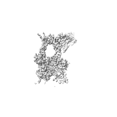
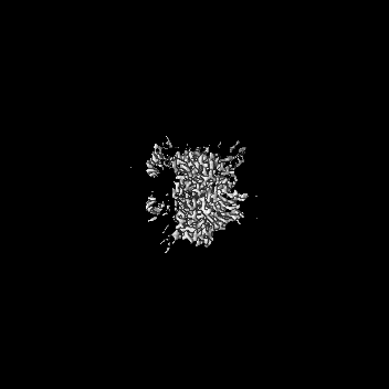
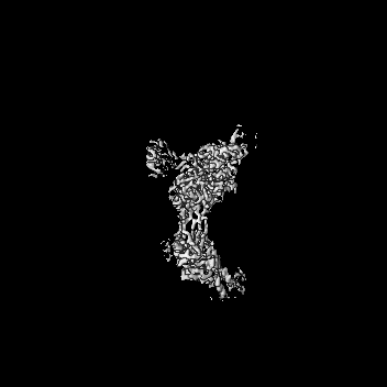
| Title | Human TOP3B-TDRD3 core complex in DNA religation state | |||||||||
 Map data Map data | ||||||||||
 Sample Sample |
| |||||||||
 Keywords Keywords | Topoisomerase / DNA / ISOMERASE-DNA complex | |||||||||
| Function / homology |  Function and homology information Function and homology informationDNA topoisomerase III-beta-TDRD3 complex / RNA topoisomerase activity / DNA topoisomerase activity / histone H3 reader activity / DNA topoisomerase / DNA topoisomerase type I (single strand cut, ATP-independent) activity / DNA topological change / histone H4 reader activity / condensed chromosome / chromosome segregation ...DNA topoisomerase III-beta-TDRD3 complex / RNA topoisomerase activity / DNA topoisomerase activity / histone H3 reader activity / DNA topoisomerase / DNA topoisomerase type I (single strand cut, ATP-independent) activity / DNA topological change / histone H4 reader activity / condensed chromosome / chromosome segregation / DNA recombination / transcription coactivator activity / DNA repair / chromatin binding / Golgi apparatus / DNA binding / RNA binding / nucleoplasm / nucleus / cytosol Similarity search - Function | |||||||||
| Biological species |  Homo sapiens (human) Homo sapiens (human) | |||||||||
| Method | single particle reconstruction / cryo EM / Resolution: 3.26 Å | |||||||||
 Authors Authors | Yang X / Chen X / Yang W / Pommier Y | |||||||||
| Funding support |  United States, 2 items United States, 2 items
| |||||||||
 Citation Citation | Journal: Nat Commun / Year: 2025 Title: Structural insights into human topoisomerase 3β DNA and RNA catalysis and nucleic acid gate dynamics. Authors: Xi Yang / Xuemin Chen / Wei Yang / Yves Pommier /  Abstract: Type IA topoisomerases (TopoIAs) are present in all living organisms. They resolve DNA/RNA catenanes, knots and supercoils by breaking and rejoining single-stranded DNA/RNA segments and allowing the ...Type IA topoisomerases (TopoIAs) are present in all living organisms. They resolve DNA/RNA catenanes, knots and supercoils by breaking and rejoining single-stranded DNA/RNA segments and allowing the passage of another nucleic acid segment through the break. Topoisomerase III-β (TOP3B), the only RNA topoisomerase in metazoans, promotes R-loop disassembly and translation of mRNAs. Defects in TOP3B lead to severe neurological diseases. We present a series of cryo-EM structures of human TOP3B with its cofactor TDRD3 during cleavage and rejoining of DNA or RNA, thus elucidating the roles of divalent metal ions and key enzyme residues in each step of the catalytic cycle. We also obtained the structure of an open-gate configuration that addresses the long-standing question of the strand-passage mechanism. Our studies reveal how TOP3B catalyzes both DNA and RNA relaxation, while TOP3A acts only on DNA. | |||||||||
| History |
|
- Structure visualization
| Supplemental images |
|---|
- Downloads & links
-EMDB archive
| Map data | emd_45379.map.gz | 144.2 MB | EMDB map data format | |
|---|---|---|---|---|
| Header (meta data) | emd-45379-v30.xmlemd-45379.xml | 16.6 KB 16.6 KB | Display Display | EMDB header |
| Images |  emd_45379.png emd_45379.png | 38.2 KB | ||
| Filedesc metadata | emd-45379.cif.gz | 6.5 KB | ||
| Others | emd_45379_half_map_1.map.gzemd_45379_half_map_2.map.gz | 154.3 MB 154.3 MB | ||
| Archive directory |  http://ftp.pdbj.org/pub/emdb/structures/EMD-45379ftp://ftp.pdbj.org/pub/emdb/structures/EMD-45379 http://ftp.pdbj.org/pub/emdb/structures/EMD-45379ftp://ftp.pdbj.org/pub/emdb/structures/EMD-45379 | HTTPS FTP |
-Related structure data
| Related structure data |  9ca1MC  9c9wC 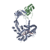 9c9yC 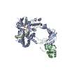 9ca0C  9ca4C  9cagC  9cahC  9cajC  9cakC  9calC M: atomic model generated by this map C: citing same article ( |
|---|---|
| Similar structure data |


-Links
| EMDB pages | EMDB (EBI/PDBe) / EMDataResource |
|---|---|
| Related items in Molecule of the Month |


-Map
| File | Download / File: emd_45379.map.gz / Format: CCP4 / Size: 166.4 MB / Type: IMAGE STORED AS FLOATING POINT NUMBER (4 BYTES) | ||||||||||||||||||||||||||||||||||||
|---|---|---|---|---|---|---|---|---|---|---|---|---|---|---|---|---|---|---|---|---|---|---|---|---|---|---|---|---|---|---|---|---|---|---|---|---|---|
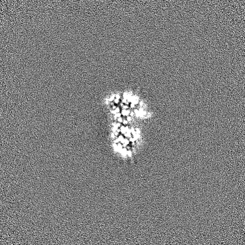
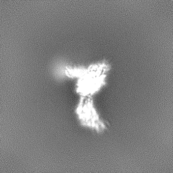
| Projections & slices | Image control
Images are generated by Spider. | ||||||||||||||||||||||||||||||||||||
| Voxel size | X=Y=Z: 0.83 Å | ||||||||||||||||||||||||||||||||||||
| Density |
| ||||||||||||||||||||||||||||||||||||
| Symmetry | Space group: 1 | ||||||||||||||||||||||||||||||||||||
| Details | EMDB XML:
|
 Z (Sec.)
 Y (Row.)
 X (Col.)


-Supplemental data
-Half map: #1
| File | emd_45379_half_map_1.map | ||||||||||||
|---|---|---|---|---|---|---|---|---|---|---|---|---|---|
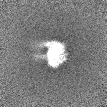
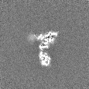
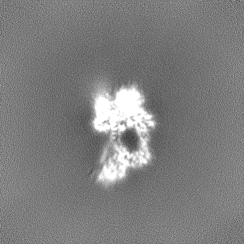
| Projections & Slices |
| ||||||||||||
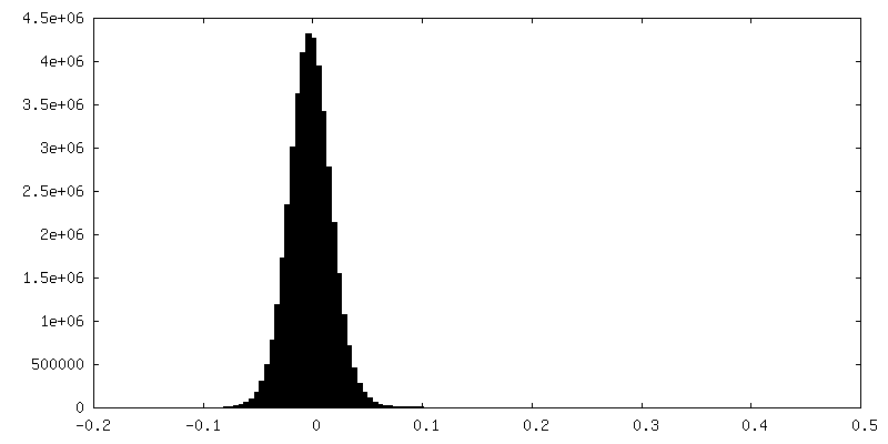
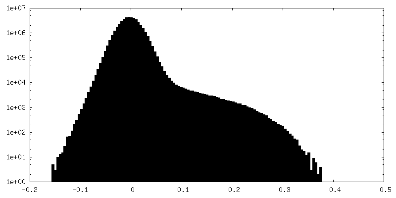
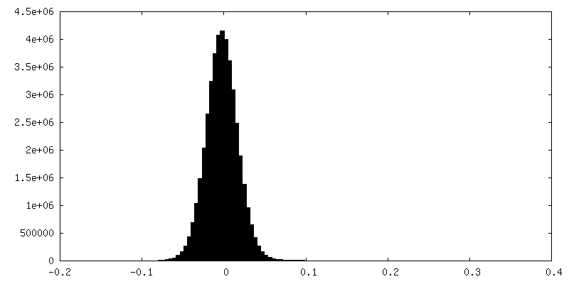
| Density Histograms |

-Half map: #2
| File | emd_45379_half_map_2.map | ||||||||||||
|---|---|---|---|---|---|---|---|---|---|---|---|---|---|
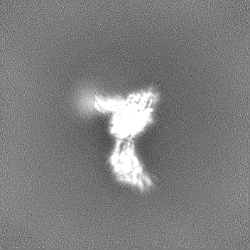
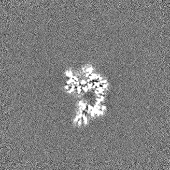
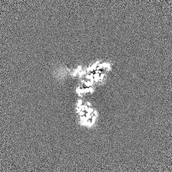
| Projections & Slices |
| ||||||||||||
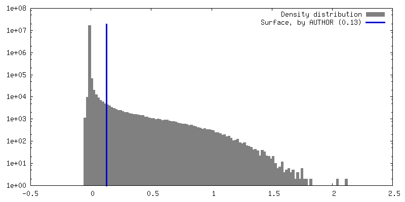
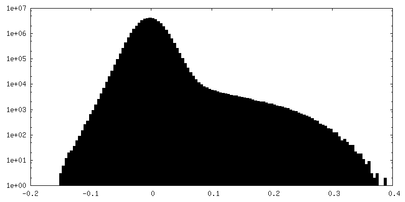
| Density Histograms |


- Sample components
-Entire : human TOP3B-TDRD3 core complex with DNA
| Entire | Name: human TOP3B-TDRD3 core complex with DNA |
|---|---|
| Components |
|
-Supramolecule #1: human TOP3B-TDRD3 core complex with DNA
| Supramolecule | Name: human TOP3B-TDRD3 core complex with DNA / type: complex / ID: 1 / Parent: 0 / Macromolecule list: #1-#4 |
|---|---|
| Source (natural) | Organism: Homo sapiens (human) |
-Macromolecule #1: DNA topoisomerase 3-beta-1
| Macromolecule | Name: DNA topoisomerase 3-beta-1 / type: protein_or_peptide / ID: 1 / Number of copies: 1 / Enantiomer: LEVO / EC number: DNA topoisomerase |
|---|---|
| Source (natural) | Organism: Homo sapiens (human) |
| Molecular weight | Theoretical: 69.119867 KDa |
| Recombinant expression | Organism: Homo sapiens (human) |
| Sequence | String: VMKTVLMVAE MPSLAQSIAK ILSRGSLSSH KGLNGACSVH EYTGTFAGQP VRFKMTSVCG HVMTLDFLGK YNKWDKVDPA ELFSQAPTE KKEANPKLNM VKFLQVEGRG CDYIVLWLDC DKEGENICFE VLDAVLPVMN KAHGGEKTVF RARFSSITDT D ICNAMACL ...String: VMKTVLMVAE MPSLAQSIAK ILSRGSLSSH KGLNGACSVH EYTGTFAGQP VRFKMTSVCG HVMTLDFLGK YNKWDKVDPA ELFSQAPTE KKEANPKLNM VKFLQVEGRG CDYIVLWLDC DKEGENICFE VLDAVLPVMN KAHGGEKTVF RARFSSITDT D ICNAMACL GEPDHNEALS VDARQELDLR IGCAFTRFQT KYFQGKYGDL DSSLISFGPC QTPTLGFCVE RHDKIQSFKP ET YWVLQAK VNTDKDRSLL LDWDRVRVFD REIAQMFLNM TKLEKEAQVE ATSRKEKAKQ RPLALNTVEM LRVASSSLGM GPQ HAMQTA ERLYTQGYIS (PTR)PRTETTHYP ENFDLKGSLR QQANHPYWAD TVKRLLAEGI NRPRKGHDAG DHPPITPMKS ATEAELGGD AWRLYEYITR HFIATVSHDC KYLQSTISFR IGPELFTCSG KTVLSPGFTE VMPWQSVPLE ESLPTCQRGD A FPVGEVKM LEKQTNPPDY LTEAELITLM EKHGIGTDAS IPVHINNICQ RNYVTVESGR RLKPTNLGIV LVHGYYKIDA EL VLPTIRS AVEKQLNLIA QGKADYRQVL GHTLDVFKRK FHYFVDSIAG MDELMEVSF UniProtKB: DNA topoisomerase 3-beta-1 |
-Macromolecule #2: Tudor domain-containing protein 3
| Macromolecule | Name: Tudor domain-containing protein 3 / type: protein_or_peptide / ID: 2 / Number of copies: 1 / Enantiomer: LEVO |
|---|---|
| Source (natural) | Organism: Homo sapiens (human) |
| Molecular weight | Theoretical: 17.428951 KDa |
| Recombinant expression | Organism: Homo sapiens (human) |
| Sequence | String: MAQVAGAALS QAGWYLSDEG IEACTSSPDK VNVNDIILIA LNTDLRTIGK KFLPSDINSG KVEKLEGPCV LQIQKIRNVA APKDNEESQ AAPRMLRLQM TDGHISCTAV EFSYMSKISL NTPPGTKVKL SGIVDIKNGF LLLNDSNTTV LGGEVEHLIE K W UniProtKB: Tudor domain-containing protein 3 |
-Macromolecule #3: DNA (5'-D(P*TP*AP*CP*TP*AP*AP*A)-3')
| Macromolecule | Name: DNA (5'-D(P*TP*AP*CP*TP*AP*AP*A)-3') / type: dna / ID: 3 / Number of copies: 1 / Classification: DNA |
|---|---|
| Source (natural) | Organism: Homo sapiens (human) |
| Molecular weight | Theoretical: 2.105437 KDa |
| Sequence | String: (DT)(DA)(DC)(DT)(DA)(DA)(DA) |
-Macromolecule #4: DNA (5'-D(*AP*TP*T)-3')
| Macromolecule | Name: DNA (5'-D(*AP*TP*T)-3') / type: dna / ID: 4 / Number of copies: 1 / Classification: DNA |
|---|---|
| Source (natural) | Organism: Homo sapiens (human) |
| Molecular weight | Theoretical: 876.635 Da |
| Sequence | String: (DA)(DT)(DT) |
-Macromolecule #5: MANGANESE (II) ION
| Macromolecule | Name: MANGANESE (II) ION / type: ligand / ID: 5 / Number of copies: 2 / Formula: MN |
|---|---|
| Molecular weight | Theoretical: 54.938 Da |
-Experimental details
-Structure determination
| Method | cryo EM |
|---|---|
 Processing Processing | single particle reconstruction |
| Aggregation state | particle |
-Sample preparation
| Buffer | pH: 6.8 |
|---|---|
| Vitrification | Cryogen name: ETHANE |
- Electron microscopy
| Microscope | FEI TALOS ARCTICA |
|---|---|
| Image recording | Film or detector model: GATAN K3 BIOQUANTUM (6k x 4k) / Average electron dose: 46.0 e/Å2 |
| Electron beam | Acceleration voltage: 200 kV / Electron source:  FIELD EMISSION GUN FIELD EMISSION GUN |
| Electron optics | Illumination mode: FLOOD BEAM / Imaging mode: BRIGHT FIELD / Nominal defocus max: 2.4 µm / Nominal defocus min: 0.8 µm |
| Experimental equipment |  Model: Talos Arctica / Image courtesy: FEI Company |
-Image processing
| Startup model | Type of model: NONE |
|---|---|
| Final reconstruction | Resolution.type: BY AUTHOR / Resolution: 3.26 Å / Resolution method: FSC 0.143 CUT-OFF / Number images used: 901517 |
| Initial angle assignment | Type: MAXIMUM LIKELIHOOD |
| Final angle assignment | Type: MAXIMUM LIKELIHOOD |
