[English] 日本語
 Yorodumi
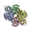
- EMDB-45298: Structure of endogenous asymmetric DPYSL2 from rat model of Alzhe... -
+ Open data
- Basic information
| Entry |  | |||||||||
|---|---|---|---|---|---|---|---|---|---|---|
| Title | Structure of endogenous asymmetric DPYSL2 from rat model of Alzheimer's disease | |||||||||
 Map data Map data | Main Sharpened Map | |||||||||
 Sample Sample |
| |||||||||
 Keywords Keywords | Dihydropyrimidinase-related protein 2 / NEUROPEPTIDE | |||||||||
| Function / homology |  Function and homology information Function and homology informationCRMPs in Sema3A signaling / hydrolase activity, acting on carbon-nitrogen (but not peptide) bonds, in cyclic amides / Recycling pathway of L1 / olfactory bulb development / positive regulation of glutamate secretion / regulation of neuron differentiation / regulation of axon extension / spinal cord development / synaptic vesicle transport / regulation of neuron projection development ...CRMPs in Sema3A signaling / hydrolase activity, acting on carbon-nitrogen (but not peptide) bonds, in cyclic amides / Recycling pathway of L1 / olfactory bulb development / positive regulation of glutamate secretion / regulation of neuron differentiation / regulation of axon extension / spinal cord development / synaptic vesicle transport / regulation of neuron projection development / regulation of postsynapse assembly / cytoskeleton organization / response to amphetamine / response to cocaine / Schaffer collateral - CA1 synapse / endocytosis / terminal bouton / myelin sheath / presynapse / growth cone / microtubule / cell differentiation / postsynaptic density / response to xenobiotic stimulus / axon / neuronal cell body / dendrite / synapse / protein kinase binding / glutamatergic synapse / protein-containing complex / identical protein binding / membrane / cytosol Similarity search - Function | |||||||||
| Biological species |  | |||||||||
| Method | single particle reconstruction / cryo EM / Resolution: 2.9 Å | |||||||||
 Authors Authors | Khalili Samani E / Keszei AFA / Mazhab-Jafari MT | |||||||||
| Funding support |  Canada, 1 items Canada, 1 items
| |||||||||
 Citation Citation |  Journal: Structure / Year: 2025 Journal: Structure / Year: 2025Title: Unveiling the structural proteome of an Alzheimer's disease rat brain model. Authors: Elnaz Khalili Samani / S M Naimul Hasan / Matthew Waas / Alexander F A Keszei / Xiaoxiao Xu / Mahtab Heydari / Mary Elizabeth Hill / JoAnne McLaurin / Thomas Kislinger / Mohammad T Mazhab-Jafari /  Abstract: Studying native protein structures at near-atomic resolution in a crowded environment presents challenges. Consequently, understanding the structural intricacies of proteins within pathologically ...Studying native protein structures at near-atomic resolution in a crowded environment presents challenges. Consequently, understanding the structural intricacies of proteins within pathologically affected tissues often relies on mass spectrometry and proteomic analysis. Here, we utilized cryoelectron microscopy (cryo-EM) and the Build and Retrieve (BaR) method to investigate protein complexes' structural characteristics such as post-translational modification, active site occupancy, and arrested conformational state in Alzheimer's disease (AD) using brain lysate from a rat model (TgF344-AD). Our findings reveal novel insights into the architecture of these complexes, corroborated through mass spectrometry analysis. Interestingly, it has been shown that the dysfunction of these protein complexes extends beyond AD, implicating them in cancer, as well as other neurodegenerative disorders such as Parkinson's disease, Huntington's disease, and schizophrenia. By elucidating these structural details, our work not only enhances our understanding of disease pathology but also suggests new avenues for future approaches in therapeutic intervention. | |||||||||
| History |
|
- Structure visualization
| Supplemental images |
|---|
- Downloads & links
-EMDB archive
| Map data |  emd_45298.map.gz emd_45298.map.gz | 15.8 MB |  EMDB map data format EMDB map data format | |
|---|---|---|---|---|
| Header (meta data) |  emd-45298-v30.xml emd-45298-v30.xml emd-45298.xml emd-45298.xml | 16.2 KB 16.2 KB | Display Display |  EMDB header EMDB header |
| Images |  emd_45298.png emd_45298.png | 57.3 KB | ||
| Filedesc metadata |  emd-45298.cif.gz emd-45298.cif.gz | 5.6 KB | ||
| Others |  emd_45298_half_map_1.map.gz emd_45298_half_map_1.map.gz emd_45298_half_map_2.map.gz emd_45298_half_map_2.map.gz | 28.3 MB 28.3 MB | ||
| Archive directory |  http://ftp.pdbj.org/pub/emdb/structures/EMD-45298 http://ftp.pdbj.org/pub/emdb/structures/EMD-45298 ftp://ftp.pdbj.org/pub/emdb/structures/EMD-45298 ftp://ftp.pdbj.org/pub/emdb/structures/EMD-45298 | HTTPS FTP |
-Related structure data
| Related structure data |  9c86MC  9c21C  9c28C  9c4eC C: citing same article ( M: atomic model generated by this map |
|---|---|
| Similar structure data | Similarity search - Function & homology  F&H Search F&H Search |
- Links
Links
| EMDB pages |  EMDB (EBI/PDBe) / EMDB (EBI/PDBe) /  EMDataResource EMDataResource |
|---|---|
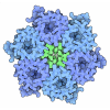
| Related items in Molecule of the Month |
- Map
Map
| File |  Download / File: emd_45298.map.gz / Format: CCP4 / Size: 30.5 MB / Type: IMAGE STORED AS FLOATING POINT NUMBER (4 BYTES) Download / File: emd_45298.map.gz / Format: CCP4 / Size: 30.5 MB / Type: IMAGE STORED AS FLOATING POINT NUMBER (4 BYTES) | ||||||||||||||||||||||||||||||||||||
|---|---|---|---|---|---|---|---|---|---|---|---|---|---|---|---|---|---|---|---|---|---|---|---|---|---|---|---|---|---|---|---|---|---|---|---|---|---|
| Annotation | Main Sharpened Map | ||||||||||||||||||||||||||||||||||||
| Projections & slices | Image control
Images are generated by Spider. | ||||||||||||||||||||||||||||||||||||
| Voxel size | X=Y=Z: 1.03 Å | ||||||||||||||||||||||||||||||||||||
| Density |
| ||||||||||||||||||||||||||||||||||||
| Symmetry | Space group: 1 | ||||||||||||||||||||||||||||||||||||
| Details | EMDB XML:
|
-Supplemental data
-Half map: Half Map A
| File | emd_45298_half_map_1.map | ||||||||||||
|---|---|---|---|---|---|---|---|---|---|---|---|---|---|
| Annotation | Half Map A | ||||||||||||
| Projections & Slices |
| ||||||||||||
| Density Histograms |
-Half map: Half Map B
| File | emd_45298_half_map_2.map | ||||||||||||
|---|---|---|---|---|---|---|---|---|---|---|---|---|---|
| Annotation | Half Map B | ||||||||||||
| Projections & Slices |
| ||||||||||||
| Density Histograms |
- Sample components
-Entire : Dihydropyrimidinase-related protein 2
| Entire | Name: Dihydropyrimidinase-related protein 2 |
|---|---|
| Components |
|
-Supramolecule #1: Dihydropyrimidinase-related protein 2
| Supramolecule | Name: Dihydropyrimidinase-related protein 2 / type: tissue / ID: 1 / Parent: 0 / Macromolecule list: #1 |
|---|---|
| Source (natural) | Organism:  |
-Macromolecule #1: Dihydropyrimidinase-related protein 2
| Macromolecule | Name: Dihydropyrimidinase-related protein 2 / type: protein_or_peptide / ID: 1 / Number of copies: 4 / Enantiomer: LEVO |
|---|---|
| Source (natural) | Organism:  |
| Molecular weight | Theoretical: 62.349352 KDa |
| Sequence | String: MSYQGKKNIP RITSDRLLIK GGKIVNDDQS FYADIYMEDG LIKQIGENLI VPGGVKTIEA HSRMVIPGGI DVHTRFQMPD QGMTSADDF FQGTKAALAG GTTMIIDHVV PEPGTSLLAA FDQWREWADS KSCCDYSLHV DITEWHKGIQ EEMEALVKDH G VNSFLVYM ...String: MSYQGKKNIP RITSDRLLIK GGKIVNDDQS FYADIYMEDG LIKQIGENLI VPGGVKTIEA HSRMVIPGGI DVHTRFQMPD QGMTSADDF FQGTKAALAG GTTMIIDHVV PEPGTSLLAA FDQWREWADS KSCCDYSLHV DITEWHKGIQ EEMEALVKDH G VNSFLVYM AFKDRFQLTD SQIYEVLSVI RDIGAIAQVH AENGDIIAEE QQRILDLGIT GPEGHVLSRP EEVEAEAVNR SI TIANQTN CPLYVTKVMS KSAAEVIAQA RKKGTVVYGE PITASLGTDG SHYWSKNWAK AAAFVTSPPL SPDPTTPDFL NSL LSCGDL QVTGSAHCTF NTAQKAVGKD NFTLIPEGTN GTEERMSVIW DKAVVTGKMD ENQFVAVTST NAAKVFNLYP RKGR ISVGS DADLVIWDPD SVKTISAKTH NSALEYNIFE GMECRGSPLV VISQGKIVLE DGTLHVTEGS GRYIPRKPFP DFVYK RIKA RSRLAELRGV PRGLYDGPVC EVSVTPKTVT PASSAKTSPA KQQAPPVRNL HQSGFSLSGA QIDDNIPRRT TQRIVA PPG GRANITSLG UniProtKB: Dihydropyrimidinase-related protein 2 |
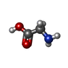
-Macromolecule #2: GLYCINE
| Macromolecule | Name: GLYCINE / type: ligand / ID: 2 / Number of copies: 1 / Formula: GLY |
|---|---|
| Source (natural) | Organism:  |
| Molecular weight | Theoretical: 75.067 Da |
| Chemical component information |  ChemComp-GLY: |
-Experimental details
-Structure determination
| Method | cryo EM |
|---|---|
 Processing Processing | single particle reconstruction |
| Aggregation state | particle |
- Sample preparation
| Buffer | pH: 7.4 |
|---|---|
| Grid | Model: Homemade / Support film - Material: GOLD / Support film - topology: HOLEY / Support film - Film thickness: 3 |
| Vitrification | Cryogen name: ETHANE / Chamber humidity: 90 % / Chamber temperature: 278 K / Instrument: FEI VITROBOT MARK IV |
| Details | Cytosolic fraction of rat brain |
- Electron microscopy
| Microscope | FEI TITAN KRIOS |
|---|---|
| Image recording | Film or detector model: FEI FALCON IV (4k x 4k) / Average electron dose: 40.0 e/Å2 |
| Electron beam | Acceleration voltage: 300 kV / Electron source:  FIELD EMISSION GUN FIELD EMISSION GUN |
| Electron optics | Illumination mode: FLOOD BEAM / Imaging mode: BRIGHT FIELD / Nominal defocus max: 5.0 µm / Nominal defocus min: 0.5 µm |
| Experimental equipment |  Model: Titan Krios / Image courtesy: FEI Company |
- Image processing
| Startup model | Type of model: NONE / Details: Ab initio reconstruction |
|---|---|
| Final reconstruction | Resolution.type: BY AUTHOR / Resolution: 2.9 Å / Resolution method: FSC 0.143 CUT-OFF / Software - Name: cryoSPARC / Number images used: 389444 |
| Initial angle assignment | Type: NOT APPLICABLE |
| Final angle assignment | Type: NOT APPLICABLE |
-Atomic model buiding 1
| Initial model | Chain - Source name: SwissModel / Chain - Initial model type: in silico model |
|---|---|
| Refinement | Space: REAL / Protocol: FLEXIBLE FIT |
| Output model |  PDB-9c86: |
 Movie
Movie Controller


 Z (Sec.)
 Y (Row.)
 X (Col.)


