 Movie
Movie Controller

[English] 日本語
 Yorodumi
- EMDB-47474: cryoEM map of endogenous V1 region of V-ATPase from rat model of ... -
+ Open data

- Basic information
| Entry |  | |||||||||
|---|---|---|---|---|---|---|---|---|---|---|
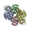
| Title | cryoEM map of endogenous V1 region of V-ATPase from rat model of Alzheimer's disease | |||||||||
 Map data Map data | map of endogenous V1 region of V-ATPase from rat model of Alzheimer's disease | |||||||||
 Sample Sample |
| |||||||||
 Keywords Keywords | V1 region / V-ATPase / PROTON TRANSPORT | |||||||||
| Biological species |  | |||||||||
| Method | single particle reconstruction / cryo EM / Resolution: 4.24 Å | |||||||||
 Authors Authors | Khalili Samani E / Keszei AFA / Mazhab-Jafari MT | |||||||||
| Funding support |  Canada, 1 items Canada, 1 items
| |||||||||
 Citation Citation | Journal: Structure / Year: 2025 Title: Unveiling the structural proteome of an Alzheimer's disease rat brain model. Authors: Elnaz Khalili Samani / S M Naimul Hasan / Matthew Waas / Alexander F A Keszei / Xiaoxiao Xu / Mahtab Heydari / Mary Elizabeth Hill / JoAnne McLaurin / Thomas Kislinger / Mohammad T Mazhab-Jafari / Abstract: Studying native protein structures at near-atomic resolution in a crowded environment presents challenges. Consequently, understanding the structural intricacies of proteins within pathologically ...Studying native protein structures at near-atomic resolution in a crowded environment presents challenges. Consequently, understanding the structural intricacies of proteins within pathologically affected tissues often relies on mass spectrometry and proteomic analysis. Here, we utilized cryoelectron microscopy (cryo-EM) and the Build and Retrieve (BaR) method to investigate protein complexes' structural characteristics such as post-translational modification, active site occupancy, and arrested conformational state in Alzheimer's disease (AD) using brain lysate from a rat model (TgF344-AD). Our findings reveal novel insights into the architecture of these complexes, corroborated through mass spectrometry analysis. Interestingly, it has been shown that the dysfunction of these protein complexes extends beyond AD, implicating them in cancer, as well as other neurodegenerative disorders such as Parkinson's disease, Huntington's disease, and schizophrenia. By elucidating these structural details, our work not only enhances our understanding of disease pathology but also suggests new avenues for future approaches in therapeutic intervention. | |||||||||
| History |
|
- Structure visualization
| Supplemental images |
|---|

- Downloads & links
-EMDB archive
| Map data | emd_47474.map.gz | 31.8 MB |  EMDB map data format EMDB map data format | |
|---|---|---|---|---|
| Header (meta data) | emd-47474-v30.xmlemd-47474.xml | 13.3 KB 13.3 KB | Display Display | EMDB header |
| Images |  emd_47474.png emd_47474.png | 62.4 KB | ||
| Filedesc metadata | emd-47474.cif.gz | 4 KB | ||
| Others | emd_47474_half_map_1.map.gzemd_47474_half_map_2.map.gz | 59.3 MB 59.3 MB | ||
| Archive directory |  http://ftp.pdbj.org/pub/emdb/structures/EMD-47474ftp://ftp.pdbj.org/pub/emdb/structures/EMD-47474 http://ftp.pdbj.org/pub/emdb/structures/EMD-47474ftp://ftp.pdbj.org/pub/emdb/structures/EMD-47474 | HTTPS FTP |
-Related structure data


-Links
| EMDB pages | EMDB (EBI/PDBe) / EMDataResource |
|---|
-Map
| File | Download / File: emd_47474.map.gz / Format: CCP4 / Size: 64 MB / Type: IMAGE STORED AS FLOATING POINT NUMBER (4 BYTES) | ||||||||||||||||||||||||||||||||||||
|---|---|---|---|---|---|---|---|---|---|---|---|---|---|---|---|---|---|---|---|---|---|---|---|---|---|---|---|---|---|---|---|---|---|---|---|---|---|
| Annotation | map of endogenous V1 region of V-ATPase from rat model of Alzheimer's disease | ||||||||||||||||||||||||||||||||||||
| Projections & slices | Image control
Images are generated by Spider. | ||||||||||||||||||||||||||||||||||||
| Voxel size | X=Y=Z: 1.03 Å | ||||||||||||||||||||||||||||||||||||
| Density |
| ||||||||||||||||||||||||||||||||||||
| Symmetry | Space group: 1 | ||||||||||||||||||||||||||||||||||||
| Details | EMDB XML:
|
 Z (Sec.)
 Y (Row.)
 X (Col.)


-Supplemental data
-Half map: Half map A
| File | emd_47474_half_map_1.map | ||||||||||||
|---|---|---|---|---|---|---|---|---|---|---|---|---|---|
| Annotation | Half map A | ||||||||||||
| Projections & Slices |
| ||||||||||||
| Density Histograms |


-Half map: Half map B
| File | emd_47474_half_map_2.map | ||||||||||||
|---|---|---|---|---|---|---|---|---|---|---|---|---|---|
| Annotation | Half map B | ||||||||||||
| Projections & Slices |
| ||||||||||||
| Density Histograms |


- Sample components
-Entire : V-ATPase
| Entire | Name: V-ATPase |
|---|---|
| Components |
|
-Supramolecule #1: V-ATPase
| Supramolecule | Name: V-ATPase / type: tissue / ID: 1 / Parent: 0 / Macromolecule list: #1 |
|---|---|
| Source (natural) | Organism: |
-Experimental details
-Structure determination
| Method | cryo EM |
|---|---|
 Processing Processing | single particle reconstruction |
| Aggregation state | particle |
-Sample preparation
| Buffer | pH: 7.4 |
|---|---|
| Grid | Model: Quantifoil R2/4 / Support film - Material: GOLD / Support film - topology: HOLEY / Support film - Film thickness: 3 / Pretreatment - Type: GLOW DISCHARGE / Pretreatment - Time: 25 sec. / Pretreatment - Atmosphere: AIR |
| Vitrification | Cryogen name: ETHANE / Chamber humidity: 90 % / Chamber temperature: 278 K / Instrument: FEI VITROBOT MARK IV |
| Details | Cytosolic fraction of rat brain |
- Electron microscopy
| Microscope | TFS KRIOS |
|---|---|
| Image recording | Film or detector model: FEI FALCON IV (4k x 4k) / Average electron dose: 40.0 e/Å2 |
| Electron beam | Acceleration voltage: 300 kV / Electron source:  FIELD EMISSION GUN FIELD EMISSION GUN |
| Electron optics | Illumination mode: FLOOD BEAM / Imaging mode: BRIGHT FIELD / Nominal defocus max: 5.0 µm / Nominal defocus min: 0.5 µm |
| Experimental equipment |  Model: Titan Krios / Image courtesy: FEI Company |
-Image processing
| Startup model | Type of model: NONE / Details: Ab initio reconstruction |
|---|---|
| Final reconstruction | Resolution.type: BY AUTHOR / Resolution: 4.24 Å / Resolution method: FSC 0.143 CUT-OFF / Software - Name: cryoSPARC / Number images used: 8906 |
| Initial angle assignment | Type: NOT APPLICABLE |
| Final angle assignment | Type: NOT APPLICABLE |
