 Movie
Movie Controller


+ Open data

- Basic information
| Entry |  | |||||||||
|---|---|---|---|---|---|---|---|---|---|---|
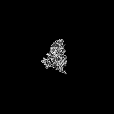
| Title | Group IIC intron embedded with the TPP riboswitch | |||||||||
 Map data Map data | ||||||||||
 Sample Sample |
| |||||||||
 Keywords Keywords | Intron / Splicing / Ribozyme / RNA | |||||||||
| Biological species |  Oceanobacillus iheyensis (bacteria) Oceanobacillus iheyensis (bacteria) | |||||||||
| Method | single particle reconstruction / cryo EM / Resolution: 2.41 Å | |||||||||
 Authors Authors | Toor N | |||||||||
| Funding support |  United States, 2 items United States, 2 items
| |||||||||
 Citation Citation | Journal: Nat Commun / Year: 2025 Title: Scaffold-enabled high-resolution cryo-EM structure determination of RNA. Authors: Daniel B Haack / Boris Rudolfs / Shouhong Jin / Alexandra Khitun / Kevin M Weeks / Navtej Toor / Abstract: Cryo-EM structure determination of protein-free RNAs has remained difficult with most attempts yielding low to moderate resolution and lacking nucleotide-level detail. These difficulties are ...Cryo-EM structure determination of protein-free RNAs has remained difficult with most attempts yielding low to moderate resolution and lacking nucleotide-level detail. These difficulties are compounded for small RNAs as cryo-EM is inherently more difficult for lower molecular weight macromolecules. Here we present a strategy for fusing small RNAs to a group II intron that yields high resolution structures of the appended RNA. We demonstrate this technology by determining the structures of the 86-nucleotide (nt) thiamine pyrophosphate (TPP) riboswitch aptamer domain and the recently described 210-nt raiA bacterial non-coding RNA involved in sporulation and biofilm formation. In the case of the TPP riboswitch aptamer domain, the scaffolding approach allowed visualization of the riboswitch ligand binding pocket at 2.5 Å resolution. We also determined the structure of the ligand-free apo state and observe that the aptamer domain of the riboswitch adopts an open Y-shaped conformation in the absence of ligand. Using this scaffold approach, we determined the structure of raiA at 2.5 Å in the core. Our versatile scaffolding strategy enables efficient RNA structure determination for a broad range of small to moderate-sized RNAs, which were previously intractable for high-resolution cryo-EM studies. | |||||||||
| History |
|
- Structure visualization
| Supplemental images |
|---|

- Downloads & links
-EMDB archive
| Map data | emd_45248.map.gz | 122.9 MB |  EMDB map data format EMDB map data format | |
|---|---|---|---|---|
| Header (meta data) | emd-45248-v30.xmlemd-45248.xml | 20.6 KB 20.6 KB | Display Display | EMDB header |
| Images |  emd_45248.png emd_45248.png | 37.5 KB | ||
| Filedesc metadata | emd-45248.cif.gz | 5.8 KB | ||
| Others | emd_45248_additional_1.map.gzemd_45248_half_map_1.map.gzemd_45248_half_map_2.map.gz | 230 MB 226.5 MB 226.5 MB | ||
| Archive directory |  http://ftp.pdbj.org/pub/emdb/structures/EMD-45248ftp://ftp.pdbj.org/pub/emdb/structures/EMD-45248 http://ftp.pdbj.org/pub/emdb/structures/EMD-45248ftp://ftp.pdbj.org/pub/emdb/structures/EMD-45248 | HTTPS FTP |
-Related structure data
| Related structure data |  9c6jMC  9c6iC  9c6kC C: citing same article ( M: atomic model generated by this map |
|---|


-Links
| EMDB pages | EMDB (EBI/PDBe) / EMDataResource |
|---|
-Map
| File | Download / File: emd_45248.map.gz / Format: CCP4 / Size: 244.1 MB / Type: IMAGE STORED AS FLOATING POINT NUMBER (4 BYTES) | ||||||||||||||||||||||||||||||||||||
|---|---|---|---|---|---|---|---|---|---|---|---|---|---|---|---|---|---|---|---|---|---|---|---|---|---|---|---|---|---|---|---|---|---|---|---|---|---|
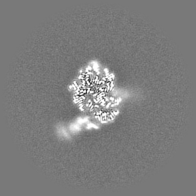
| Projections & slices | Image control
Images are generated by Spider. | ||||||||||||||||||||||||||||||||||||
| Voxel size | X=Y=Z: 0.811 Å | ||||||||||||||||||||||||||||||||||||
| Density |
| ||||||||||||||||||||||||||||||||||||
| Symmetry | Space group: 1 | ||||||||||||||||||||||||||||||||||||
| Details | EMDB XML:
|
 Z (Sec.)
 Y (Row.)
 X (Col.)


-Supplemental data
-Additional map: #1
| File | emd_45248_additional_1.map | ||||||||||||
|---|---|---|---|---|---|---|---|---|---|---|---|---|---|
| Projections & Slices |
| ||||||||||||
| Density Histograms |


-Half map: #1
| File | emd_45248_half_map_1.map | ||||||||||||
|---|---|---|---|---|---|---|---|---|---|---|---|---|---|
| Projections & Slices |
| ||||||||||||
| Density Histograms |


-Half map: #2
| File | emd_45248_half_map_2.map | ||||||||||||
|---|---|---|---|---|---|---|---|---|---|---|---|---|---|
| Projections & Slices |
| ||||||||||||
| Density Histograms |


- Sample components
-Entire : Group IIC intron embedded with the TPP riboswitch
| Entire | Name: Group IIC intron embedded with the TPP riboswitch |
|---|---|
| Components |
|
-Supramolecule #1: Group IIC intron embedded with the TPP riboswitch
| Supramolecule | Name: Group IIC intron embedded with the TPP riboswitch / type: complex / ID: 1 / Parent: 0 / Macromolecule list: #1 |
|---|---|
| Source (natural) | Organism: Oceanobacillus iheyensis (bacteria) |
| Molecular weight | Theoretical: 160 KDa |
-Macromolecule #1: Group IIC intron embedded with the TPP riboswitch
| Macromolecule | Name: Group IIC intron embedded with the TPP riboswitch / type: rna / ID: 1 / Number of copies: 1 |
|---|---|
| Source (natural) | Organism: Oceanobacillus iheyensis (bacteria) |
| Molecular weight | Theoretical: 160.467078 KDa |
| Sequence | String: GUUAUGUGUG CCCGGCAUGG GUGCAGUCUA UAGGGUGAGA GUCCCGAACU GUGAAGGCAG AAGUAACAGU UAGCCUAACG CAAGGGUGU CCGUGGCGAC AUGGAAUCUG AAGGAAGCGG ACGGCAAACC UUCGGUCUGA GGAACACGAA CUUCAUAUGA G GCUAGGUA ...String: GUUAUGUGUG CCCGGCAUGG GUGCAGUCUA UAGGGUGAGA GUCCCGAACU GUGAAGGCAG AAGUAACAGU UAGCCUAACG CAAGGGUGU CCGUGGCGAC AUGGAAUCUG AAGGAAGCGG ACGGCAAACC UUCGGUCUGA GGAACACGAA CUUCAUAUGA G GCUAGGUA UCAAUGGAUG AGUUUGCAUA ACAAAACAAA GUCCUUUCUG CCAAAGUUGG UACAGAGUAA AUGAAGCAGA UU GAUGAAG GGAAAGACUG CAUUCUUACC CGGGGAGGUC UGGAAACAGA AGUCAGCAGA AGUCAUAGUA CCCUCAGUAC UCG GGGUGC CCUUCUGCGU GAAGGCUGAG AAAUACCCGU AUCACCUGAU CUGGAUAAUG CCAGCGUAGG GAAGUGCUGA GGGG AAGGA CGGAACAAGU AUGGCGUUCG CGCCUAAGCU UGAACCACCG UAUACCGAAC GGUACGUACG GUGGUGUGAG AGGAG UUCG CUCUACUCUA U |
-Macromolecule #2: MAGNESIUM ION
| Macromolecule | Name: MAGNESIUM ION / type: ligand / ID: 2 / Number of copies: 21 / Formula: MG |
|---|---|
| Molecular weight | Theoretical: 24.305 Da |
-Experimental details
-Structure determination
| Method | cryo EM |
|---|---|
 Processing Processing | single particle reconstruction |
| Aggregation state | particle |
-Sample preparation
| Buffer | pH: 6.5 |
|---|---|
| Vitrification | Cryogen name: ETHANE-PROPANE |
- Electron microscopy
| Microscope | FEI TITAN KRIOS |
|---|---|
| Image recording | Film or detector model: GATAN K3 BIOQUANTUM (6k x 4k) / Average electron dose: 50.0 e/Å2 |
| Electron beam | Acceleration voltage: 300 kV / Electron source:  FIELD EMISSION GUN FIELD EMISSION GUN |
| Electron optics | Illumination mode: SPOT SCAN / Imaging mode: BRIGHT FIELD / Nominal defocus max: 2.0 µm / Nominal defocus min: 1.0 µm |
| Experimental equipment |  Model: Titan Krios / Image courtesy: FEI Company |
