[English] 日本語
Yorodumi
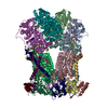
- EMDB-4481: Complex III2 focused refinement from Ovine respiratory supercompl... -

+
Open data


ID or keywords:

Loading...

-
Basic information

Entry
Database: EMDB / ID: EMD-4481
TitleComplex III2 focused refinement from Ovine respiratory supercomplex I+III2
Map dataFocused refinement around complex III2 from ovine mitochondrial supercomplex I III2
Sample
  • Complex: Ovine mitochondrial SC I+III2
    • Protein or peptide: x 10 types
  • Ligand: x 6 types
Keywordscomplex III / cellular respiration / mitochondria / ELECTRON TRANSPORT
Function / homology
Function and homology information


: / : / quinol-cytochrome-c reductase / ubiquinol-cytochrome-c reductase activity / mitochondrial electron transport, ubiquinol to cytochrome c / membrane => GO:0016020 / : / metalloendopeptidase activity / 2 iron, 2 sulfur cluster binding / mitochondrial inner membrane ...: / : / quinol-cytochrome-c reductase / ubiquinol-cytochrome-c reductase activity / mitochondrial electron transport, ubiquinol to cytochrome c / membrane => GO:0016020 / : / metalloendopeptidase activity / 2 iron, 2 sulfur cluster binding / mitochondrial inner membrane / electron transfer activity / ubiquitin protein ligase binding / heme binding / proteolysis / nucleoplasm / metal ion binding
Similarity search - Function
Ubiquinol-cytochrome c reductase 8kDa, N-terminal / Ubiquinol-cytochrome c reductase 8 kDa, N-terminal / Cytochrome b-c1 complex, subunit 6 / Globular protein, non-globular alpha/beta subunit / Cytochrome b-c1 complex subunit 8 / UcrQ family / Cytochrome bc1 complex subunit Rieske, transmembrane domain superfamily / Cytochrome b-c1 complex subunit 7 / Cytochrome b-c1 complex subunit 7 superfamily / Ubiquinol-cytochrome C reductase complex 14kD subunit ...Ubiquinol-cytochrome c reductase 8kDa, N-terminal / Ubiquinol-cytochrome c reductase 8 kDa, N-terminal / Cytochrome b-c1 complex, subunit 6 / Globular protein, non-globular alpha/beta subunit / Cytochrome b-c1 complex subunit 8 / UcrQ family / Cytochrome bc1 complex subunit Rieske, transmembrane domain superfamily / Cytochrome b-c1 complex subunit 7 / Cytochrome b-c1 complex subunit 7 superfamily / Ubiquinol-cytochrome C reductase complex 14kD subunit / Cytochrome b-c1 complex subunit 9 / Cytochrome b-c1 complex subunit 8 superfamily / Cytochrome b-c1 complex subunit 9 superfamily / Ubiquinol-cytochrome C reductase, UQCRX/QCR9 like / Cytochrome b-c1 complex subunit Rieske, transmembrane domain / Ubiquinol cytochrome reductase transmembrane region / Ubiquinol-cytochrome C reductase hinge domain / Ubiquinol-cytochrome C reductase hinge domain superfamily / Ubiquinol-cytochrome C reductase hinge protein / Cytochrome c1, transmembrane anchor, C-terminal / : / Cytochrome b / : / Ubiquinol-cytochrome c reductase, iron-sulphur subunit / Cytochrome c1 / Cytochrome C1 family / : / Cytochrome b/b6, C-terminal / Cytochrome b(C-terminal)/b6/petD / Cytochrome b/b6 C-terminal region profile. / Cytochrome b/b6, C-terminal domain superfamily / Cytochrome b/b6/petB / Peptidase M16, zinc-binding site / Insulinase family, zinc-binding region signature. / Rieske iron-sulphur protein, C-terminal / Cytochrome b/b6, N-terminal / Cytochrome b/b6-like domain superfamily / Cytochrome b/b6 N-terminal region profile. / Di-haem cytochrome, transmembrane / Rieske iron-sulphur protein / Peptidase M16, C-terminal / Peptidase M16 inactive domain / Peptidase M16, N-terminal / Insulinase (Peptidase family M16) / Metalloenzyme, LuxS/M16 peptidase-like / Rieske [2Fe-2S] domain / Rieske [2Fe-2S] iron-sulphur domain / Rieske [2Fe-2S] iron-sulfur domain profile. / Rieske [2Fe-2S] iron-sulphur domain superfamily / Cytochrome c family profile. / Cytochrome c-like domain / Cytochrome c-like domain superfamily
Similarity search - Domain/homology
Cytochrome b-c1 complex subunit 6 / Cytochrome b / Cytochrome b-c1 complex subunit Rieske, mitochondrial / Cytochrome b-c1 complex subunit 7 / Complex III subunit 9 / Cytochrome b-c1 complex subunit 8 / Cytochrome b-c1 complex subunit 6 / cytochrome c1, heme protein, mitochondrial / Cytochrome b-c1 complex subunit 2, mitochondrial / Cytochrome b-c1 complex subunit 1, mitochondrial
Similarity search - Component
Biological speciesOvis aries (sheep)
Methodsingle particle reconstruction / cryo EM / Resolution: 3.9 Å
AuthorsLetts JA / Sazanov LA
Funding support Austria, 1 items
OrganizationGrant numberCountry
European Research Council701309 Austria
CitationJournal: Mol Cell / Year: 2019
Title: Structures of Respiratory Supercomplex I+III Reveal Functional and Conformational Crosstalk.
Authors: James A Letts / Karol Fiedorczuk / Gianluca Degliesposti / Mark Skehel / Leonid A Sazanov /
Abstract: The mitochondrial electron transport chain complexes are organized into supercomplexes (SCs) of defined stoichiometry, which have been proposed to regulate electron flux via substrate channeling. We ...The mitochondrial electron transport chain complexes are organized into supercomplexes (SCs) of defined stoichiometry, which have been proposed to regulate electron flux via substrate channeling. We demonstrate that CoQ trapping in the isolated SC I+III limits complex (C)I turnover, arguing against channeling. The SC structure, resolved at up to 3.8 Å in four distinct states, suggests that CoQ oxidation may be rate limiting because of unequal access of CoQ to the active sites of CIII. CI shows a transition between "closed" and "open" conformations, accompanied by the striking rotation of a key transmembrane helix. Furthermore, the state of CI affects the conformational flexibility within CIII, demonstrating crosstalk between the enzymes. CoQ was identified at only three of the four binding sites in CIII, suggesting that interaction with CI disrupts CIII symmetry in a functionally relevant manner. Together, these observations indicate a more nuanced functional role for the SCs.
History
DepositionDec 18, 2018-
Header (metadata) releaseAug 21, 2019-
Map releaseAug 21, 2019-
UpdateNov 13, 2024-
Current statusNov 13, 2024Processing site: PDBe / Status: Released

-
Structure visualization

Movie
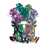
  • Surface view with section colored by density value
  • Surface level: 0.14
  • Imaged by UCSF Chimera
  • Download
  • Surface view colored by height
  • Surface level: 0.14
  • Imaged by UCSF Chimera
  • Download
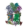
  • Surface view with fitted model
  • Atomic models: PDB-6q9e
  • Surface level: 0.14
  • Imaged by UCSF Chimera
  • Download
Movie viewer
Structure viewerEM map:
SurfViewMolmilJmol/JSmol
Supplemental images

Downloads & links

-
Map

FileDownload / File: emd_4481.map.gz / Format: CCP4 / Size: 184 MB / Type: IMAGE STORED AS FLOATING POINT NUMBER (4 BYTES)
AnnotationFocused refinement around complex III2 from ovine mitochondrial supercomplex I III2
Projections & slices

Image control

Size
Brightness
Contrast
Others
AxesZ (Sec.)Y (Row.)X (Col.)
1.4 Å/pix.
x 364 pix.
= 509.6 Å
1.4 Å/pix.
x 364 pix.
= 509.6 Å
1.4 Å/pix.
x 364 pix.
= 509.6 Å

Surface

Projections

Slices (1/3)

Slices (1/2)

Slices (2/3)

Images are generated by Spider.

Voxel sizeX=Y=Z: 1.4 Å
Density
Contour LevelBy AUTHOR: 0.14 / Movie #1: 0.14
Minimum - Maximum-0.32649752 - 0.7996147
Average (Standard dev.)-0.000016271637 (±0.017880779)
SymmetrySpace group: 1
Details

EMDB XML:

Map geometry
Axis orderXYZ
Origin000
Dimensions364364364
Spacing364364364
CellA=B=C: 509.6 Å
α=β=γ: 90.0 °

CCP4 map header:

modeImage stored as Reals
Å/pix. X/Y/Z1.41.41.4
M x/y/z364364364
origin x/y/z0.0000.0000.000
length x/y/z509.600509.600509.600
α/β/γ90.00090.00090.000
MAP C/R/S123
start NC/NR/NS000
NC/NR/NS364364364
D min/max/mean-0.3260.800-0.000

-
Supplemental data

-
Mask #1

Fileemd_4481_msk_1.map
Projections & Slices
AxesZYX

Projections

Slices (1/2)
Density Histograms

-
Half map: Focused refinement around complex III2 from ovine mitochondrial...

Fileemd_4481_half_map_1.map
AnnotationFocused refinement around complex III2 from ovine mitochondrial supercomplex I III2. Half map 1
Projections & Slices
AxesZYX

Projections

Slices (1/2)
Density Histograms

-
Half map: Focused refinement around complex III2 from ovine mitochondrial...

Fileemd_4481_half_map_2.map
AnnotationFocused refinement around complex III2 from ovine mitochondrial supercomplex I III2. Half map 2
Projections & Slices
AxesZYX

Projections

Slices (1/2)
Density Histograms

-
Sample components

+
Entire : Ovine mitochondrial SC I+III2

EntireName: Ovine mitochondrial SC I+III2
Components
  • Complex: Ovine mitochondrial SC I+III2
    • Protein or peptide: Ubiquinol-cytochrome c reductase core protein 1
    • Protein or peptide: Ubiquinol-cytochrome c reductase core protein 2
    • Protein or peptide: Cytochrome b
    • Protein or peptide: Cytochrome c1
    • Protein or peptide: Cytochrome b-c1 complex subunit Rieske, mitochondrial
    • Protein or peptide: Cytochrome b-c1 complex subunit 7
    • Protein or peptide: Ubiquinol-cytochrome c reductase complex III subunit VII
    • Protein or peptide: Cytochrome b-c1 complex subunit 6
    • Protein or peptide: Cytochrome b-c1 complex subunit Rieske, mitochondrial
    • Protein or peptide: Ubiquinol-cytochrome c reductase, complex III subunit X
  • Ligand: PROTOPORPHYRIN IX CONTAINING FE
  • Ligand: 1,2-Distearoyl-sn-glycerophosphoethanolamine
  • Ligand: CARDIOLIPIN
  • Ligand: UBIQUINONE-10
  • Ligand: HEME C
  • Ligand: FE2/S2 (INORGANIC) CLUSTER

+
Supramolecule #1: Ovine mitochondrial SC I+III2

SupramoleculeName: Ovine mitochondrial SC I+III2 / type: complex / ID: 1 / Parent: 0 / Macromolecule list: #1-#10
Details: Complex III2 focused refinement from ovine respiratory SC I+III2
Source (natural)Organism: Ovis aries (sheep) / Organ: Heart / Tissue: Cardiac / Organelle: Mitochondria / Location in cell: Mitochondrial inner membrane
Molecular weightTheoretical: 1.4 MDa

+
Macromolecule #1: Ubiquinol-cytochrome c reductase core protein 1

MacromoleculeName: Ubiquinol-cytochrome c reductase core protein 1 / type: protein_or_peptide / ID: 1 / Number of copies: 2 / Enantiomer: LEVO
Source (natural)Organism: Ovis aries (sheep) / Organ: Heart / Tissue: Cardiac
Molecular weightTheoretical: 49.385414 KDa
SequenceString: TATYAQALQS VPETQVSQLD NGLRVASEQS SQPTCTVGVW IDAGSRYETE KNNGAGYFVE HLAFKGTKNR PGNALEKEVE SMGAHLNAY STREHTAYYI KALSKDLPKA VELLADIVQN CSLEDSQIEK ERDVILQELQ ENDTSMRDVV FNYLHATAFQ G TPLAQSVE ...String:
TATYAQALQS VPETQVSQLD NGLRVASEQS SQPTCTVGVW IDAGSRYETE KNNGAGYFVE HLAFKGTKNR PGNALEKEVE SMGAHLNAY STREHTAYYI KALSKDLPKA VELLADIVQN CSLEDSQIEK ERDVILQELQ ENDTSMRDVV FNYLHATAFQ G TPLAQSVE GPSENVRKLS RADLTEYLSR HYKAPRMVLA AAGGLEHRQL LDLAQKHFSG LSGTYDEDAV PTLTPCRFTG SE IRHREDG LPLAHVAIAV EGPGWANPDN VALQVANAII GHYDCTYGGG MHLSSPLASV AVTNKLCQSF QTFNICYADT GLL GAHFVC DHMSIDDMMF VLQGQWMRLC TSATESEVVR GKNLLRNALV SHLDGTTPVC EDIGRSLLTY GRRIPLAEWE SRIA EVDAR VVREVCSKYF YDQCPAVAGF GPIEQLPDYN RIRSGMFWLR F

UniProtKB: Cytochrome b-c1 complex subunit 1, mitochondrial

+
Macromolecule #2: Ubiquinol-cytochrome c reductase core protein 2

MacromoleculeName: Ubiquinol-cytochrome c reductase core protein 2 / type: protein_or_peptide / ID: 2 / Number of copies: 2 / Enantiomer: LEVO
Source (natural)Organism: Ovis aries (sheep) / Organ: Heart / Tissue: Cardiac
Molecular weightTheoretical: 46.655531 KDa
SequenceString: SLKVAPKAKA TAAPAGVPQH PQDLEFTRLP NGLVIASLEN YAPASRIGLF IKAGSRYENF NNLGTSHLLR LASSLTTKGA SSFKITRGI EAVGGKLSVT STRENMAYTV ECLRDDVDIL MEFLLNVTTA PEFRRWEVAA LQSQLRIDKA VAFQNPQAHV I ENLHAAAY ...String:
SLKVAPKAKA TAAPAGVPQH PQDLEFTRLP NGLVIASLEN YAPASRIGLF IKAGSRYENF NNLGTSHLLR LASSLTTKGA SSFKITRGI EAVGGKLSVT STRENMAYTV ECLRDDVDIL MEFLLNVTTA PEFRRWEVAA LQSQLRIDKA VAFQNPQAHV I ENLHAAAY RNALANSLYC PDYRIGKVTP DELHDYVQNH FTSARMALIG LGVSHPVLKQ VAEQFLNIRG GLGLSGAKAK YR GGEIREQ NGDSLVHAAL VAESAAIGSA EANAFSVLQH VLGAGPHVKR GSNATSSLYQ AVAKGVHQPF DVSAFNASYS DSG LFGFYT ISQAASAGDV IKAAYNQVKT IAQGNLSNPD VQAAKNKLKA GYLMSVESSE GFLDEVGCQA LAAGSYTPPS TVLQ QIDAV ADADVINAAK KFVSGRKSMA ASGNLGHTPF IDEL

UniProtKB: Cytochrome b-c1 complex subunit 2, mitochondrial

+
Macromolecule #3: Cytochrome b

MacromoleculeName: Cytochrome b / type: protein_or_peptide / ID: 3 / Number of copies: 2 / Enantiomer: LEVO
Source (natural)Organism: Ovis aries (sheep) / Organ: Heart / Tissue: Cardiac
Molecular weightTheoretical: 42.87784 KDa
SequenceString: MINIRKTHPL MKIVNNAFID LPAPSNISSW WNFGSLLGIC LILQILTGLF LAMHYTPDTT TAFSSVTHIC RDVNYGWIIR YMHANGASM FFICLFMHVG RGLYYGSYTF LETWNIGVIL LFATMATAFM GYVLPWGQMS FWGATVITNL LSAIPYIGTN L VEWIWGGF ...String:
MINIRKTHPL MKIVNNAFID LPAPSNISSW WNFGSLLGIC LILQILTGLF LAMHYTPDTT TAFSSVTHIC RDVNYGWIIR YMHANGASM FFICLFMHVG RGLYYGSYTF LETWNIGVIL LFATMATAFM GYVLPWGQMS FWGATVITNL LSAIPYIGTN L VEWIWGGF SVDKATLTRF FAFHFIFPFI IAALAMVHLL FLHETGSNNP TGIPSDTDKI PFHPYYTIKD ILGAILLILI LM LLVLFTP DLLGDPDNYT PANPLNTPPH IKPEWYFLFA YAILRSIPNK LGGVLALVLS ILVLVIMPLL HTSKQRSMMF RPI SQCMFW ILVADLLTLT WIGGQPVEHP YIIIGQLASI MYFLIILVMM PVASIIENNL LKW

UniProtKB: Cytochrome b

+
Macromolecule #4: Cytochrome c1

MacromoleculeName: Cytochrome c1 / type: protein_or_peptide / ID: 4 / Number of copies: 2 / Enantiomer: LEVO
Source (natural)Organism: Ovis aries (sheep) / Organ: Heart / Tissue: Cardiac
Molecular weightTheoretical: 27.322316 KDa
SequenceString: SDLELHPPSY PWSHRGLLSS LDHTSIRRGF QVYKQVCSSC HSMDYVAYRH LVGVCYTEDE AKALAEEVEV QDGPNEDGEM FMRPGKLSD YFPKPYPNPE AARAANNGAL PPDLSYIVRA RHGGEDYVFS LLTGYCEPPR VSLREGLYFN PYFPGQAIGM A PPIYNEVL ...String:
SDLELHPPSY PWSHRGLLSS LDHTSIRRGF QVYKQVCSSC HSMDYVAYRH LVGVCYTEDE AKALAEEVEV QDGPNEDGEM FMRPGKLSD YFPKPYPNPE AARAANNGAL PPDLSYIVRA RHGGEDYVFS LLTGYCEPPR VSLREGLYFN PYFPGQAIGM A PPIYNEVL EFDDGTPATM SQVAKDVCTF LRWAAEPEHD HRKRMGLKML LMMGLLLPLV YAMKRHKWSV LKSRKLAYRP PK

UniProtKB: cytochrome c1, heme protein, mitochondrial

+
Macromolecule #5: Cytochrome b-c1 complex subunit Rieske, mitochondrial

MacromoleculeName: Cytochrome b-c1 complex subunit Rieske, mitochondrial / type: protein_or_peptide / ID: 5 / Number of copies: 2 / Enantiomer: LEVO / EC number: quinol-cytochrome-c reductase
Source (natural)Organism: Ovis aries (sheep) / Organ: Heart / Tissue: Cardiac
Molecular weightTheoretical: 21.654607 KDa
SequenceString: SHTDIKVPDF SDYRRPEVLD STKSSKESSE ARKGFSYLIT ATTTVGVAYA AKNVVSQFVS SMSASADVLA MSKIEIKLSD IPEGKNMAF KWRGKPLFVR HRTKKEIDQE AAVEVSQLRD PQHDLERVKK PEWVILIGVC THLGCVPIAN AGDFGGYYCP C HGSHYDAS ...String:
SHTDIKVPDF SDYRRPEVLD STKSSKESSE ARKGFSYLIT ATTTVGVAYA AKNVVSQFVS SMSASADVLA MSKIEIKLSD IPEGKNMAF KWRGKPLFVR HRTKKEIDQE AAVEVSQLRD PQHDLERVKK PEWVILIGVC THLGCVPIAN AGDFGGYYCP C HGSHYDAS GRIRKGPAPL NLEVPSYEFT SDDMVIVG

UniProtKB: Cytochrome b-c1 complex subunit Rieske, mitochondrial

+
Macromolecule #6: Cytochrome b-c1 complex subunit 7

MacromoleculeName: Cytochrome b-c1 complex subunit 7 / type: protein_or_peptide / ID: 6 / Number of copies: 2 / Enantiomer: LEVO
Source (natural)Organism: Ovis aries (sheep) / Organ: Heart / Tissue: Cardiac
Molecular weightTheoretical: 13.432314 KDa
SequenceString:
AGRPAVSASS KWLEGIRKWY YNAAGFNKLG LMRDDTIHEN DDVKEAIRRL PENLYNDRVF RIKRALDLSM RQQILPKEQW TKYEEDKFY LEPYLKEVIR ERKEREEWTK K

UniProtKB: Cytochrome b-c1 complex subunit 7

+
Macromolecule #7: Ubiquinol-cytochrome c reductase complex III subunit VII

MacromoleculeName: Ubiquinol-cytochrome c reductase complex III subunit VII
type: protein_or_peptide / ID: 7 / Number of copies: 2 / Enantiomer: LEVO
Source (natural)Organism: Ovis aries (sheep) / Organ: Heart / Tissue: Cardiac
Molecular weightTheoretical: 9.606067 KDa
SequenceString:
GRQFGHLTRV RHVITYSLSP FEQRAFPHYF SKGIPNVLRR TRACILRVAP PFVVFYLVYT WGTQEFEKSK RKNPAAYEND K

UniProtKB: Cytochrome b-c1 complex subunit 8

+
Macromolecule #8: Cytochrome b-c1 complex subunit 6

MacromoleculeName: Cytochrome b-c1 complex subunit 6 / type: protein_or_peptide / ID: 8 / Number of copies: 2 / Enantiomer: LEVO
Source (natural)Organism: Ovis aries (sheep) / Organ: Heart / Tissue: Cardiac
Molecular weightTheoretical: 9.223133 KDa
SequenceString:
GDPKEEEEEE EELVDPLTTV REQCEQLEKC VKARERLELC DERVSSRSQT EEDCTEELFD FLHARDHCVA HKLFNSLK

UniProtKB: Cytochrome b-c1 complex subunit 6

+
Macromolecule #9: Cytochrome b-c1 complex subunit Rieske, mitochondrial

MacromoleculeName: Cytochrome b-c1 complex subunit Rieske, mitochondrial / type: protein_or_peptide / ID: 9 / Details: Modeled as poly-alanine due to poor density. / Number of copies: 2 / Enantiomer: LEVO
Source (natural)Organism: Ovis aries (sheep) / Organ: Heart / Tissue: Cardiac
Molecular weightTheoretical: 2.826475 KDa
SequenceString:
(UNK)(UNK)(UNK)(UNK)(UNK)(UNK)(UNK)(UNK)(UNK)(UNK) (UNK)(UNK)(UNK)(UNK)(UNK)(UNK) (UNK)(UNK)(UNK) (UNK)(UNK)(UNK)(UNK)(UNK)(UNK)(UNK)(UNK)(UNK)(UNK) (UNK)(UNK)(UNK) (UNK)

+
Macromolecule #10: Ubiquinol-cytochrome c reductase, complex III subunit X

MacromoleculeName: Ubiquinol-cytochrome c reductase, complex III subunit X
type: protein_or_peptide / ID: 10 / Number of copies: 2 / Enantiomer: LEVO
Source (natural)Organism: Ovis aries (sheep) / Organ: Heart / Tissue: Cardiac
Molecular weightTheoretical: 7.310372 KDa
SequenceString:
AAPTLTARLY SLLFRRTSTF ALTIVVGALF FERAFDQGAD AIYEHINEGK LWKHIKHKYE NKE

UniProtKB: Complex III subunit 9

+
Macromolecule #11: PROTOPORPHYRIN IX CONTAINING FE

MacromoleculeName: PROTOPORPHYRIN IX CONTAINING FE / type: ligand / ID: 11 / Number of copies: 4 / Formula: HEM
Molecular weightTheoretical: 616.487 Da
Chemical component information

ChemComp-HEM:
PROTOPORPHYRIN IX CONTAINING FE

+
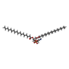
Macromolecule #12: 1,2-Distearoyl-sn-glycerophosphoethanolamine

MacromoleculeName: 1,2-Distearoyl-sn-glycerophosphoethanolamine / type: ligand / ID: 12 / Number of copies: 4 / Formula: 3PE
Molecular weightTheoretical: 748.065 Da
Chemical component information

ChemComp-3PE:
1,2-Distearoyl-sn-glycerophosphoethanolamine / phospholipid*YM

+
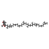
Macromolecule #13: CARDIOLIPIN

MacromoleculeName: CARDIOLIPIN / type: ligand / ID: 13 / Number of copies: 5 / Formula: CDL
Molecular weightTheoretical: 1.464043 KDa
Chemical component information

ChemComp-CDL:
CARDIOLIPIN / phospholipid*YM

+
Macromolecule #14: UBIQUINONE-10

MacromoleculeName: UBIQUINONE-10 / type: ligand / ID: 14 / Number of copies: 3 / Formula: U10
Molecular weightTheoretical: 863.343 Da
Chemical component information

ChemComp-U10:
UBIQUINONE-10

+
Macromolecule #15: HEME C

MacromoleculeName: HEME C / type: ligand / ID: 15 / Number of copies: 2 / Formula: HEC
Molecular weightTheoretical: 618.503 Da
Chemical component information

ChemComp-HEC:
HEME C

+
Macromolecule #16: FE2/S2 (INORGANIC) CLUSTER

MacromoleculeName: FE2/S2 (INORGANIC) CLUSTER / type: ligand / ID: 16 / Number of copies: 2 / Formula: FES
Molecular weightTheoretical: 175.82 Da
Chemical component information

ChemComp-FES:
FE2/S2 (INORGANIC) CLUSTER

-
Experimental details

-
Structure determination

Methodcryo EM
Processingsingle particle reconstruction
Aggregation stateparticle

-
Sample preparation

Concentration2 mg/mL
BufferpH: 7.4
Component:
ConcentrationFormulaName
0.25 MNaClsodium chloride
0.02 MHEPES
0.02 %Brij-35

Details: 250 mM NaCl, 20 mM HEPES, pH 7.7, 0.02% Brij-35
VitrificationCryogen name: PROPANE / Chamber humidity: 95 % / Chamber temperature: 277 K / Instrument: FEI VITROBOT MARK IV
Details: blotting for 30 seconds at 4 degrees Celsius, 95% humidity and flash freezing.

-
Electron microscopy

MicroscopeFEI TITAN KRIOS
Image recordingFilm or detector model: FEI FALCON II (4k x 4k) / Number grids imaged: 1 / Number real images: 1854 / Average exposure time: 2.0 sec. / Average electron dose: 51.0 e/Å2
Electron beamAcceleration voltage: 300 kV / Electron source: FIELD EMISSION GUN
Electron opticsIllumination mode: OTHER / Imaging mode: BRIGHT FIELD
Sample stageSpecimen holder model: FEI TITAN KRIOS AUTOGRID HOLDER / Cooling holder cryogen: NITROGEN
Experimental equipment
Model: Titan Krios / Image courtesy: FEI Company

+
Image processing

Particle selectionNumber selected: 400000
Startup modelType of model: PDB ENTRY
PDB model - PDB ID:
Final reconstructionNumber classes used: 1 / Applied symmetry - Point group: C1 (asymmetric) / Algorithm: FOURIER SPACE / Resolution.type: BY AUTHOR / Resolution: 3.9 Å / Resolution method: FSC 0.143 CUT-OFF / Software - Name: RELION (ver. 2) / Number images used: 102314
Initial angle assignmentType: MAXIMUM LIKELIHOOD / Software - Name: RELION (ver. 2)
Final angle assignmentType: MAXIMUM LIKELIHOOD / Software - Name: RELION (ver. 2)
Final 3D classificationNumber classes: 3 / Software - Name: RELION (ver. 2)
FSC plot (resolution estimation)

-
Atomic model buiding 1

Initial modelPDB ID:

Chain - Source name: PDB / Chain - Initial model type: experimental model
RefinementSpace: REAL / Protocol: OTHER / Overall B value: 90
Output model

PDB-6q9e:
Complex III2 focused refinement from Ovine respiratory supercomplex I+III2

+
About Yorodumi

-
News

-
Feb 9, 2022. New format data for meta-information of EMDB entries

New format data for meta-information of EMDB entries

  • Version 3 of the EMDB header file is now the official format.
  • The previous official version 1.9 will be removed from the archive.

Related info.:EMDB header

External links:wwPDB to switch to version 3 of the EMDB data model

-
Aug 12, 2020. Covid-19 info

Covid-19 info

URL: https://pdbj.org/emnavi/covid19.php

New page: Covid-19 featured information page in EM Navigator.

Related info.:Covid-19 info / Mar 5, 2020. Novel coronavirus structure data

+
Mar 5, 2020. Novel coronavirus structure data

Novel coronavirus structure data

Related info.:Yorodumi Speices / Aug 12, 2020. Covid-19 info

External links:COVID-19 featured content - PDBj / Molecule of the Month (242):Coronavirus Proteases

+
Jan 31, 2019. EMDB accession codes are about to change! (news from PDBe EMDB page)

EMDB accession codes are about to change! (news from PDBe EMDB page)

  • The allocation of 4 digits for EMDB accession codes will soon come to an end. Whilst these codes will remain in use, new EMDB accession codes will include an additional digit and will expand incrementally as the available range of codes is exhausted. The current 4-digit format prefixed with “EMD-” (i.e. EMD-XXXX) will advance to a 5-digit format (i.e. EMD-XXXXX), and so on. It is currently estimated that the 4-digit codes will be depleted around Spring 2019, at which point the 5-digit format will come into force.
  • The EM Navigator/Yorodumi systems omit the EMD- prefix.

Related info.:Q: What is EMD? / ID/Accession-code notation in Yorodumi/EM Navigator

External links:EMDB Accession Codes are Changing Soon! / Contact to PDBj

+
Jul 12, 2017. Major update of PDB

Major update of PDB

  • wwPDB released updated PDB data conforming to the new PDBx/mmCIF dictionary.
  • This is a major update changing the version number from 4 to 5, and with Remediation, in which all the entries are updated.
  • In this update, many items about electron microscopy experimental information are reorganized (e.g. em_software).
  • Now, EM Navigator and Yorodumi are based on the updated data.

External links:wwPDB Remediation / Enriched Model Files Conforming to OneDep Data Standards Now Available in the PDB FTP Archive

-
Yorodumi

Thousand views of thousand structures

  • Yorodumi is a browser for structure data from EMDB, PDB, SASBDB, etc.
  • This page is also the successor to EM Navigator detail page, and also detail information page/front-end page for Omokage search.
  • The word "yorodu" (or yorozu) is an old Japanese word meaning "ten thousand". "mi" (miru) is to see.

Related info.:EMDB / PDB / SASBDB / Comparison of 3 databanks / Yorodumi Search / Aug 31, 2016. New EM Navigator & Yorodumi / Yorodumi Papers / Jmol/JSmol / Function and homology information / Changes in new EM Navigator and Yorodumi

Read more