angiogenin-PRI complex / tRNA-specific ribonuclease activity / negative regulation of translation in response to stress / tRNA-derived small RNA (tsRNA or tRNA-related fragment, tRF) biogenesis / tRNA decay / signaling / cell communication / Hydrolases; Acting on ester bonds; Endoribonucleases producing 3'-phosphomonoesters / Adherens junctions interactions / oocyte maturation ...angiogenin-PRI complex / tRNA-specific ribonuclease activity / negative regulation of translation in response to stress / tRNA-derived small RNA (tsRNA or tRNA-related fragment, tRF) biogenesis / tRNA decay / signaling / cell communication / Hydrolases; Acting on ester bonds; Endoribonucleases producing 3'-phosphomonoesters / Adherens junctions interactions / oocyte maturation / hematopoietic stem cell proliferation / homeostatic process / rRNA transcription / basement membrane / positive regulation of phosphorylation / RNA nuclease activity / endocytic vesicle / 90S preribosome / ubiquitin ligase inhibitor activity / positive regulation of signal transduction by p53 class mediator / ovarian follicle development / phagocytic cup / protein-RNA complex assembly / translation regulator activity / actin filament polymerization / peptide binding / rough endoplasmic reticulum / RNA endonuclease activity / ribosomal small subunit export from nucleus / response to hormone / positive regulation of endothelial cell proliferation / gastrulation / MDM2/MDM4 family protein binding / class I DNA-(apurinic or apyrimidinic site) endonuclease activity / cytosolic ribosome / placenta development / stress granule assembly / DNA-(apurinic or apyrimidinic site) lyase / ribosomal large subunit biogenesis / maturation of LSU-rRNA from tricistronic rRNA transcript (SSU-rRNA, 5.8S rRNA, LSU-rRNA) / positive regulation of protein secretion / maturation of SSU-rRNA from tricistronic rRNA transcript (SSU-rRNA, 5.8S rRNA, LSU-rRNA) / positive regulation of apoptotic signaling pathway / negative regulation of smooth muscle cell proliferation / maturation of SSU-rRNA / small-subunit processome / spindle / cytoplasmic stress granule / rRNA processing / rhythmic process / regulation of translation / positive regulation of canonical Wnt signaling pathway / antimicrobial humoral immune response mediated by antimicrobial peptide / cell migration / heparin binding / chromosome / antibacterial humoral response / actin cytoskeleton / large ribosomal subunit / ribosomal small subunit assembly / growth cone / ribosome binding / ribosomal small subunit biogenesis / actin binding / 5S rRNA binding / ribosomal large subunit assembly / small ribosomal subunit / endonuclease activity / small ribosomal subunit rRNA binding / angiogenesis / cytosolic small ribosomal subunit / large ribosomal subunit rRNA binding / killing of cells of another organism / cytosolic large ribosomal subunit / defense response to Gram-negative bacterium / perikaryon / response to hypoxia / cell differentiation / cytoplasmic translation / tRNA binding / mitochondrial inner membrane / negative regulation of translation / postsynaptic density / rRNA binding / defense response to Gram-positive bacterium / structural constituent of ribosome / ribosome / translation / ribonucleoprotein complex / receptor ligand activity / copper ion binding / signaling receptor binding / innate immune response / cell division / DNA repair / hydrolase activity / mRNA binding / neuronal cell body / apoptotic process / centrosome Similarity search - Function
Pancreatic ribonuclease / Ribonuclease A, active site / Ribonuclease A-domain / Ribonuclease A-like domain superfamily / Pancreatic ribonuclease / Pancreatic ribonuclease family signature. / Pancreatic ribonuclease / Ribosomal protein L6, N-terminal / Ribosomal protein L6, N-terminal domain / 40S ribosomal protein SA, C-terminal domain ...Pancreatic ribonuclease / Ribonuclease A, active site / Ribonuclease A-domain / Ribonuclease A-like domain superfamily / Pancreatic ribonuclease / Pancreatic ribonuclease family signature. / Pancreatic ribonuclease / Ribosomal protein L6, N-terminal / Ribosomal protein L6, N-terminal domain / 40S ribosomal protein SA, C-terminal domain / 40S ribosomal protein SA C-terminus / Ubiquitin-like protein FUBI / Ribosomal protein L30e / Ribosomal L15/L27a, N-terminal / Ribosomal protein L28e / : / Ribosomal protein L23 / Ribosomal protein L2, archaeal-type / Ribosomal L28e/Mak16 / Ribosomal L28e protein family / metallochaperone-like domain / TRASH domain / : / Ribosomal protein S26e signature. / Ribosomal protein L41 / Ribosomal protein L41 / Ribosomal protein S21e, conserved site / Ribosomal protein S21e signature. / : / Ribosomal protein S12e signature. / Ribosomal protein S12e / Ribosomal protein S26e / Ribosomal protein S26e superfamily / Ribosomal protein S26e / Ribosomal protein L29e / Ribosomal L29e protein family / Ribosomal protein L27e, conserved site / Ribosomal protein L27e signature. / Ribosomal protein L22e / Ribosomal protein L22e superfamily / Ribosomal L22e protein family / Small (40S) ribosomal subunit Asc1/RACK1 / Ribosomal protein L13e / Ribosomal protein S5, eukaryotic/archaeal / Ribosomal protein L13e / Ribosomal protein S21e / Ribosomal protein L38e / Ribosomal protein L38e superfamily / Ribosomal protein S21e superfamily / Ribosomal protein S21e / Ribosomal L38e protein family / Ribosomal protein S19e, conserved site / Ribosomal protein S19e signature. / Ribosomal protein S2, eukaryotic / Ribosomal protein L19, eukaryotic / : / Ribosomal protein L10e, conserved site / Ribosomal protein L6e signature. / Ribosomal protein L10e signature. / Ribosomal protein L19/L19e conserved site / Ribosomal protein L19e signature. / Ribosomal protein L44e signature. / 60S ribosomal protein L18a/ L20, eukaryotes / 40S Ribosomal protein S10 / Ribosomal protein L10e / Ribosomal protein L24e, conserved site / Ribosomal protein L24e signature. / Ribosomal protein L34e, conserved site / Ribosomal protein L34e signature. / Plectin/S10, N-terminal / Plectin/S10 domain / Ribosomal protein L5 eukaryotic, C-terminal / Ribosomal L18 C-terminal region / Ribosomal protein L23/L25, N-terminal / Ribosomal protein L23, N-terminal domain / : / Ribosomal L40e family / Ribosomal protein L36e signature. / Ribosomal protein S30 / Ribosomal protein S30 / Ribosomal protein L30e signature 1. / Ribosomal protein L44e / Ribosomal protein S10, eukaryotic/archaeal / Ribosomal protein L44 / Ribosomal protein S25 / 50S ribosomal protein L18Ae/60S ribosomal protein L20 and L18a / Ribosomal protein S8e subdomain, eukaryotes / S25 ribosomal protein / : / Ribosomal protein L35Ae, conserved site / Ribosomal protein 50S-L18Ae/60S-L20/60S-L18A / Ribosomal proteins 50S-L18Ae/60S-L20/60S-L18A / Ribosomal protein 60S L18 and 50S L18e / Ribosomal protein L35Ae signature. / Eukaryotic Ribosomal Protein L27, KOW domain / Ribosomal protein S7e signature. / Ribosomal_L40e / Ribosomal protein L40e / Ribosomal protein L40e superfamily / Ribosomal protein L27e Similarity search - Domain/homology
Small ribosomal subunit protein eS32 / Ribosomal protein L36a / Large ribosomal subunit protein uL4 / Large ribosomal subunit protein uL16 / Small ribosomal subunit protein uS4 / Large ribosomal subunit protein uL22 / Large ribosomal subunit protein eL24 / Large ribosomal subunit protein uL23 / Large ribosomal subunit protein eL33 / Small ribosomal subunit protein eS12 ...Small ribosomal subunit protein eS32 / Ribosomal protein L36a / Large ribosomal subunit protein uL4 / Large ribosomal subunit protein uL16 / Small ribosomal subunit protein uS4 / Large ribosomal subunit protein uL22 / Large ribosomal subunit protein eL24 / Large ribosomal subunit protein uL23 / Large ribosomal subunit protein eL33 / Small ribosomal subunit protein eS12 / Large ribosomal subunit protein eL29 / Small ribosomal subunit protein uS9 / Large ribosomal subunit protein eL31 / Large ribosomal subunit protein eL21 / Large ribosomal subunit protein uL29 / Small ribosomal subunit protein uS10 / Small ribosomal subunit protein RACK1 / Large ribosomal subunit protein eL6 / Large ribosomal subunit protein uL15 / Small ribosomal subunit protein uS15 / Large ribosomal subunit protein uL24 / Small ribosomal subunit protein eS1 / Large ribosomal subunit protein eL8 / Large ribosomal subunit protein uL30 / Small ribosomal subunit protein eS7 / Large ribosomal subunit protein uL6 / Large ribosomal subunit protein eL43 / Large ribosomal subunit protein eL14 / Small ribosomal subunit protein uS12 / Large ribosomal subunit protein eL15 / Small ribosomal subunit protein uS11 / 40S ribosomal protein S24 / Large ribosomal subunit protein uL14 / Ubiquitin-like FUBI-ribosomal protein eS30 fusion protein / 40S ribosomal protein S4 / Small ribosomal subunit protein eS25 / Large ribosomal subunit protein eL30 / Large ribosomal subunit protein eL18 / Small ribosomal subunit protein eS26 / Small ribosomal subunit protein uS7 / Small ribosomal subunit protein uS8 / Small ribosomal subunit protein eS28 / 40S ribosomal protein SA / Small ribosomal subunit protein eS8 / Large ribosomal subunit protein eL13 / Large ribosomal subunit protein uL3 / Small ribosomal subunit protein eS6 / Small ribosomal subunit protein eS21 / Small ribosomal subunit protein eS19 / Small ribosomal subunit protein uS3 / Small ribosomal subunit protein uS13 / Small ribosomal subunit protein eS10 / Small ribosomal subunit protein uS17 / Large ribosomal subunit protein eL22 / Large ribosomal subunit protein uL2 / Large ribosomal subunit protein eL39 / Large ribosomal subunit protein eL36 / Large ribosomal subunit protein eL20 / Small ribosomal subunit protein eS17 / Large ribosomal subunit protein uL5 / Large ribosomal subunit protein eL32 / Large ribosomal subunit protein uL13 / Large ribosomal subunit protein eL27 / Large ribosomal subunit protein eL34 / Large ribosomal subunit protein eL19 / Small ribosomal subunit protein eS27 / Large ribosomal subunit protein eL38 / Small ribosomal subunit protein uS19 / Large ribosomal subunit protein eL28 / Small ribosomal subunit protein uS14 / Small ribosomal subunit protein uS5 / Angiogenin / Ubiquitin-ribosomal protein eL40 fusion protein / Large ribosomal subunit protein uL18 / Large ribosomal subunit protein eL37 Similarity search - Component
National Institutes of Health/National Institute of General Medical Sciences (NIH/NIGMS)
R01 GM127094
United States
Citation
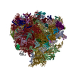
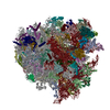
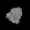
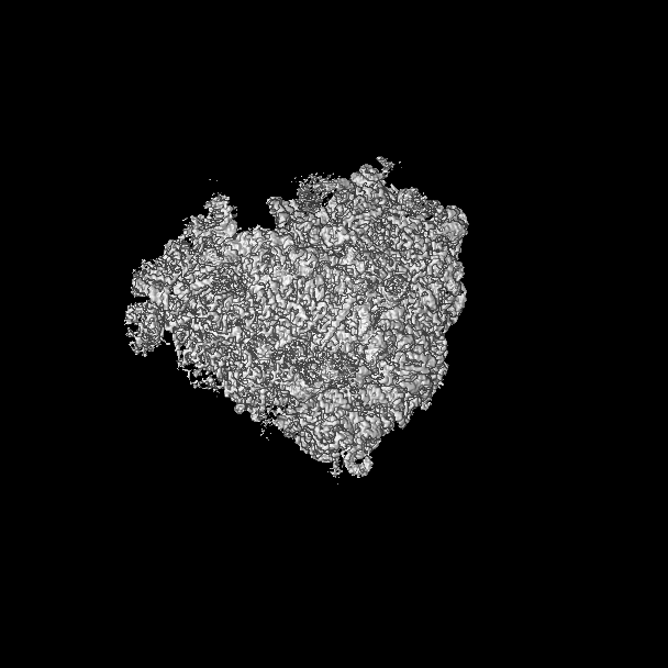
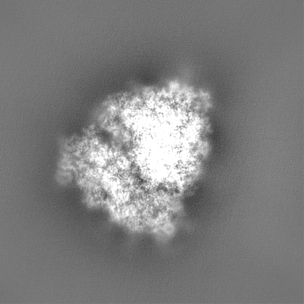
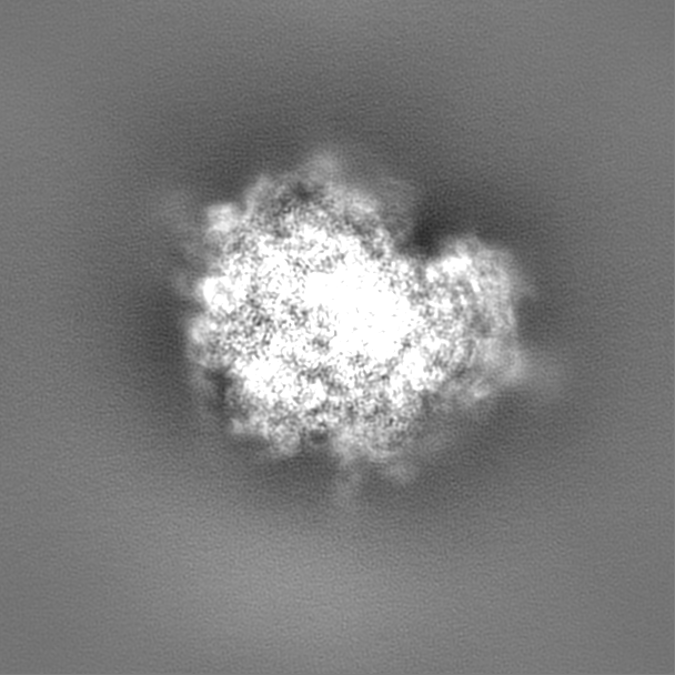
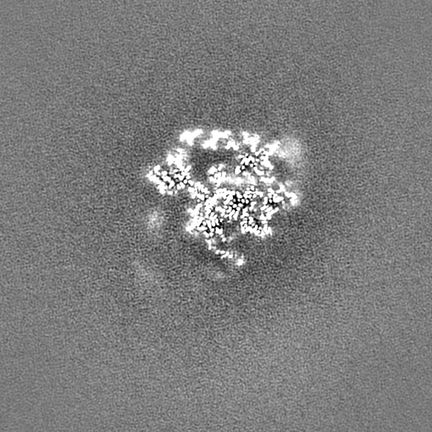
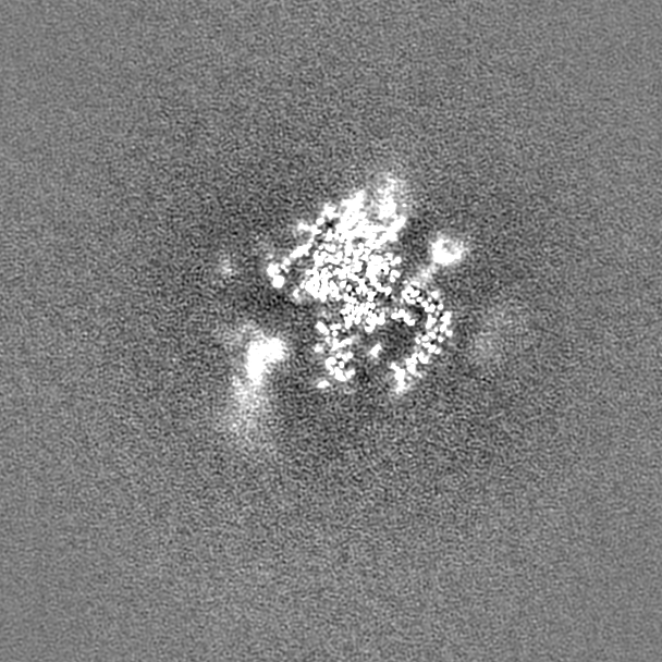
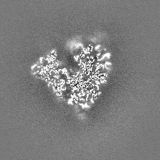
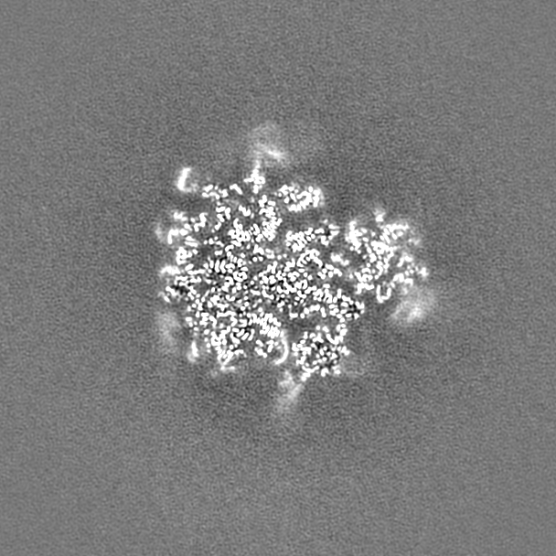
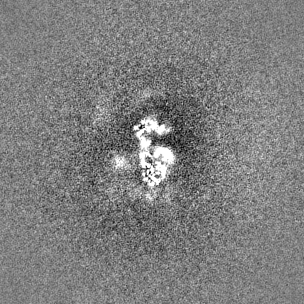
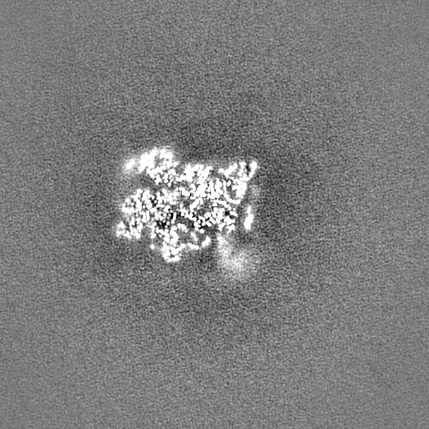
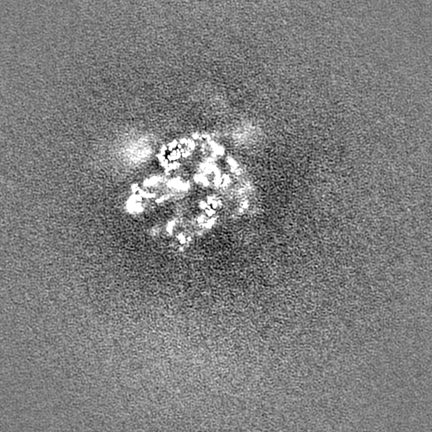
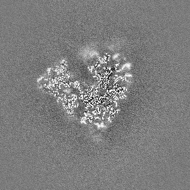
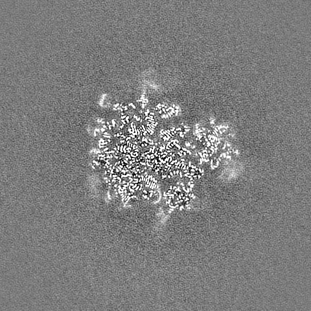
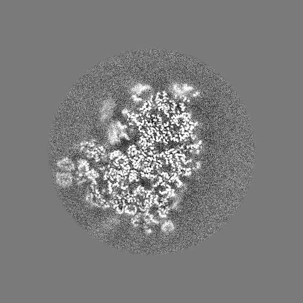
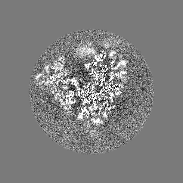
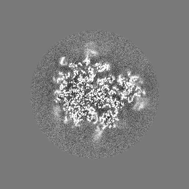
Journal: Nature / Year: 2024 Title: Structural mechanism of angiogenin activation by the ribosome. Authors: Anna B Loveland / Cha San Koh / Robin Ganesan / Allan Jacobson / Andrei A Korostelev / Abstract: Angiogenin, an RNase-A-family protein, promotes angiogenesis and has been implicated in cancer, neurodegenerative diseases and epigenetic inheritance. After activation during cellular stress, ...Angiogenin, an RNase-A-family protein, promotes angiogenesis and has been implicated in cancer, neurodegenerative diseases and epigenetic inheritance. After activation during cellular stress, angiogenin cleaves tRNAs at the anticodon loop, resulting in translation repression. However, the catalytic activity of isolated angiogenin is very low, and the mechanisms of the enzyme activation and tRNA specificity have remained a puzzle. Here we identify these mechanisms using biochemical assays and cryogenic electron microscopy (cryo-EM). Our study reveals that the cytosolic ribosome is the activator of angiogenin. A cryo-EM structure features angiogenin bound in the A site of the 80S ribosome. The C-terminal tail of angiogenin is rearranged by interactions with the ribosome to activate the RNase catalytic centre, making the enzyme several orders of magnitude more efficient in tRNA cleavage. Additional 80S-angiogenin structures capture how tRNA substrate is directed by the ribosome into angiogenin's active site, demonstrating that the ribosome acts as the specificity factor. Our findings therefore suggest that angiogenin is activated by ribosomes with a vacant A site, the abundance of which increases during cellular stress. These results may facilitate the development of therapeutics to treat cancer and neurodegenerative diseases.
In the structure databanks used in Yorodumi, some data are registered as the other names, "COVID-19 virus" and "2019-nCoV". Here are the details of the virus and the list of structure data.
Jan 31, 2019. EMDB accession codes are about to change! (news from PDBe EMDB page)
EMDB accession codes are about to change! (news from PDBe EMDB page)
The allocation of 4 digits for EMDB accession codes will soon come to an end. Whilst these codes will remain in use, new EMDB accession codes will include an additional digit and will expand incrementally as the available range of codes is exhausted. The current 4-digit format prefixed with “EMD-” (i.e. EMD-XXXX) will advance to a 5-digit format (i.e. EMD-XXXXX), and so on. It is currently estimated that the 4-digit codes will be depleted around Spring 2019, at which point the 5-digit format will come into force.
The EM Navigator/Yorodumi systems omit the EMD- prefix.
Related info.:Q: What is EMD? / ID/Accession-code notation in Yorodumi/EM Navigator
Yorodumi is a browser for structure data from EMDB, PDB, SASBDB, etc.
This page is also the successor to EM Navigator detail page, and also detail information page/front-end page for Omokage search.
The word "yorodu" (or yorozu) is an old Japanese word meaning "ten thousand". "mi" (miru) is to see.
Related info.:EMDB / PDB / SASBDB / Comparison of 3 databanks / Yorodumi Search / Aug 31, 2016. New EM Navigator & Yorodumi / Yorodumi Papers / Jmol/JSmol / Function and homology information / Changes in new EM Navigator and Yorodumi
 Movie
Movie Controller


 Open data
 Basic information
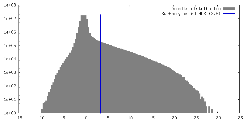
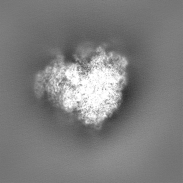
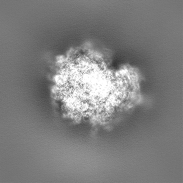
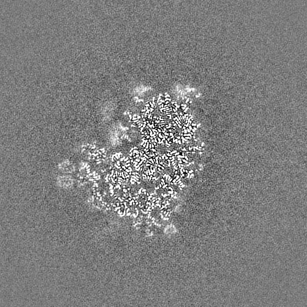
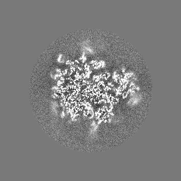
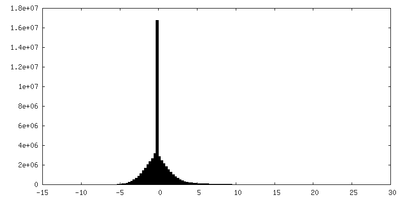
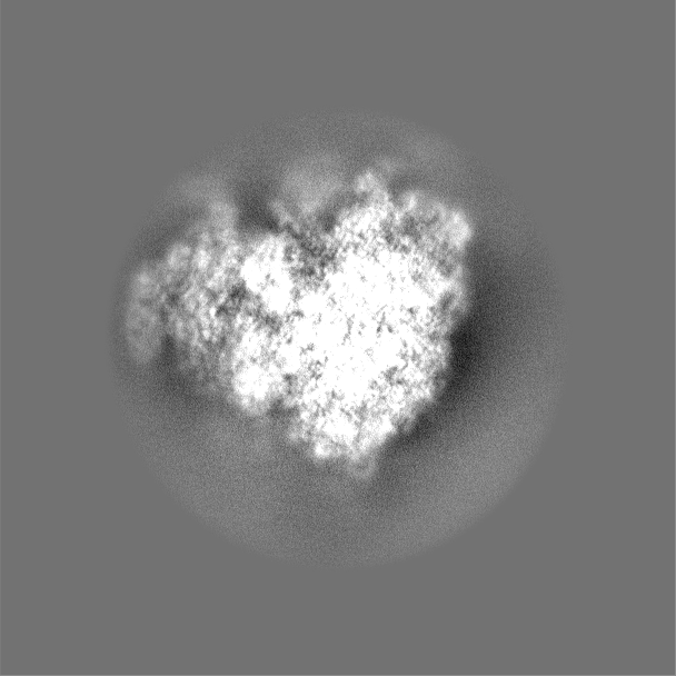
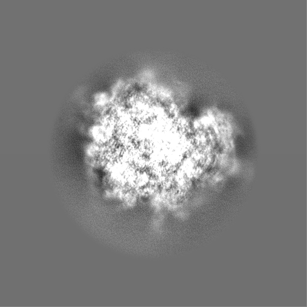
 Map data
 Sample
 Keywords
 Function and homology information
 Homo sapiens (human) /
 Authors
 United States, 1 items
  Citation
 Structure visualization
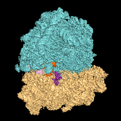
 Downloads & links
 emd_44461.png
 http://ftp.pdbj.org/pub/emdb/structures/EMD-44461
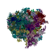


 Z (Sec.)
 Y (Row.)
 X (Col.)


 Sample components
 Processing
 Electron microscopy
 FIELD EMISSION GUN