 Movie
Movie Controller


+ Open data

- Basic information
| Entry |  | |||||||||
|---|---|---|---|---|---|---|---|---|---|---|
| Title | The structure of DSR2-Tail tube complex | |||||||||
 Map data Map data | ||||||||||
 Sample Sample |
| |||||||||
 Keywords Keywords | Complex / IMMUNE SYSTEM | |||||||||
| Function / homology | : / Uncharacterized protein Function and homology information Function and homology information | |||||||||
| Biological species |   Bacillus phage SPR (virus) Bacillus phage SPR (virus) | |||||||||
| Method | single particle reconstruction / cryo EM / Resolution: 3.53 Å | |||||||||
 Authors Authors | Zheng J / Yang X | |||||||||
| Funding support |  China, 1 items China, 1 items
| |||||||||
 Citation Citation | Journal: Int J Biol Macromol / Year: 2024 Title: Structural insights into autoinhibition and activation of defense-associated sirtuin protein. Authors: Xu Yang / Yiqun Wang / Jianting Zheng / Abstract: Bacterial defense-associated sirtuin 2 (DSR2) proteins harbor an N-terminal sirtuin (SIR2) domain degrading NAD. DSR2 from Bacillus subtilis 29R is autoinhibited and unable to hydrolyze NAD in the ...Bacterial defense-associated sirtuin 2 (DSR2) proteins harbor an N-terminal sirtuin (SIR2) domain degrading NAD. DSR2 from Bacillus subtilis 29R is autoinhibited and unable to hydrolyze NAD in the absence of phage infection. A tail tube protein (TTP) of phage SPR activates the DSR2 while a DSR2-inhibiting protein of phage SPbeta, known as DSAD1 (DSR anti-defense 1), inactivates the DSR2. Although DSR2 structures in complexed with TTP and DSAD1, respectively, have been reported recently, the autoinhibition and activation mechanisms remain incompletely understood. Here, we present cryo-electron microscopy structures of the DSR2-NAD complex in autoinhibited state and the in vitro assembled DSR2-TFD (TTP tube-forming domain) complex in activated state. The DSR2-NAD complex reveals that the autoinhibited DSR2 assembles into an inactive tetramer, binding NAD through a distinct pocket situated outside active site. Binding of TFD into cavities within the sensor domains of DSR2 triggers a conformational change in SIR2 regions, activating its NADase activity, whereas the TTP β-sandwich domain (BSD) is flexible and does not contribute to the activation process. The activated form of DSR2 exists as tetramers and dimers, with the tetramers exhibiting more NADase activity. Overall, our results extend the current understanding of autoinhibition and activation of DSR2 immune proteins. | |||||||||
| History |
|
- Structure visualization
| Supplemental images |
|---|

- Downloads & links
-EMDB archive
| Map data | emd_39385.map.gz | 3.3 MB | EMDB map data format | |
|---|---|---|---|---|
| Header (meta data) | emd-39385-v30.xmlemd-39385.xml | 20 KB 20 KB | Display Display | EMDB header |
| FSC (resolution estimation) | emd_39385_fsc.xml | 8.5 KB | Display | FSC data file |
| Images |  emd_39385.png emd_39385.png | 110.6 KB | ||
| Masks | emd_39385_msk_1.map | 64 MB | Mask map | |
| Filedesc metadata | emd-39385.cif.gz | 6.8 KB | ||
| Others | emd_39385_half_map_1.map.gzemd_39385_half_map_2.map.gz | 59.5 MB 59.5 MB | ||
| Archive directory |  http://ftp.pdbj.org/pub/emdb/structures/EMD-39385ftp://ftp.pdbj.org/pub/emdb/structures/EMD-39385 http://ftp.pdbj.org/pub/emdb/structures/EMD-39385ftp://ftp.pdbj.org/pub/emdb/structures/EMD-39385 | HTTPS FTP |
-Related structure data
| Related structure data | 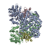 8ylnMC 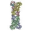 8ykfC 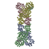 8yl5C  8yltC 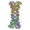 8z18C 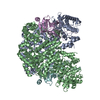 8ztrC M: atomic model generated by this map C: citing same article ( |
|---|---|
| Similar structure data |


-Links
| EMDB pages | EMDB (EBI/PDBe) / EMDataResource |
|---|
-Map
| File | Download / File: emd_39385.map.gz / Format: CCP4 / Size: 64 MB / Type: IMAGE STORED AS FLOATING POINT NUMBER (4 BYTES) | ||||||||||||||||||||||||||||||||||||
|---|---|---|---|---|---|---|---|---|---|---|---|---|---|---|---|---|---|---|---|---|---|---|---|---|---|---|---|---|---|---|---|---|---|---|---|---|---|
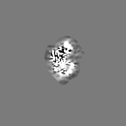
| Projections & slices | Image control
Images are generated by Spider. | ||||||||||||||||||||||||||||||||||||
| Voxel size | X=Y=Z: 0.96 Å | ||||||||||||||||||||||||||||||||||||
| Density |
| ||||||||||||||||||||||||||||||||||||
| Symmetry | Space group: 1 | ||||||||||||||||||||||||||||||||||||
| Details | EMDB XML:
|
 Z (Sec.)
 Y (Row.)
 X (Col.)


-Supplemental data
-Mask #1
| File | emd_39385_msk_1.map | ||||||||||||
|---|---|---|---|---|---|---|---|---|---|---|---|---|---|
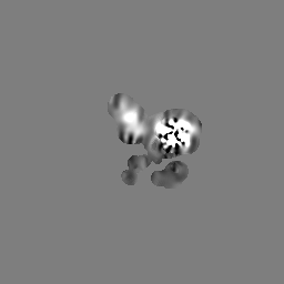
| Projections & Slices |
| ||||||||||||
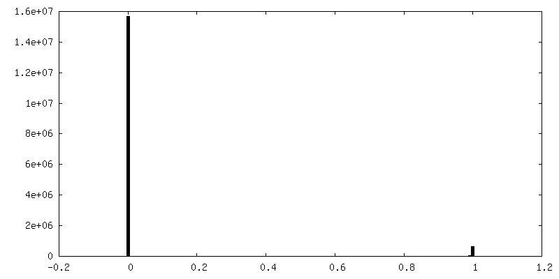
| Density Histograms |


-Half map: #2
| File | emd_39385_half_map_1.map | ||||||||||||
|---|---|---|---|---|---|---|---|---|---|---|---|---|---|
| Projections & Slices |
| ||||||||||||
| Density Histograms |


-Half map: #1
| File | emd_39385_half_map_2.map | ||||||||||||
|---|---|---|---|---|---|---|---|---|---|---|---|---|---|
| Projections & Slices |
| ||||||||||||
| Density Histograms |


- Sample components
-Entire : The complex of DSR2 and Tail tube
| Entire | Name: The complex of DSR2 and Tail tube |
|---|---|
| Components |
|
-Supramolecule #1: The complex of DSR2 and Tail tube
| Supramolecule | Name: The complex of DSR2 and Tail tube / type: complex / ID: 1 / Parent: 0 / Macromolecule list: all |
|---|---|
| Source (natural) | Organism: |
-Macromolecule #1: SIR2-like domain-containing protein
| Macromolecule | Name: SIR2-like domain-containing protein / type: protein_or_peptide / ID: 1 / Number of copies: 2 / Enantiomer: LEVO |
|---|---|
| Source (natural) | Organism: |
| Molecular weight | Theoretical: 118.635789 KDa |
| Recombinant expression | Organism: |
| Sequence | String: MVKVDLESKR YGEKLKEVFL MLDNNVVECI KEITESSRNG KLVFFVGAGV STLSDYPQWW RLVDKYHEEL YGSPKKGNYS SDEYLRIPQ IFYNVKGEMA FDGILKDFFQ VDKPTNPIHD KILAMNPAHV ITTNYDNLID TACWKRGKYF SVISAEEDVA N ATSSRYLL ...String: MVKVDLESKR YGEKLKEVFL MLDNNVVECI KEITESSRNG KLVFFVGAGV STLSDYPQWW RLVDKYHEEL YGSPKKGNYS SDEYLRIPQ IFYNVKGEMA FDGILKDFFQ VDKPTNPIHD KILAMNPAHV ITTNYDNLID TACWKRGKYF SVISAEEDVA N ATSSRYLL KVHGDFRKGF KGENVVLKED DYLNYDQNYP LISNLMKTII ATHTIVFIGY GLGDYNINML LNWVRKLQKD SF HKPFFIR TDPSPIENET LIYYENKGLR IIDAASLIDS NEYDYLERYS AVMDLLIESQ ENKFITKDDE VIDYIYGKIS PLF ALQYIR KIDLKHVFEY DYHFEVNGTV VRHKNKGFGY MERFFELKES CDERSKLSKK QYERFNALFN FFEKNGVICM AKDA GTLNT SIEINSLAYH GKYDVMKKFI EEQSVSIEDD YKKAFFLACL GRWEESYDLY SNIILNSIDE SNGCVYYLSQ INRYR IYQS ITQAVTQFNG LGLLTFGRHY KPFTDEFLAR IEREMTNFNI DDLFNGMPFE FQKKYKILEF LSDNQFLYDD TVKLFE LTN KVRSEMSEGS YSFGMSSDIV VLLRLYDNLR FLYENCLWSV SFHEFHQYIR NSMSLLIEKA EYERTRDIDE LGFSFFG KK SGFFMEYYDF VNISRHFKID DIKNLERSCS IDKIRFGEQE KIEEYLVGIA EEITKQFSAN GMNVVFYTQF ISEAKAAL Y FAKYVKLSEE GLGKIVKALL FYFPERDLDI GKRYVWLERL TKCNELPKSI ISIIDDFLVL QAEKHIDQNY SEVSSNGLY SRDYGALIKH FEKNFISKRL SEITLCLTQD KQKQIDFLFK LLPLLSTNAK SHLLSFKSVE NINDLMNGIR IGLIDEFTPE HEELIIEYL ETRKVNYIVE KEKGIQTFSS NDYMSTFGIW YFLEEINNSK MEEFIGMDDQ YDFFVDPENF DYKKFIPSWL K NYNDKLLG KIAGNKHMKH HVIEVLKERV KNSNDKRYLE ILMNYFI UniProtKB: UNIPROTKB: A0A162TTM4 |
-Macromolecule #2: Bacillus phage SPR Tube protein
| Macromolecule | Name: Bacillus phage SPR Tube protein / type: protein_or_peptide / ID: 2 / Number of copies: 2 / Enantiomer: LEVO |
|---|---|
| Source (natural) | Organism: Bacillus phage SPR (virus) |
| Molecular weight | Theoretical: 29.304701 KDa |
| Recombinant expression | Organism: |
| Sequence | String: MKTVIQDTAD VYFKRKSDGK LVFTAEAQTA SFSQAISEEK LRGGIGNKPL YILKSEKEIN LTVKNAFFDL EWLAMTQGET IQEETKVKV FDREHGLIVD DTNKVTLKGK PVSDVTFYNK KGLTYKIAVS TDGTYTIPTA FAAAKDKLTA VYQIEKVGRR L AIKASKFS ...String: MKTVIQDTAD VYFKRKSDGK LVFTAEAQTA SFSQAISEEK LRGGIGNKPL YILKSEKEIN LTVKNAFFDL EWLAMTQGET IQEETKVKV FDREHGLIVD DTNKVTLKGK PVSDVTFYNK KGLTYKIAVS TDGTYTIPTA FAAAKDKLTA VYQIEKVGRR L AIKASKFS ERYEVEYRTI AYNPDTEEVY SDIYIQFPNV SPSGEFEMSL ENGNALAPEI KFEALADTDT DEMAVVIEAS RD ENTAAPV EDTTGSTQSS DLGGTTE UniProtKB: Uncharacterized protein |
-Experimental details
-Structure determination
| Method | cryo EM |
|---|---|
 Processing Processing | single particle reconstruction |
| Aggregation state | particle |
-Sample preparation
| Buffer | pH: 8 Component:
| |||||||||||||||
|---|---|---|---|---|---|---|---|---|---|---|---|---|---|---|---|---|
| Grid | Model: Quantifoil R1.2/1.3 / Material: GOLD / Mesh: 300 / Support film - Material: CARBON / Support film - topology: HOLEY / Pretreatment - Type: GLOW DISCHARGE / Pretreatment - Time: 60 sec. / Pretreatment - Atmosphere: AIR | |||||||||||||||
| Vitrification | Cryogen name: ETHANE / Chamber humidity: 100 % / Chamber temperature: 289 K / Instrument: FEI VITROBOT MARK IV |
- Electron microscopy
| Microscope | FEI TITAN KRIOS |
|---|---|
| Image recording | Film or detector model: FEI FALCON IV (4k x 4k) / Average electron dose: 40.0 e/Å2 |
| Electron beam | Acceleration voltage: 300 kV / Electron source:  FIELD EMISSION GUN FIELD EMISSION GUN |
| Electron optics | Illumination mode: FLOOD BEAM / Imaging mode: BRIGHT FIELD / Nominal defocus max: 2.0 µm / Nominal defocus min: 1.0 µm |
| Sample stage | Specimen holder model: FEI TITAN KRIOS AUTOGRID HOLDER |
| Experimental equipment |  Model: Titan Krios / Image courtesy: FEI Company |

