National Natural Science Foundation of China (NSFC)
32070040, 32370071
China
Citation
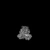
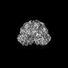
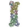
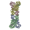
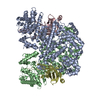
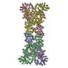
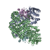
Journal: Int J Biol Macromol / Year: 2024 Title: Structural insights into autoinhibition and activation of defense-associated sirtuin protein. Authors: Xu Yang / Yiqun Wang / Jianting Zheng / Abstract: Bacterial defense-associated sirtuin 2 (DSR2) proteins harbor an N-terminal sirtuin (SIR2) domain degrading NAD. DSR2 from Bacillus subtilis 29R is autoinhibited and unable to hydrolyze NAD in the ...Bacterial defense-associated sirtuin 2 (DSR2) proteins harbor an N-terminal sirtuin (SIR2) domain degrading NAD. DSR2 from Bacillus subtilis 29R is autoinhibited and unable to hydrolyze NAD in the absence of phage infection. A tail tube protein (TTP) of phage SPR activates the DSR2 while a DSR2-inhibiting protein of phage SPbeta, known as DSAD1 (DSR anti-defense 1), inactivates the DSR2. Although DSR2 structures in complexed with TTP and DSAD1, respectively, have been reported recently, the autoinhibition and activation mechanisms remain incompletely understood. Here, we present cryo-electron microscopy structures of the DSR2-NAD complex in autoinhibited state and the in vitro assembled DSR2-TFD (TTP tube-forming domain) complex in activated state. The DSR2-NAD complex reveals that the autoinhibited DSR2 assembles into an inactive tetramer, binding NAD through a distinct pocket situated outside active site. Binding of TFD into cavities within the sensor domains of DSR2 triggers a conformational change in SIR2 regions, activating its NADase activity, whereas the TTP β-sandwich domain (BSD) is flexible and does not contribute to the activation process. The activated form of DSR2 exists as tetramers and dimers, with the tetramers exhibiting more NADase activity. Overall, our results extend the current understanding of autoinhibition and activation of DSR2 immune proteins.
In the structure databanks used in Yorodumi, some data are registered as the other names, "COVID-19 virus" and "2019-nCoV". Here are the details of the virus and the list of structure data.
Jan 31, 2019. EMDB accession codes are about to change! (news from PDBe EMDB page)
EMDB accession codes are about to change! (news from PDBe EMDB page)
The allocation of 4 digits for EMDB accession codes will soon come to an end. Whilst these codes will remain in use, new EMDB accession codes will include an additional digit and will expand incrementally as the available range of codes is exhausted. The current 4-digit format prefixed with “EMD-” (i.e. EMD-XXXX) will advance to a 5-digit format (i.e. EMD-XXXXX), and so on. It is currently estimated that the 4-digit codes will be depleted around Spring 2019, at which point the 5-digit format will come into force.
The EM Navigator/Yorodumi systems omit the EMD- prefix.
Related info.:Q: What is EMD? / ID/Accession-code notation in Yorodumi/EM Navigator
Yorodumi is a browser for structure data from EMDB, PDB, SASBDB, etc.
This page is also the successor to EM Navigator detail page, and also detail information page/front-end page for Omokage search.
The word "yorodu" (or yorozu) is an old Japanese word meaning "ten thousand". "mi" (miru) is to see.
Related info.:EMDB / PDB / SASBDB / Comparison of 3 databanks / Yorodumi Search / Aug 31, 2016. New EM Navigator & Yorodumi / Yorodumi Papers / Jmol/JSmol / Function and homology information / Changes in new EM Navigator and Yorodumi
 Movie
Movie Controller


 Open data
 Basic information
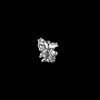
 Map data
 Sample
 Keywords
 Authors
 China, 1 items
  Citation
 Structure visualization
 Downloads & links
 EMDB map data format
 emd_39386.png
 http://ftp.pdbj.org/pub/emdb/structures/EMD-39386


 Z (Sec.)
 Y (Row.)
 X (Col.)


 Sample components
 Processing
 Electron microscopy
 FIELD EMISSION GUN
