 Movie
Movie Controller


+ Open data

- Basic information
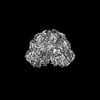
| Entry | Database: PDB / ID: 8ylt | ||||||
|---|---|---|---|---|---|---|---|
| Title | The structure of DSR2 and NAD+ complex | ||||||
 Components Components | SIR2-like domain-containing protein | ||||||
 Keywords Keywords | IMMUNE SYSTEM / Complex | ||||||
| Function / homology | SIR2-like domain / SIR2-like domain / DHS-like NAD/FAD-binding domain superfamily / NICOTINAMIDE-ADENINE-DINUCLEOTIDE / SIR2-like domain-containing protein Function and homology information Function and homology information | ||||||
| Biological species |  | ||||||
| Method | ELECTRON MICROSCOPY / single particle reconstruction / cryo EM / Resolution: 3.09 Å | ||||||
 Authors Authors | Zheng, J. / Yang, X. | ||||||
| Funding support |  China, 1items China, 1items
| ||||||
 Citation Citation | Journal: Int J Biol Macromol / Year: 2024 Title: Structural insights into autoinhibition and activation of defense-associated sirtuin protein. Authors: Xu Yang / Yiqun Wang / Jianting Zheng / Abstract: Bacterial defense-associated sirtuin 2 (DSR2) proteins harbor an N-terminal sirtuin (SIR2) domain degrading NAD. DSR2 from Bacillus subtilis 29R is autoinhibited and unable to hydrolyze NAD in the ...Bacterial defense-associated sirtuin 2 (DSR2) proteins harbor an N-terminal sirtuin (SIR2) domain degrading NAD. DSR2 from Bacillus subtilis 29R is autoinhibited and unable to hydrolyze NAD in the absence of phage infection. A tail tube protein (TTP) of phage SPR activates the DSR2 while a DSR2-inhibiting protein of phage SPbeta, known as DSAD1 (DSR anti-defense 1), inactivates the DSR2. Although DSR2 structures in complexed with TTP and DSAD1, respectively, have been reported recently, the autoinhibition and activation mechanisms remain incompletely understood. Here, we present cryo-electron microscopy structures of the DSR2-NAD complex in autoinhibited state and the in vitro assembled DSR2-TFD (TTP tube-forming domain) complex in activated state. The DSR2-NAD complex reveals that the autoinhibited DSR2 assembles into an inactive tetramer, binding NAD through a distinct pocket situated outside active site. Binding of TFD into cavities within the sensor domains of DSR2 triggers a conformational change in SIR2 regions, activating its NADase activity, whereas the TTP β-sandwich domain (BSD) is flexible and does not contribute to the activation process. The activated form of DSR2 exists as tetramers and dimers, with the tetramers exhibiting more NADase activity. Overall, our results extend the current understanding of autoinhibition and activation of DSR2 immune proteins. | ||||||
| History |
|
- Structure visualization
| Structure viewer | Molecule: MolmilJmol/JSmol |
|---|
- Downloads & links
-Download
| PDBx/mmCIF format | 8ylt.cif.gz | 1.4 MB | Display | PDBx/mmCIF format |
|---|---|---|---|---|
| PDB format | pdb8ylt.ent.gz | Display | PDB format | |
| PDBx/mmJSON format | 8ylt.json.gz | Tree view | PDBx/mmJSON format | |
| Others |  Other downloads Other downloads |
-Validation report
| Arichive directory | https://data.pdbj.org/pub/pdb/validation_reports/yl/8yltftp://data.pdbj.org/pub/pdb/validation_reports/yl/8ylt | HTTPS FTP |
|---|
-Related structure data
| Related structure data |  39390MC 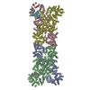 8ykfC 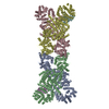 8yl5C 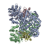 8ylnC 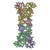 8z18C 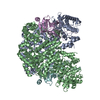 8ztrC M: map data used to model this data C: citing same article ( |
|---|---|
| Similar structure data |


-Links
 PDBj
PDBj

- Assembly
| Deposited unit | 
|
|---|---|
| 1 |
|
-Components
| #1: Protein | Mass: 118635.789 Da / Num. of mol.: 4 Source method: isolated from a genetically manipulated source Source: (gene. exp.) Gene: BSNT_07056 / Production host: #2: Chemical | ChemComp-NAD /   Mass: 663.425 Da / Num. of mol.: 4 / Source method: obtained synthetically / Formula: C21H27N7O14P2 / Feature type: SUBJECT OF INVESTIGATION / Comment: NAD*YM Mass: 663.425 Da / Num. of mol.: 4 / Source method: obtained synthetically / Formula: C21H27N7O14P2 / Feature type: SUBJECT OF INVESTIGATION / Comment: NAD*YMHas ligand of interest | Y | |
|---|
-Experimental details
-Experiment
| Experiment | Method: ELECTRON MICROSCOPY |
|---|---|
| EM experiment | Aggregation state: PARTICLE / 3D reconstruction method: single particle reconstruction |
- Sample preparation
| Component | Name: the complex of DSR2 with NAD / Type: COMPLEX / Entity ID: #1 / Source: RECOMBINANT | |||||||||||||||||||||||||
|---|---|---|---|---|---|---|---|---|---|---|---|---|---|---|---|---|---|---|---|---|---|---|---|---|---|---|
| Molecular weight | Experimental value: NO | |||||||||||||||||||||||||
| Source (natural) | Organism: | |||||||||||||||||||||||||
| Source (recombinant) | Organism: | |||||||||||||||||||||||||
| Buffer solution | pH: 8 | |||||||||||||||||||||||||
| Buffer component |
| |||||||||||||||||||||||||
| Specimen | Conc.: 2.5 mg/ml / Embedding applied: NO / Shadowing applied: NO / Staining applied: NO / Vitrification applied: YES | |||||||||||||||||||||||||
| Specimen support | Grid material: GOLD / Grid mesh size: 300 divisions/in. / Grid type: Quantifoil R1.2/1.3 | |||||||||||||||||||||||||
| Vitrification | Instrument: FEI VITROBOT MARK IV / Cryogen name: ETHANE / Humidity: 100 % / Chamber temperature: 289 K |
- Electron microscopy imaging
| Microscopy | Model: FEI TITAN |
|---|---|
| Electron gun | Electron source:  FIELD EMISSION GUN / Accelerating voltage: 300 kV / Illumination mode: FLOOD BEAM FIELD EMISSION GUN / Accelerating voltage: 300 kV / Illumination mode: FLOOD BEAM |
| Electron lens | Mode: BRIGHT FIELD / Nominal defocus max: 2000 nm / Nominal defocus min: 1000 nm / Cs: 2.7 mm / C2 aperture diameter: 70 µm |
| Specimen holder | Cryogen: NITROGEN / Specimen holder model: FEI TITAN KRIOS AUTOGRID HOLDER |
| Image recording | Electron dose: 40 e/Å2 / Film or detector model: FEI FALCON IV (4k x 4k) |
- Processing
| EM software |
| ||||||||||||||||||||||||
|---|---|---|---|---|---|---|---|---|---|---|---|---|---|---|---|---|---|---|---|---|---|---|---|---|---|
| CTF correction | Type: NONE | ||||||||||||||||||||||||
| 3D reconstruction | Resolution: 3.09 Å / Resolution method: FSC 0.143 CUT-OFF / Num. of particles: 259267 / Symmetry type: POINT | ||||||||||||||||||||||||
| Refine LS restraints |
|
