Movie
Movie Controller

[English] 日本語
 Yorodumi
- EMDB-35301: Structure of mammalian spectrin-actin junctional complex of membr... -
+ Open data

- Basic information
| Entry |  | |||||||||
|---|---|---|---|---|---|---|---|---|---|---|
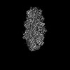
| Title | Structure of mammalian spectrin-actin junctional complex of membrane skeleton, State I, Global map | |||||||||
 Map data Map data | ||||||||||
 Sample Sample |
| |||||||||
 Keywords Keywords | Macrocomplex / membrane skeleton / spectrin-actin junction / MEMBRANE PROTEIN | |||||||||
| Function / homology |  Function and homology information Function and homology informationNCAM signaling for neurite out-growth / RAF/MAP kinase cascade / regulation of cellular component size / lens fiber cell development / endoplasmic reticulum tubular network organization / smooth endoplasmic reticulum calcium ion homeostasis / pointed-end actin filament capping / negative regulation of protein targeting to membrane / regulation of multicellular organismal development / positive regulation of developmental process ...NCAM signaling for neurite out-growth / RAF/MAP kinase cascade / regulation of cellular component size / lens fiber cell development / endoplasmic reticulum tubular network organization / smooth endoplasmic reticulum calcium ion homeostasis / pointed-end actin filament capping / negative regulation of protein targeting to membrane / regulation of multicellular organismal development / positive regulation of developmental process / spectrin / Regulation of actin dynamics for phagocytic cup formation / EPHB-mediated forward signaling / Adherens junctions interactions / VEGFA-VEGFR2 Pathway / Cell-extracellular matrix interactions / RHO GTPases Activate WASPs and WAVEs / MAP2K and MAPK activation / RHOF GTPase cycle / Formation of the canonical BAF (cBAF) complex / Formation of the polybromo-BAF (pBAF) complex / Formation of the embryonic stem cell BAF (esBAF) complex / Formation of the non-canonical BAF (ncBAF) complex / GBP-mediated host defense / Platelet degranulation / Striated Muscle Contraction / Gap junction degradation / Formation of annular gap junctions / UCH proteinases / spectrin-associated cytoskeleton / negative regulation of substrate adhesion-dependent cell spreading / Clathrin-mediated endocytosis / cuticular plate / Regulation of CDH1 Function / Formation of the dystrophin-glycoprotein complex (DGC) / myofibril assembly / positive regulation of cellular component organization / platelet dense tubular network membrane / cell projection membrane / negative regulation of focal adhesion assembly / regulation of filopodium assembly / cellular response to cytochalasin B / COP9 signalosome / regulation of transepithelial transport / morphogenesis of a polarized epithelium / structural constituent of postsynaptic actin cytoskeleton / protein localization to adherens junction / dense body / regulation of lamellipodium assembly / Tat protein binding / actin filament capping / postsynaptic actin cytoskeleton / positive regulation of fibroblast migration / apical protein localization / adherens junction assembly / RHO GTPases activate IQGAPs / RHO GTPases Activate Formins / tight junction / COPI-mediated anterograde transport / ankyrin binding / cortical actin cytoskeleton / positive regulation of wound healing / apical junction complex / spectrin binding / tropomyosin binding / erythrocyte development / NuA4 histone acetyltransferase complex / regulation of norepinephrine uptake / transporter regulator activity / smooth endoplasmic reticulum / cortical cytoskeleton / establishment or maintenance of cell polarity / myofibril / nitric-oxide synthase binding / striated muscle thin filament / brush border / regulation of synaptic vesicle endocytosis / kinesin binding / regulation of protein localization to plasma membrane / microtubule-based process / positive regulation of double-strand break repair via homologous recombination / axonogenesis / cytoskeleton organization / cell projection / muscle contraction / cellular response to cAMP / calyx of Held / cell motility / nitric-oxide synthase regulator activity / actin filament organization / actin filament / adherens junction / SH3 domain binding / synapse organization / cell junction / Schaffer collateral - CA1 synapse / Hydrolases; Acting on acid anhydrides; Acting on acid anhydrides to facilitate cellular and subcellular movement / structural constituent of cytoskeleton / cytoplasmic ribonucleoprotein granule / regulation of cell shape Similarity search - Function | |||||||||
| Biological species |   Sus scrofa (pig) Sus scrofa (pig) | |||||||||
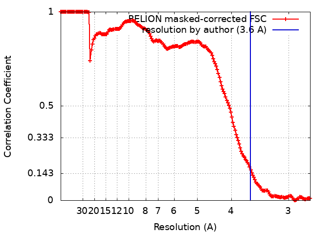
| Method | single particle reconstruction / cryo EM / Resolution: 3.6 Å | |||||||||
 Authors Authors | Li N / Chen S / Gao N | |||||||||
| Funding support |  China, 1 items China, 1 items
| |||||||||
 Citation Citation | Journal: Cell / Year: 2023 Title: Structural basis of membrane skeleton organization in red blood cells. Authors: Ningning Li / Siyi Chen / Kui Xu / Meng-Ting He / Meng-Qiu Dong / Qiangfeng Cliff Zhang / Ning Gao / Abstract: The spectrin-based membrane skeleton is a ubiquitous membrane-associated two-dimensional cytoskeleton underneath the lipid membrane of metazoan cells. Mutations of skeleton proteins impair the ...The spectrin-based membrane skeleton is a ubiquitous membrane-associated two-dimensional cytoskeleton underneath the lipid membrane of metazoan cells. Mutations of skeleton proteins impair the mechanical strength and functions of the membrane, leading to several different types of human diseases. Here, we report the cryo-EM structures of the native spectrin-actin junctional complex (from porcine erythrocytes), which is a specialized short F-actin acting as the central organizational unit of the membrane skeleton. While an α-/β-adducin hetero-tetramer binds to the barbed end of F-actin as a flexible cap, tropomodulin and SH3BGRL2 together create an absolute cap at the pointed end. The junctional complex is strengthened by ring-like structures of dematin in the middle actin layers and by patterned periodic interactions with tropomyosin over its entire length. This work serves as a structural framework for understanding the assembly and dynamics of membrane skeleton and offers insights into mechanisms of various ubiquitous F-actin-binding factors in other F-actin systems. | |||||||||
| History |
|
- Structure visualization
| Supplemental images |
|---|

- Downloads & links
-EMDB archive
| Map data | emd_35301.map.gz | 398.8 MB | EMDB map data format | |
|---|---|---|---|---|
| Header (meta data) | emd-35301-v30.xmlemd-35301.xml | 31.4 KB 31.4 KB | Display Display | EMDB header |
| FSC (resolution estimation) | emd_35301_fsc.xml | 16.9 KB | Display | FSC data file |
| Images |  emd_35301.png emd_35301.png | 36.7 KB | ||
| Filedesc metadata | emd-35301.cif.gz | 11 KB | ||
| Others | emd_35301_half_map_1.map.gzemd_35301_half_map_2.map.gz | 392 MB 392 MB | ||
| Archive directory |  https://data.pdbj.org/pub/emdb/structures/EMD-35301ftp://data.pdbj.org/pub/emdb/structures/EMD-35301 https://data.pdbj.org/pub/emdb/structures/EMD-35301ftp://data.pdbj.org/pub/emdb/structures/EMD-35301 | HTTPS FTP |
-Related structure data
| Related structure data |  8iahMC  8iaiC 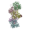 8ib2C M: atomic model generated by this map C: citing same article ( |
|---|---|
| Similar structure data |




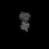


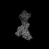

-Links
| EMDB pages | EMDB (EBI/PDBe) / EMDataResource |
|---|---|
| Related items in Molecule of the Month |




-Map
| File | Download / File: emd_35301.map.gz / Format: CCP4 / Size: 421.9 MB / Type: IMAGE STORED AS FLOATING POINT NUMBER (4 BYTES) | ||||||||||||||||||||||||||||||||||||
|---|---|---|---|---|---|---|---|---|---|---|---|---|---|---|---|---|---|---|---|---|---|---|---|---|---|---|---|---|---|---|---|---|---|---|---|---|---|
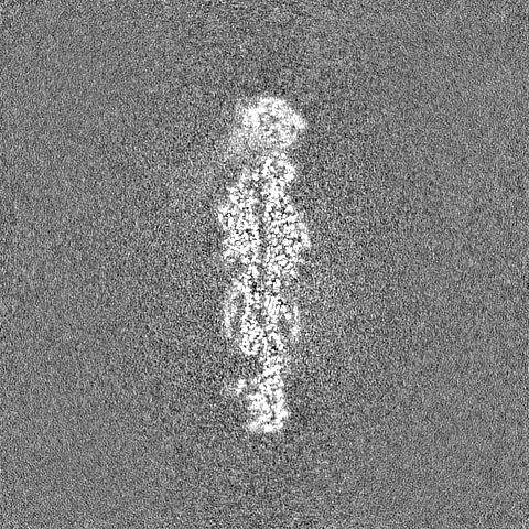
| Projections & slices | Image control
Images are generated by Spider. | ||||||||||||||||||||||||||||||||||||
| Voxel size | X=Y=Z: 1.37 Å | ||||||||||||||||||||||||||||||||||||
| Density |
| ||||||||||||||||||||||||||||||||||||
| Symmetry | Space group: 1 | ||||||||||||||||||||||||||||||||||||
| Details | EMDB XML:
|
 Z (Sec.)
 Y (Row.)
 X (Col.)


-Supplemental data
-Half map: #2
| File | emd_35301_half_map_1.map | ||||||||||||
|---|---|---|---|---|---|---|---|---|---|---|---|---|---|
| Projections & Slices |
| ||||||||||||
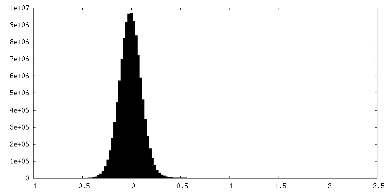
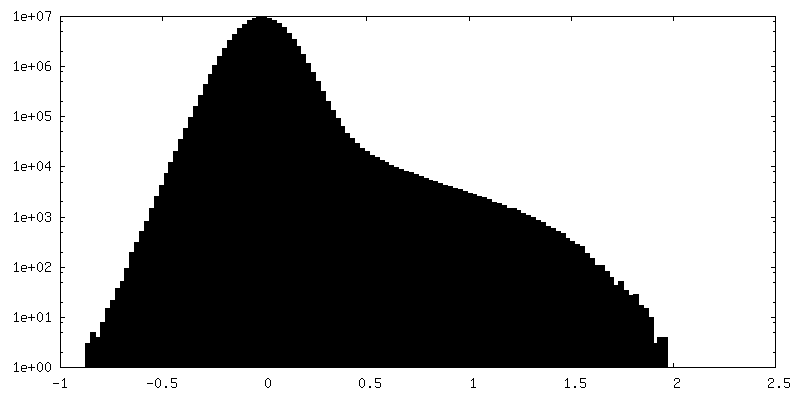
| Density Histograms |

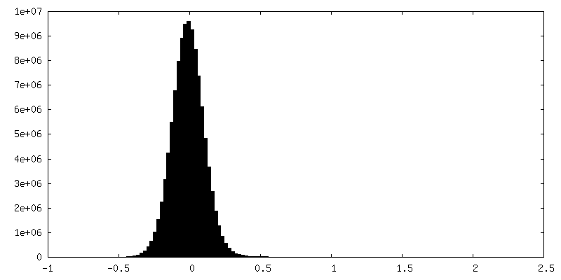
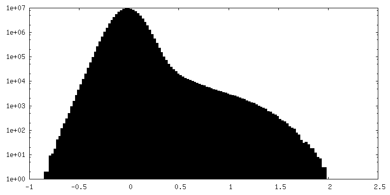
-Half map: #1
| File | emd_35301_half_map_2.map | ||||||||||||
|---|---|---|---|---|---|---|---|---|---|---|---|---|---|
| Projections & Slices |
| ||||||||||||
| Density Histograms |

- Sample components
+Entire : Spectrin-actin junctional complex
+Supramolecule #1: Spectrin-actin junctional complex
+Macromolecule #1: Adducin 1
+Macromolecule #2: Beta-adducin
+Macromolecule #3: Dematin actin binding protein
+Macromolecule #4: Spectrin alpha, erythrocytic 1
+Macromolecule #5: Actin, cytoplasmic 1
+Macromolecule #6: Spectrin beta chain
+Macromolecule #7: Tropomyosin-1.9
+Macromolecule #8: Tropomyosin 3
+Macromolecule #9: Tropomodulin-1
+Macromolecule #10: SH3 domain-binding glutamic acid-rich-like protein
+Macromolecule #11: ADENOSINE-5'-DIPHOSPHATE

-Experimental details
-Structure determination
| Method | cryo EM |
|---|---|
 Processing Processing | single particle reconstruction |
| Aggregation state | particle |
-Sample preparation
| Buffer | pH: 7.5 |
|---|---|
| Vitrification | Cryogen name: ETHANE / Chamber humidity: 100 % |
- Electron microscopy
| Microscope | FEI TITAN KRIOS |
|---|---|
| Image recording | Film or detector model: GATAN K2 QUANTUM (4k x 4k) / Detector mode: SUPER-RESOLUTION / Average electron dose: 34.4 e/Å2 |
| Electron beam | Acceleration voltage: 300 kV / Electron source:  FIELD EMISSION GUN FIELD EMISSION GUN |
| Electron optics | Illumination mode: FLOOD BEAM / Imaging mode: BRIGHT FIELD / Nominal defocus max: 3.0 µm / Nominal defocus min: 2.0 µm |
| Experimental equipment |  Model: Titan Krios / Image courtesy: FEI Company |