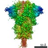
Journal: RSC Adv / Year: 2025 Title: Cryo-EM reveals conformational variability in the SARS-CoV-2 spike protein RBD induced by two broadly neutralizing monoclonal antibodies. Authors: Clayton Fernando Rencilin / Arnab Chatterjee / Mohammad Yousuf Ansari / Suprit Deshpande / Sohini Mukherjee / Randhir Singh / Sowrabha B Jayatheertha / Poorvi M Reddy / Nitin Hingankar / ...Authors: Clayton Fernando Rencilin / Arnab Chatterjee / Mohammad Yousuf Ansari / Suprit Deshpande / Sohini Mukherjee / Randhir Singh / Sowrabha B Jayatheertha / Poorvi M Reddy / Nitin Hingankar / Raghavan Varadarajan / Jayanta Bhattacharya / Somnath Dutta / Abstract: SARS-CoV-2 spike proteins play a critical role in infection by interacting with the ACE2 receptors. Their receptor-binding domains and N-terminal domains exhibit remarkable flexibility and can adopt ...SARS-CoV-2 spike proteins play a critical role in infection by interacting with the ACE2 receptors. Their receptor-binding domains and N-terminal domains exhibit remarkable flexibility and can adopt various conformations that facilitate receptor engagement. Previous structural studies have reported the RBD of the spike protein in "up", "down", and various intermediate states, as well as its different conformational changes during ACE2 binding. This flexibility also influences its interactions with the neutralizing antibodies, yet its role in the antibody complexes remains understudied. In this study, we used cryo-electron microscopy to investigate the structural properties of two broadly neutralizing monoclonal antibodies, THSC20.HVTR04 and THSC20.HVTR26. These antibodies were isolated from an unvaccinated individual and demonstrated potent neutralization of multiple SARS-CoV-2 variants. Our analysis revealed distinct binding characteristics and conformational changes in the spike RBD upon binding with the monoclonal antibodies. The structural characterization of the spike protein-monoclonal antibody complexes provided valuable insights into the structural variability of the spike protein and the possible mechanisms for antibody-mediated neutralization.
In the structure databanks used in Yorodumi, some data are registered as the other names, "COVID-19 virus" and "2019-nCoV". Here are the details of the virus and the list of structure data.
Jan 31, 2019. EMDB accession codes are about to change! (news from PDBe EMDB page)
EMDB accession codes are about to change! (news from PDBe EMDB page)
The allocation of 4 digits for EMDB accession codes will soon come to an end. Whilst these codes will remain in use, new EMDB accession codes will include an additional digit and will expand incrementally as the available range of codes is exhausted. The current 4-digit format prefixed with “EMD-” (i.e. EMD-XXXX) will advance to a 5-digit format (i.e. EMD-XXXXX), and so on. It is currently estimated that the 4-digit codes will be depleted around Spring 2019, at which point the 5-digit format will come into force.
The EM Navigator/Yorodumi systems omit the EMD- prefix.
Related info.:Q: What is EMD? / ID/Accession-code notation in Yorodumi/EM Navigator
Yorodumi is a browser for structure data from EMDB, PDB, SASBDB, etc.
This page is also the successor to EM Navigator detail page, and also detail information page/front-end page for Omokage search.
The word "yorodu" (or yorozu) is an old Japanese word meaning "ten thousand". "mi" (miru) is to see.
Related info.:EMDB / PDB / SASBDB / Comparison of 3 databanks / Yorodumi Search / Aug 31, 2016. New EM Navigator & Yorodumi / Yorodumi Papers / Jmol/JSmol / Function and homology information / Changes in new EM Navigator and Yorodumi
 Movie
Movie Controller


 Open data
 Basic information
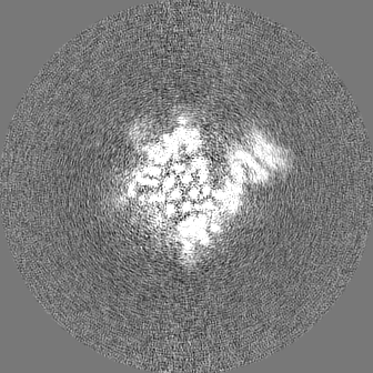
 Map data
 Sample
 Keywords
 Function and homology information
 Severe acute respiratory syndrome coronavirus 2 /
  Homo sapiens (human)
 Authors
 India,
  United States, 4 items
  Citation
 Structure visualization
 Downloads & links
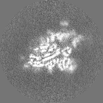
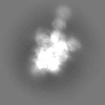
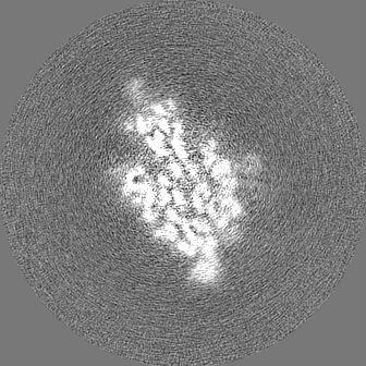
 emd_34546.png
 http://ftp.pdbj.org/pub/emdb/structures/EMD-34546


 X (Sec.)
 Y (Row.)
 Z (Col.)


 Sample components
 Processing
 Electron microscopy
 FIELD EMISSION GUN