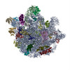
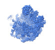
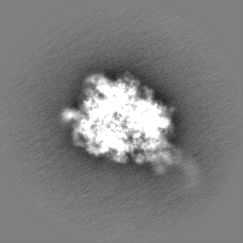
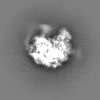
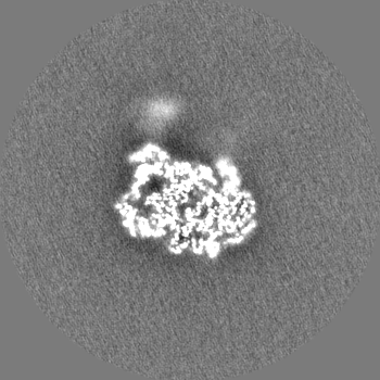
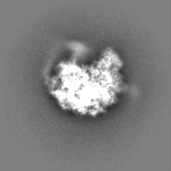
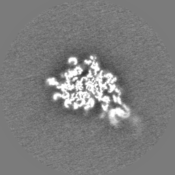
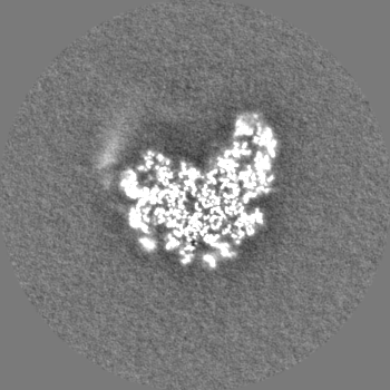
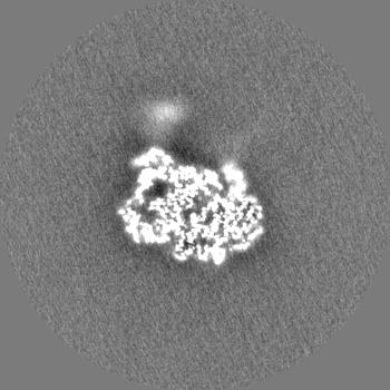
Mycobacterium smegmatis 50S ribosomal subunit from Stationary phase of growth
マップデータ
Mycobacterium smegmatis 50S ribosomal subunit from Stationary Phase of growth
試料
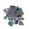
複合体: Mycobacterium smegmatis 50S ribosomal subunit from Stationary Phase of growth
タンパク質・ペプチド: x 32種
RNA: x 2種
リガンド: x 2種
キーワード
50S Subunit / Domain IV of 23S rRNA / Alternate conformation / Helix 68 / RIBOSOME
機能・相同性
機能・相同性情報
large ribosomal subunit / transferase activity / 5S rRNA binding / large ribosomal subunit rRNA binding / cytosolic large ribosomal subunit / tRNA binding / cytoplasmic translation / rRNA binding / negative regulation of translation / ribosome ...large ribosomal subunit / transferase activity / 5S rRNA binding / large ribosomal subunit rRNA binding / cytosolic large ribosomal subunit / tRNA binding / cytoplasmic translation / rRNA binding / negative regulation of translation / ribosome / structural constituent of ribosome / ribonucleoprotein complex / translation / mRNA binding / metal ion binding / cytoplasm 類似検索 - 分子機能
Ribosomal protein L25, long-form / Ribosomal protein L25, beta domain / Ribosomal protein L25, C-terminal / Ribosomal protein TL5, C-terminal domain / Ribosomal protein L10, eubacterial, conserved site / Ribosomal protein L10 signature. / Ribosomal protein L10 / : / Ribosomal protein L11, bacterial-type / Ribosomal protein L31 type A ...Ribosomal protein L25, long-form / Ribosomal protein L25, beta domain / Ribosomal protein L25, C-terminal / Ribosomal protein TL5, C-terminal domain / Ribosomal protein L10, eubacterial, conserved site / Ribosomal protein L10 signature. / Ribosomal protein L10 / : / Ribosomal protein L11, bacterial-type / Ribosomal protein L31 type A / Ribosomal protein L31 signature. / Ribosomal protein L31 / Ribosomal protein L31 superfamily / Ribosomal protein L31 / Ribosomal protein L21, conserved site / Ribosomal protein L21 signature. / Ribosomal protein L11, conserved site / Ribosomal protein L11 signature. / Ribosomal protein L16 signature 1. / Ribosomal protein L10-like domain superfamily / : / Ribosomal protein L10 / Ribosomal protein L10P / Ribosomal protein L6, conserved site / Ribosomal protein L6 signature 1. / Ribosomal protein L16, conserved site / Ribosomal protein L16 signature 2. / Ribosomal protein L9 signature. / Ribosomal protein L9, bacteria/chloroplast / Ribosomal protein L9, C-terminal / Ribosomal protein L9, C-terminal domain / Ribosomal protein L9, C-terminal domain superfamily / Ribosomal protein L17 signature. / Ribosomal L25p family / Ribosomal protein L25 / Ribosomal protein L11, N-terminal / Ribosomal protein L11, N-terminal domain / Ribosomal protein L11/L12 / Ribosomal protein L11, C-terminal / Ribosomal protein L11, C-terminal domain superfamily / Ribosomal protein L11/L12, N-terminal domain superfamily / Ribosomal protein L11, RNA binding domain / Ribosomal protein L11/L12 / Ribosomal protein L36 signature. / Ribosomal protein L28/L24 superfamily / Ribosomal protein L25/Gln-tRNA synthetase, N-terminal / Ribosomal protein L25/Gln-tRNA synthetase, anti-codon-binding domain superfamily / Ribosomal protein L9, N-terminal domain superfamily / Ribosomal protein L9 / Ribosomal protein L9, N-terminal / Ribosomal protein L9, N-terminal domain / Ribosomal protein L28 / Ribosomal protein L35, conserved site / Ribosomal protein L35 signature. / Ribosomal protein L33, conserved site / Ribosomal protein L33 signature. / Ribosomal protein L35, non-mitochondrial / Ribosomal protein L5, bacterial-type / Ribosomal protein L18, bacterial-type / Ribosomal protein L6, bacterial-type / Ribosomal protein L19, conserved site / Ribosomal protein L19 signature. / Ribosomal protein L36 / Ribosomal protein L36 superfamily / Ribosomal protein L36 / Ribosomal protein L9/RNase H1, N-terminal / Ribosomal protein L20 signature. / Ribosomal protein L27, conserved site / Ribosomal protein L27 signature. / Ribosomal protein L14P, bacterial-type / Ribosomal protein L34, conserved site / Ribosomal protein L34 signature. / Ribosomal protein L22, bacterial/chloroplast-type / Ribosomal protein L2, bacterial/organellar-type / Ribosomal protein L35 / Ribosomal protein L35 superfamily / Ribosomal protein L35 / Ribosomal L28 family / Ribosomal protein L33 / Ribosomal protein L33 / Ribosomal protein L28/L24 / Ribosomal protein L18 / Ribosomal L18 of archaea, bacteria, mitoch. and chloroplast / Ribosomal protein L33 superfamily / Ribosomal protein L30, bacterial-type / : / Ribosomal protein L16 / L28p-like / Ribosomal protein L20 / Ribosomal protein L20 / Ribosomal protein L20, C-terminal / Ribosomal protein L21 / Ribosomal protein L27 / Ribosomal L27 protein / Ribosomal protein L19 / Ribosomal protein L19 superfamily / Ribosomal protein L19 / Ribosomal proteins 50S L24/mitochondrial 39S L24 / Ribosomal protein L17 / Ribosomal protein L17 superfamily 類似検索 - ドメイン・相同性
Large ribosomal subunit protein bL33A / Large ribosomal subunit protein uL11 / Large ribosomal subunit protein uL10 / Large ribosomal subunit protein uL3 / Large ribosomal subunit protein uL4 / Large ribosomal subunit protein uL23 / Large ribosomal subunit protein uL2 / Large ribosomal subunit protein uL22 / Large ribosomal subunit protein uL16 / Large ribosomal subunit protein uL29 ...Large ribosomal subunit protein bL33A / Large ribosomal subunit protein uL11 / Large ribosomal subunit protein uL10 / Large ribosomal subunit protein uL3 / Large ribosomal subunit protein uL4 / Large ribosomal subunit protein uL23 / Large ribosomal subunit protein uL2 / Large ribosomal subunit protein uL22 / Large ribosomal subunit protein uL16 / Large ribosomal subunit protein uL29 / Large ribosomal subunit protein uL14 / Large ribosomal subunit protein uL24 / Large ribosomal subunit protein uL5 / Large ribosomal subunit protein uL6 / Large ribosomal subunit protein uL18 / Large ribosomal subunit protein uL30 / Large ribosomal subunit protein uL15 / Large ribosomal subunit protein bL36 / Large ribosomal subunit protein bL17 / Large ribosomal subunit protein uL13 / Uncharacterized protein / Large ribosomal subunit protein bL28 / Large ribosomal subunit protein bL19 / Large ribosomal subunit protein bL20 / Large ribosomal subunit protein bL35 / Large ribosomal subunit protein bL27 / Large ribosomal subunit protein bL21 / Large ribosomal subunit protein bL31 / Large ribosomal subunit protein bL25 / Large ribosomal subunit protein bL32 / Large ribosomal subunit protein bL9 / Large ribosomal subunit protein bL34 類似検索 - 構成要素
Council of Scientific & Industrial Research (CSIR)
MLP-139
インド
Department of Science & Technology (DST, India)
SB/SO/BB-0025/2014
インド
Department of Science & Technology (DST, India)
CRG/2019/001788
インド
Department of Biotechnology (DBT, India)
BT/PR15017/BRB/10/1445
インド
引用
ジャーナル: Int J Biol Macromol / 年: 2023 タイトル: Cryo-EM captures a unique conformational rearrangement in 23S rRNA helices of the Mycobacterium 50S subunit. 著者: Priya Baid / Jayati Sengupta / 要旨: Structural investigations of the ribosomes isolated from pathogenic and non-pathogenic Mycobacterium species have identified several mycobacteria-specific structural features of ribosomal RNA and ...Structural investigations of the ribosomes isolated from pathogenic and non-pathogenic Mycobacterium species have identified several mycobacteria-specific structural features of ribosomal RNA and proteins. Here, we report structural evidence of a hitherto unknown conformational switch of mycobacterium 23S rRNA helices (H54a and H67-H71). Cryo-electron microscopy (cryo-EM) structures (~3-4 Å) of the M. smegmatis (Msm) log-phase 50S ribosomal subunit revealed conformational variability in H67-H71 region of the 23S rRNA, and manifested that, while H68 possesses the usual stretched conformation in one class of the maps, another one exhibits a bulge-out, fused density of H68-H69 at the inter-subunit surface, indicating an intrinsic dynamics of these rRNA helices. Remarkably, altered conformation of H68 forming a more prominent bulge-out structure at the inter-subunit surface of the 50S subunit due to the conformational rearrangements of 23S rRNA H67-H71 region was clearly visualized in a 3 Å cryo-EM map of the 50S subunit obtained from the stationary phase ribosome dataset. The Msm50S subunit having such bulge-out conformation at the intersubunit surface would be incompatible for associating with the 30S subunit due to its inability to form major inter-subunit bridges. Evidently, availability of active 70S ribosome pool can be modulated by stabilizing either one of the H68 conformation.
 ムービー
ムービー コントローラー


 データを開く
 基本情報
基本情報
 マップデータ
 試料
試料 キーワード
キーワード 機能・相同性情報
 Mycolicibacterium smegmatis MC2 155 (バクテリア)
 データ登録者
 インド, 4件
  引用
引用 構造の表示
構造の表示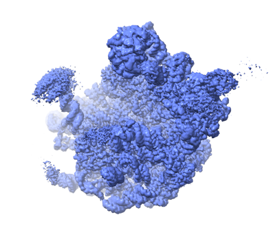
 ダウンロードとリンク
 emd_33096.png
 http://ftp.pdbj.org/pub/emdb/structures/EMD-33096
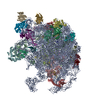


 Z
Z Y
Y X
X


 試料の構成要素
 解析
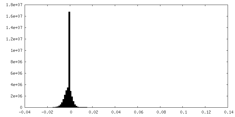
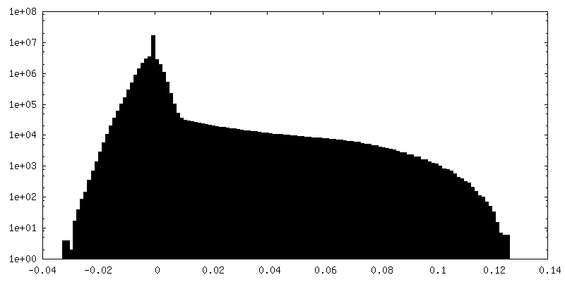
解析 電子顕微鏡法
 FIELD EMISSION GUN