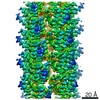
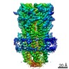
ジャーナル: Cell / 年: 2016 タイトル: Structure of a Chaperone-Usher Pilus Reveals the Molecular Basis of Rod Uncoiling. 著者: Manuela K Hospenthal / Adam Redzej / Karen Dodson / Marta Ukleja / Brandon Frenz / Catarina Rodrigues / Scott J Hultgren / Frank DiMaio / Edward H Egelman / Gabriel Waksman / 要旨: Types 1 and P pili are prototypical bacterial cell-surface appendages playing essential roles in mediating adhesion of bacteria to the urinary tract. These pili, assembled by the chaperone-usher ...Types 1 and P pili are prototypical bacterial cell-surface appendages playing essential roles in mediating adhesion of bacteria to the urinary tract. These pili, assembled by the chaperone-usher pathway, are polymers of pilus subunits assembling into two parts: a thin, short tip fibrillum at the top, mounted on a long pilus rod. The rod adopts a helical quaternary structure and is thought to play essential roles: its formation may drive pilus extrusion by preventing backsliding of the nascent growing pilus within the secretion pore; the rod also has striking spring-like properties, being able to uncoil and recoil depending on the intensity of shear forces generated by urine flow. Here, we present an atomic model of the P pilus generated from a 3.8 Å resolution cryo-electron microscopy reconstruction. This structure provides the molecular basis for the rod's remarkable mechanical properties and illuminates its role in pilus secretion.
 ムービー
ムービー コントローラー


 データを開く
 基本情報
基本情報 マップデータ
 試料
試料 キーワード
キーワード 機能・相同性情報
 データ登録者
 引用
引用

 構造の表示
構造の表示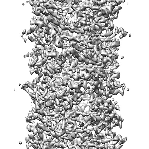
 ダウンロードとリンク
 EMD-3222_EMDB.jpg
 http://ftp.pdbj.org/pub/emdb/structures/EMD-3222


 試料の構成要素
 解析
解析 電子顕微鏡法
 FIELD EMISSION GUN
