 Movie
Movie Controller

[English] 日本語
 Yorodumi
- PDB-5flu: Structure of a Chaperone-Usher pilus reveals the molecular basis ... -
+ Open data

- Basic information
| Entry | Database: PDB / ID: 5flu | ||||||
|---|---|---|---|---|---|---|---|
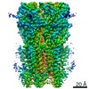
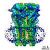
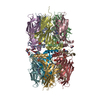
| Title | Structure of a Chaperone-Usher pilus reveals the molecular basis of rod uncoilin | ||||||
 Components Components | PAP FIMBRIAL MAJOR PILIN PROTEIN | ||||||
 Keywords Keywords | STRUCTURAL PROTEIN / HELICAL POLYMER / STRAND DONATION | ||||||
| Function / homology |  Function and homology information Function and homology informationcell adhesion involved in single-species biofilm formation / pilus / extracellular region Similarity search - Function | ||||||
| Biological species |  | ||||||
| Method | ELECTRON MICROSCOPY / helical reconstruction / cryo EM / Resolution: 3.8 Å | ||||||
 Authors Authors | Hospenthal, M.K. / Redzej, A. / Dodson, K. / Ukleja, M. / Frenz, B. / Hultgren, S.J. / DiMaio, F. / Egelman, E.H. / Waksman, G. | ||||||
 Citation Citation | Journal: Cell / Year: 2016 Title: Structure of a Chaperone-Usher Pilus Reveals the Molecular Basis of Rod Uncoiling. Authors: Manuela K Hospenthal / Adam Redzej / Karen Dodson / Marta Ukleja / Brandon Frenz / Catarina Rodrigues / Scott J Hultgren / Frank DiMaio / Edward H Egelman / Gabriel Waksman /   Abstract: Types 1 and P pili are prototypical bacterial cell-surface appendages playing essential roles in mediating adhesion of bacteria to the urinary tract. These pili, assembled by the chaperone-usher ...Types 1 and P pili are prototypical bacterial cell-surface appendages playing essential roles in mediating adhesion of bacteria to the urinary tract. These pili, assembled by the chaperone-usher pathway, are polymers of pilus subunits assembling into two parts: a thin, short tip fibrillum at the top, mounted on a long pilus rod. The rod adopts a helical quaternary structure and is thought to play essential roles: its formation may drive pilus extrusion by preventing backsliding of the nascent growing pilus within the secretion pore; the rod also has striking spring-like properties, being able to uncoil and recoil depending on the intensity of shear forces generated by urine flow. Here, we present an atomic model of the P pilus generated from a 3.8 Å resolution cryo-electron microscopy reconstruction. This structure provides the molecular basis for the rod's remarkable mechanical properties and illuminates its role in pilus secretion. | ||||||
| History |
|
- Structure visualization
| Movie |
Movie viewer |
|---|---|
| Structure viewer | Molecule: MolmilJmol/JSmol |
- Downloads & links
-Download
| PDBx/mmCIF format | 5flu.cif.gz | 363.3 KB | Display | PDBx/mmCIF format |
|---|---|---|---|---|
| PDB format | pdb5flu.ent.gz | 299.6 KB | Display | PDB format |
| PDBx/mmJSON format | 5flu.json.gz | Tree view | PDBx/mmJSON format | |
| Others |  Other downloads Other downloads |
-Validation report
| Arichive directory | https://data.pdbj.org/pub/pdb/validation_reports/fl/5fluftp://data.pdbj.org/pub/pdb/validation_reports/fl/5flu | HTTPS FTP |
|---|
-Related structure data
| Related structure data |  3222MC M: map data used to model this data C: citing same article ( |
|---|---|
| Similar structure data |


-Links
 PDBj
PDBj- Assembly
| Deposited unit | 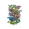
|
|---|---|
| 1 |
|
-Components
| #1: Protein | Mass: 16568.346 Da / Num. of mol.: 13 Source method: isolated from a genetically manipulated source Source: (gene. exp.) Has protein modification | Y | |
|---|
-Experimental details
-Experiment
| Experiment | Method: ELECTRON MICROSCOPY |
|---|---|
| EM experiment | Aggregation state: FILAMENT / 3D reconstruction method: helical reconstruction |
- Sample preparation
| Component | Name: P PILUS / Type: COMPLEX |
|---|---|
| Specimen | Embedding applied: NO / Shadowing applied: NO / Staining applied: NO / Vitrification applied: YES |
| Specimen support | Details: CARBON |
| Vitrification | Instrument: FEI VITROBOT MARK IV / Cryogen name: ETHANE |
- Electron microscopy imaging
| Experimental equipment |  Model: Titan Krios / Image courtesy: FEI Company |
|---|---|
| Microscopy | Model: FEI TITAN KRIOS / Date: Jul 8, 2015 |
| Electron gun | Electron source:  FIELD EMISSION GUN / Accelerating voltage: 300 kV / Illumination mode: FLOOD BEAM FIELD EMISSION GUN / Accelerating voltage: 300 kV / Illumination mode: FLOOD BEAM |
| Electron lens | Mode: BRIGHT FIELD / Nominal defocus max: 3000 nm / Nominal defocus min: 1000 nm / Cs: 2.7 mm |
| Image recording | Electron dose: 17 e/Å2 / Film or detector model: FEI FALCON II (4k x 4k) |
| Image scans | Num. digital images: 90 |
- Processing
| EM software | Name: SPIDER / Category: 3D reconstruction | ||||||||||||
|---|---|---|---|---|---|---|---|---|---|---|---|---|---|
| CTF correction | Details: EACH IMAGE | ||||||||||||
| 3D reconstruction | Method: IHRSR / Resolution: 3.8 Å / Num. of particles: 56341 / Nominal pixel size: 1.1 Å / Actual pixel size: 1.1 Å Details: SUBMISSION BASED ON EXPERIMENTAL DATA FROM EMDB EMD-3222. (DEPOSITION ID: 13990). Symmetry type: HELICAL | ||||||||||||
| Atomic model building | Protocol: OTHER / Space: REAL / Details: REFINEMENT PROTOCOL--EM | ||||||||||||
| Refinement | Highest resolution: 3.8 Å | ||||||||||||
| Refinement step | Cycle: LAST / Highest resolution: 3.8 Å
|
