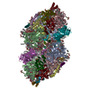
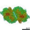
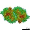
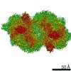
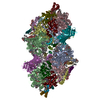
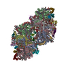
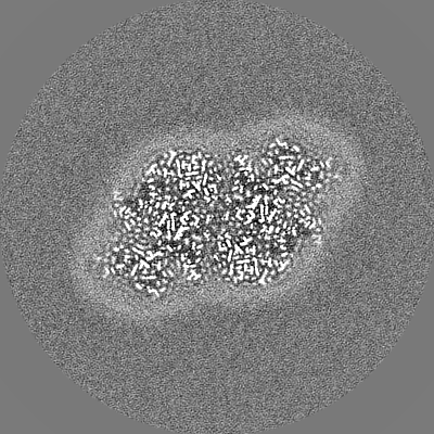
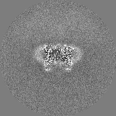
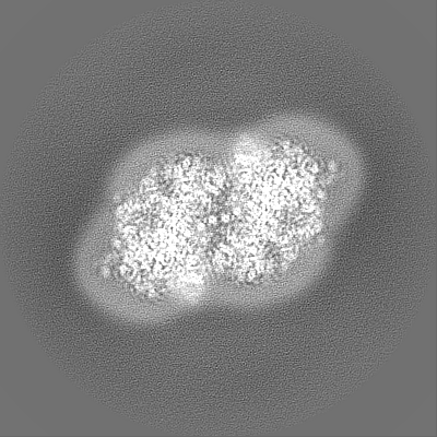
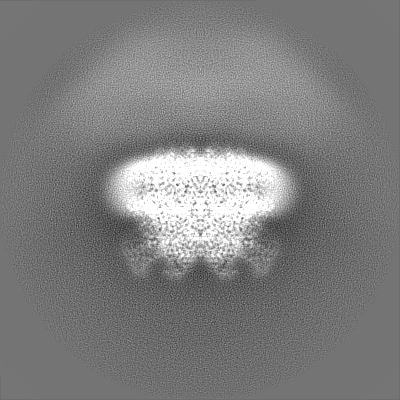
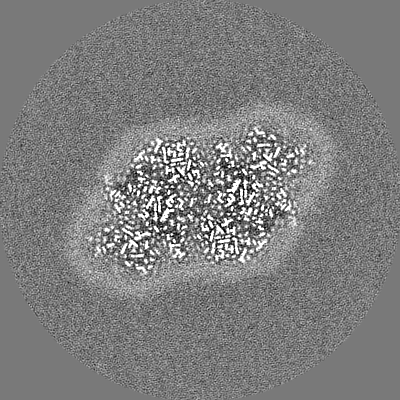
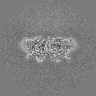
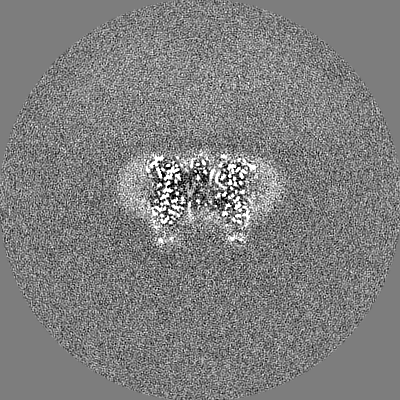
- EMDB-30547: Cryo-EM Structure of PSII at 1.95 angstrom resolution -
+
Open data
ID or keywords:
Loading...
-
Basic information
Entry
Database: EMDB / ID: EMD-30547
Title
Cryo-EM Structure of PSII at 1.95 angstrom resolution
Map data
Sample
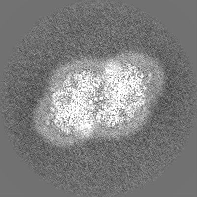
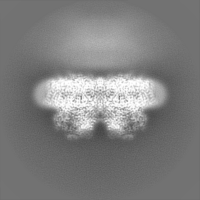
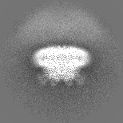
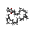
Complex: PSII dimer
Protein or peptide: x 20 types
Ligand: x 19 types
Function / homology
Function and homology information
photosystem II oxygen evolving complex / photosystem II assembly / oxygen evolving activity / photosystem II stabilization / photosystem II / photosystem II reaction center / oxidoreductase activity, acting on diphenols and related substances as donors, oxygen as acceptor / photosynthetic electron transport chain / response to herbicide / photosystem II ...photosystem II oxygen evolving complex / photosystem II assembly / oxygen evolving activity / photosystem II stabilization / photosystem II / photosystem II reaction center / oxidoreductase activity, acting on diphenols and related substances as donors, oxygen as acceptor / photosynthetic electron transport chain / response to herbicide / photosystem II / extrinsic component of membrane / plasma membrane-derived thylakoid membrane / chlorophyll binding / photosynthesis, light reaction / electron transporter, transferring electrons within the cyclic electron transport pathway of photosynthesis activity / phosphate ion binding / photosynthetic electron transport in photosystem II / photosynthesis / respiratory electron transport chain / manganese ion binding / protein stabilization / electron transfer activity / iron ion binding / heme binding / metal ion binding Similarity search - Function
Photosystem II protein Y (PsbY) / Photosystem II PsbY / Photosystem II PsbU, oxygen evolving complex / Photosystem II 12 kDa extrinsic protein (PsbU) / Photosystem II PsbV, cytochrome c-550 precursor / Photosystem II cytochrome c-550 precursor / Cytochrome c-550 domain / Cytochrome c-550 domain / Photosystem II PsbJ / Photosystem II PsbJ superfamily ...Photosystem II protein Y (PsbY) / Photosystem II PsbY / Photosystem II PsbU, oxygen evolving complex / Photosystem II 12 kDa extrinsic protein (PsbU) / Photosystem II PsbV, cytochrome c-550 precursor / Photosystem II cytochrome c-550 precursor / Cytochrome c-550 domain / Cytochrome c-550 domain / Photosystem II PsbJ / Photosystem II PsbJ superfamily / PsbJ / Photosystem II PsbO, manganese-stabilising / Manganese-stabilising protein / photosystem II polypeptide / Photosystem II reaction centre protein Ycf12 / Photosystem II complex subunit Ycf12 / Photosystem II PsbX, type 1 subfamily / Photosystem II reaction centre M protein (PsbM) / Photosystem II PsbM superfamily / Photosystem II PsbM / Photosystem II PsbZ, reaction centre / Photosystem II PsbZ superfamily / YCF9 / Photosystem II PsbX / Photosystem II reaction centre X protein (PsbX) / Photosystem II CP43 reaction centre protein superfamily / Photosystem II PsbT / Photosystem II PsbL / Photosystem II PsbL superfamily / Photosystem II PsbT superfamily / Photosystem II reaction centre T protein / PsbL protein / Photosystem II CP43 reaction centre protein / Photosystem II PsbK / Photosystem II PsbK superfamily / Photosystem II 4 kDa reaction centre component / Photosystem II CP47 reaction centre protein / Photosystem II PsbI / Photosystem II PsbI superfamily / Photosystem II reaction centre I protein (PSII 4.8 kDa protein) / Photosystem II reaction centre protein H / Photosystem II protein D1 / Photosystem II D2 protein / Photosystem II cytochrome b559, conserved site / Photosystem II cytochrome b559, alpha subunit / Photosystem II cytochrome b559, beta subunit / Photosystem II cytochrome b559, N-terminal / Photosystem II cytochrome b559, alpha subunit, lumenal region / Photosystem II reaction centre protein H superfamily / Photosystem II cytochrome b559, alpha subunit superfamily / Cytochrome b559, alpha (gene psbE) and beta (gene psbF)subunits / Lumenal portion of Cytochrome b559, alpha (gene psbE) subunit / Photosystem II 10 kDa phosphoprotein / Cytochrome b559 subunits heme-binding site signature. / Photosystem antenna protein-like / Photosystem antenna protein-like superfamily / Photosystem II protein / Outer membrane protein/outer membrane enzyme PagP, beta-barrel / Photosynthetic reaction centre, L/M / Photosystem II protein D1/D2 superfamily / Photosynthetic reaction centre protein / Photosynthetic reaction center proteins signature. / Cytochrome c family profile. / Cytochrome c-like domain / Cytochrome c-like domain superfamily Similarity search - Domain/homology
Photosystem II CP47 reaction center protein / Photosystem II extrinsic protein O / Photosystem II reaction center protein Psb30 / Photosystem II reaction center protein X / Photosystem II reaction center protein Z / Photosystem II CP43 reaction center protein / Photosystem II D2 protein / Photosystem II extrinsic protein V / Photosystem II reaction center protein Y / Cytochrome b559 subunit alpha ...Photosystem II CP47 reaction center protein / Photosystem II extrinsic protein O / Photosystem II reaction center protein Psb30 / Photosystem II reaction center protein X / Photosystem II reaction center protein Z / Photosystem II CP43 reaction center protein / Photosystem II D2 protein / Photosystem II extrinsic protein V / Photosystem II reaction center protein Y / Cytochrome b559 subunit alpha / Cytochrome b559 subunit beta / Photosystem II reaction center protein I / Photosystem II reaction center protein L / Photosystem II reaction center protein M / Photosystem II reaction center protein T / Photosystem II reaction center protein H / Photosystem II reaction center protein K / Photosystem II protein D1 / Photosystem II extrinsic protein U / Photosystem II reaction center protein J Similarity search - Component
Biological species
Thermosynechococcus vulcanus (bacteria)
Method
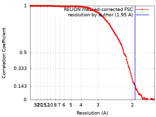
single particle reconstruction / cryo EM / Resolution: 1.95 Å
Journal: Commun Biol / Year: 2021 Title: High-resolution cryo-EM structure of photosystem II reveals damage from high-dose electron beams. Authors: Koji Kato / Naoyuki Miyazaki / Tasuku Hamaguchi / Yoshiki Nakajima / Fusamichi Akita / Koji Yonekura / Jian-Ren Shen / Abstract: Photosystem II (PSII) plays a key role in water-splitting and oxygen evolution. X-ray crystallography has revealed its atomic structure and some intermediate structures. However, these structures are ...Photosystem II (PSII) plays a key role in water-splitting and oxygen evolution. X-ray crystallography has revealed its atomic structure and some intermediate structures. However, these structures are in the crystalline state and its final state structure has not been solved. Here we analyzed the structure of PSII in solution at 1.95 Å resolution by single-particle cryo-electron microscopy (cryo-EM). The structure obtained is similar to the crystal structure, but a PsbY subunit was visible in the cryo-EM structure, indicating that it represents its physiological state more closely. Electron beam damage was observed at a high-dose in the regions that were easily affected by redox states, and reducing the beam dosage by reducing frames from 50 to 2 yielded a similar resolution but reduced the damage remarkably. This study will serve as a good indicator for determining damage-free cryo-EM structures of not only PSII but also all biological samples, especially redox-active metalloproteins.
History
Deposition
Sep 15, 2020
-
Header (metadata) release
Mar 31, 2021
-
Map release
Mar 31, 2021
-
Update
Apr 7, 2021
-
Current status
Apr 7, 2021
Processing site: PDBj / Status: Released
-
Structure visualization
Movie
Surface view with section colored by density value
EMPIAR-10556 (Title: Cryo-EM Structure of PSII at 1.95 angstrom resolution Data size: 427.6 Data #1: Unaligned multi-frame micrographs of PSII recorded by CRYO ARM 300 [micrographs - multiframe])
In the structure databanks used in Yorodumi, some data are registered as the other names, "COVID-19 virus" and "2019-nCoV". Here are the details of the virus and the list of structure data.
Jan 31, 2019. EMDB accession codes are about to change! (news from PDBe EMDB page)
EMDB accession codes are about to change! (news from PDBe EMDB page)
The allocation of 4 digits for EMDB accession codes will soon come to an end. Whilst these codes will remain in use, new EMDB accession codes will include an additional digit and will expand incrementally as the available range of codes is exhausted. The current 4-digit format prefixed with “EMD-” (i.e. EMD-XXXX) will advance to a 5-digit format (i.e. EMD-XXXXX), and so on. It is currently estimated that the 4-digit codes will be depleted around Spring 2019, at which point the 5-digit format will come into force.
The EM Navigator/Yorodumi systems omit the EMD- prefix.
Related info.:Q: What is EMD? / ID/Accession-code notation in Yorodumi/EM Navigator
Yorodumi is a browser for structure data from EMDB, PDB, SASBDB, etc.
This page is also the successor to EM Navigator detail page, and also detail information page/front-end page for Omokage search.
The word "yorodu" (or yorozu) is an old Japanese word meaning "ten thousand". "mi" (miru) is to see.
Related info.:EMDB / PDB / SASBDB / Comparison of 3 databanks / Yorodumi Search / Aug 31, 2016. New EM Navigator & Yorodumi / Yorodumi Papers / Jmol/JSmol / Function and homology information / Changes in new EM Navigator and Yorodumi
 Movie
Movie Controller


 Open data
 Basic information
 Map data
 Sample
 Function and homology information
 Thermosynechococcus vulcanus (bacteria)
 Authors
 Citation
 Structure visualization
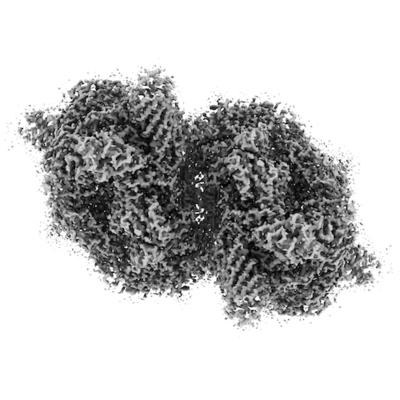
 Downloads & links
 emd_30547.png
 http://ftp.pdbj.org/pub/emdb/structures/EMD-30547
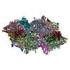


 Z
Z Y
Y X
X


 Sample components


 Processing
 Electron microscopy
 FIELD EMISSION GUN