 Movie
Movie Controller


+ Open data

- Basic information
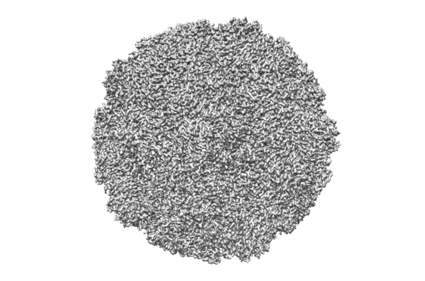
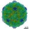
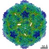
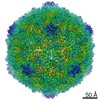
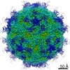
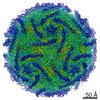
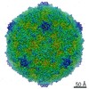
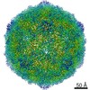
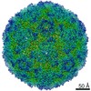
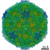
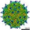
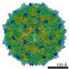
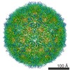
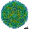
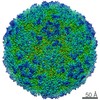
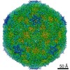
| Entry | Database: EMDB / ID: EMD-30320 | |||||||||
|---|---|---|---|---|---|---|---|---|---|---|
| Title | Echovirus 3 F-particle | |||||||||
 Map data Map data | ||||||||||
 Sample Sample |
| |||||||||
 Keywords Keywords | Echovirus B / mature / VIRUS | |||||||||
| Function / homology |  Function and homology information Function and homology informationsymbiont-mediated suppression of host cytoplasmic pattern recognition receptor signaling pathway via inhibition of RIG-I activity / picornain 2A / symbiont-mediated suppression of host mRNA export from nucleus / symbiont genome entry into host cell via pore formation in plasma membrane / picornain 3C / T=pseudo3 icosahedral viral capsid / host cell cytoplasmic vesicle membrane / viral capsid / ribonucleoside triphosphate phosphatase activity / host cell ...symbiont-mediated suppression of host cytoplasmic pattern recognition receptor signaling pathway via inhibition of RIG-I activity / picornain 2A / symbiont-mediated suppression of host mRNA export from nucleus / symbiont genome entry into host cell via pore formation in plasma membrane / picornain 3C / T=pseudo3 icosahedral viral capsid / host cell cytoplasmic vesicle membrane / viral capsid / ribonucleoside triphosphate phosphatase activity / host cell / nucleoside-triphosphate phosphatase / channel activity / monoatomic ion transmembrane transport / DNA replication / RNA helicase activity / endocytosis involved in viral entry into host cell / symbiont-mediated suppression of host gene expression / symbiont-mediated activation of host autophagy / RNA-directed RNA polymerase / cysteine-type endopeptidase activity / viral RNA genome replication / RNA-directed RNA polymerase activity / symbiont entry into host cell / DNA-templated transcription / virion attachment to host cell / host cell nucleus / structural molecule activity / proteolysis / RNA binding / zinc ion binding / ATP binding Similarity search - Function | |||||||||
| Biological species |  Echovirus E3 Echovirus E3 | |||||||||
| Method | single particle reconstruction / cryo EM / Resolution: 3.4 Å | |||||||||
 Authors Authors | Wang K / Rao Z | |||||||||
| Funding support |  China, 1 items China, 1 items
| |||||||||
 Citation Citation | Journal: Nat Commun / Year: 2020 Title: Structures of Echovirus 30 in complex with its receptors inform a rational prediction for enterovirus receptor usage. Authors: Kang Wang / Ling Zhu / Yao Sun / Minhao Li / Xin Zhao / Lunbiao Cui / Li Zhang / George F Gao / Weiwei Zhai / Fengcai Zhu / Zihe Rao / Xiangxi Wang / Abstract: Receptor usage that determines cell tropism and drives viral classification closely correlates with the virus structure. Enterovirus B (EV-B) consists of several subgroups according to receptor ...Receptor usage that determines cell tropism and drives viral classification closely correlates with the virus structure. Enterovirus B (EV-B) consists of several subgroups according to receptor usage, among which echovirus 30 (E30), a leading causative agent for human aseptic meningitis, utilizes FcRn as an uncoating receptor. However, receptors for many EVs remain unknown. Here we analyzed the atomic structures of E30 mature virion, empty- and A-particles, which reveals serotype-specific epitopes and striking conformational differences between the subgroups within EV-Bs. Of these, the VP1 BC loop markedly distinguishes E30 from other EV-Bs, indicative of a role as a structural marker for EV-B. By obtaining cryo-electron microscopy structures of E30 in complex with its receptor FcRn and CD55 and comparing its homologs, we deciphered the underlying molecular basis for receptor recognition. Together with experimentally derived viral receptor identifications, we developed a structure-based in silico algorithm to inform a rational prediction for EV receptor usage. | |||||||||
| History |
|
- Structure visualization
| Movie |
Movie viewer |
|---|---|
| Structure viewer | EM map: SurfViewMolmilJmol/JSmol |
| Supplemental images |
- Downloads & links
-EMDB archive
| Map data | emd_30320.map.gz | 115.3 MB | EMDB map data format | |
|---|---|---|---|---|
| Header (meta data) | emd-30320-v30.xmlemd-30320.xml | 15.1 KB 15.1 KB | Display Display | EMDB header |
| Images |  emd_30320.png emd_30320.png | 116.9 KB | ||
| Filedesc metadata | emd-30320.cif.gz | 6 KB | ||
| Archive directory |  http://ftp.pdbj.org/pub/emdb/structures/EMD-30320ftp://ftp.pdbj.org/pub/emdb/structures/EMD-30320 http://ftp.pdbj.org/pub/emdb/structures/EMD-30320ftp://ftp.pdbj.org/pub/emdb/structures/EMD-30320 | HTTPS FTP |
-Related structure data
| Related structure data | 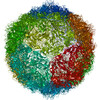 7c9xMC 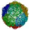 7c9sC 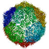 7c9tC 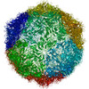 7c9uC  7c9vC  7c9wC 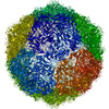 7c9yC 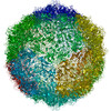 7c9zC M: atomic model generated by this map C: citing same article ( |
|---|---|
| Similar structure data |

-Links
| EMDB pages | EMDB (EBI/PDBe) / EMDataResource |
|---|---|
| Related items in Molecule of the Month |


-Map
| File | Download / File: emd_30320.map.gz / Format: CCP4 / Size: 125 MB / Type: IMAGE STORED AS FLOATING POINT NUMBER (4 BYTES) | ||||||||||||||||||||||||||||||||||||||||||||||||||||||||||||||||||||
|---|---|---|---|---|---|---|---|---|---|---|---|---|---|---|---|---|---|---|---|---|---|---|---|---|---|---|---|---|---|---|---|---|---|---|---|---|---|---|---|---|---|---|---|---|---|---|---|---|---|---|---|---|---|---|---|---|---|---|---|---|---|---|---|---|---|---|---|---|---|
| Projections & slices | Image control
Images are generated by Spider. | ||||||||||||||||||||||||||||||||||||||||||||||||||||||||||||||||||||
| Voxel size | X=Y=Z: 1.347 Å | ||||||||||||||||||||||||||||||||||||||||||||||||||||||||||||||||||||
| Density |
| ||||||||||||||||||||||||||||||||||||||||||||||||||||||||||||||||||||
| Symmetry | Space group: 1 | ||||||||||||||||||||||||||||||||||||||||||||||||||||||||||||||||||||
| Details | EMDB XML:
CCP4 map header:
| ||||||||||||||||||||||||||||||||||||||||||||||||||||||||||||||||||||
 Z (Sec.)
 Y (Row.)
 X (Col.)


-Supplemental data
- Sample components
-Entire : Echovirus E3
| Entire | Name: Echovirus E3 |
|---|---|
| Components |
|
-Supramolecule #1: Echovirus E3
| Supramolecule | Name: Echovirus E3 / type: virus / ID: 1 / Parent: 0 / Macromolecule list: #1-#4 Details: Particles purified from the cell cultures innoculated with the live E3. NCBI-ID: 47516 / Sci species name: Echovirus E3 / Virus type: VIRION / Virus isolate: SEROTYPE / Virus enveloped: No / Virus empty: No |
|---|---|
| Host (natural) | Organism:  Homo sapiens (human) Homo sapiens (human) |
| Virus shell | Shell ID: 1 / Diameter: 30.0 Å / T number (triangulation number): 3 |
-Macromolecule #1: VP1
| Macromolecule | Name: VP1 / type: protein_or_peptide / ID: 1 / Number of copies: 1 / Enantiomer: LEVO |
|---|---|
| Source (natural) | Organism: Echovirus E3 |
| Molecular weight | Theoretical: 31.761631 KDa |
| Sequence | String: GDVEEAIDRA VARVADTMPT GPRNTESVPA LTAVETGHTS QVVPGDTMQT RHVKNYHSRT ESSIENFLCR AACVYIATYK SAGGTPTER YASWRINTRQ MVQLRRKFEL FTYLRFDMEI TFVITSTQDP GTQLAQDMPV LTHQIMYIPP GGPVPNSATD F AWQSSTNP ...String: GDVEEAIDRA VARVADTMPT GPRNTESVPA LTAVETGHTS QVVPGDTMQT RHVKNYHSRT ESSIENFLCR AACVYIATYK SAGGTPTER YASWRINTRQ MVQLRRKFEL FTYLRFDMEI TFVITSTQDP GTQLAQDMPV LTHQIMYIPP GGPVPNSATD F AWQSSTNP SIFWTEGCAP ARMSVPFISI GNAYSNFYDG WSHFTQEGVY GFNSLNNMGH IYVRHVNEQS LGVSTSTLRV YF KPKHVRA WVPRPPRLSP YVKSSNVNFK PTAVTTERKD INDVGT UniProtKB: Genome polyprotein |
-Macromolecule #2: VP2
| Macromolecule | Name: VP2 / type: protein_or_peptide / ID: 2 / Number of copies: 1 / Enantiomer: LEVO |
|---|---|
| Source (natural) | Organism: Echovirus E3 |
| Molecular weight | Theoretical: 29.221725 KDa |
| Sequence | String: SPTVEECGFS DRVRSITLGN STITTQECAN VVVGYGVWPS YLQDNEATAE DQPTQPDVAT CRFYTLDSIQ WQKESDGWWW KFPEALKNM GLFGQNMEYH YLGRSGYTIH VQCNASKFHQ GCLLVVCVPE AEMGCSDVER EVVAASLSSE DTAKSFSRTE S NGQHTVQT ...String: SPTVEECGFS DRVRSITLGN STITTQECAN VVVGYGVWPS YLQDNEATAE DQPTQPDVAT CRFYTLDSIQ WQKESDGWWW KFPEALKNM GLFGQNMEYH YLGRSGYTIH VQCNASKFHQ GCLLVVCVPE AEMGCSDVER EVVAASLSSE DTAKSFSRTE S NGQHTVQT VVYNAGMGVG VGNLTIFPHQ WINLRTNNSA TIVMPYINSV PMDNMFRHYN FTLMIIPFAK LEYTEQASNY VP ITVTVAP MCAEYNGLRL ASHQ UniProtKB: Genome polyprotein |
-Macromolecule #3: VP3
| Macromolecule | Name: VP3 / type: protein_or_peptide / ID: 3 / Number of copies: 1 / Enantiomer: LEVO |
|---|---|
| Source (natural) | Organism: Echovirus E3 |
| Molecular weight | Theoretical: 26.281973 KDa |
| Sequence | String: GLPTMLTPGS NQFLTSDDFQ SPSAMPQFDV TPEMKIPGEV HNLMEIAEVD SVVPVNNTKE NINSMEAYRI PVTGGDQLHT QVFGFQMQP GLNSVFKRTL LGEILNYYAH WSGSVKLTFV FCGSAMATGK FLLAYSPPGA SPPQNRKQAM LGTHVIWDVG L QSSCVLCI ...String: GLPTMLTPGS NQFLTSDDFQ SPSAMPQFDV TPEMKIPGEV HNLMEIAEVD SVVPVNNTKE NINSMEAYRI PVTGGDQLHT QVFGFQMQP GLNSVFKRTL LGEILNYYAH WSGSVKLTFV FCGSAMATGK FLLAYSPPGA SPPQNRKQAM LGTHVIWDVG L QSSCVLCI PWISQTHYRL VQQDEYTSAG YVTCWYQTGL IVPPGAPPSC TILCFASACN DFSVRMLRDT PFIEQTQLLQ UniProtKB: Genome polyprotein |
-Macromolecule #4: VP4
| Macromolecule | Name: VP4 / type: protein_or_peptide / ID: 4 / Number of copies: 1 / Enantiomer: LEVO |
|---|---|
| Source (natural) | Organism: Echovirus E3 |
| Molecular weight | Theoretical: 7.402076 KDa |
| Sequence | String: GAQVSTQKTG AHETSLTASG NSTIHYTNIN YYKDAASNSA NRQDFTQDPS KFTEPMKDVM IKSLPALN UniProtKB: Genome polyprotein |
-Macromolecule #5: SPHINGOSINE
| Macromolecule | Name: SPHINGOSINE / type: ligand / ID: 5 / Number of copies: 1 / Formula: SPH |
|---|---|
| Molecular weight | Theoretical: 299.492 Da |
| Chemical component information |  ChemComp-SPH: |
-Macromolecule #6: MYRISTIC ACID
| Macromolecule | Name: MYRISTIC ACID / type: ligand / ID: 6 / Number of copies: 1 / Formula: MYR |
|---|---|
| Molecular weight | Theoretical: 228.371 Da |
| Chemical component information |  ChemComp-MYR: |
-Experimental details
-Structure determination
| Method | cryo EM |
|---|---|
 Processing Processing | single particle reconstruction |
| Aggregation state | particle |
-Sample preparation
| Buffer | pH: 7.4 |
|---|---|
| Grid | Model: Quantifoil R1.2/1.3 / Support film - Material: CARBON / Support film - topology: HOLEY / Pretreatment - Type: PLASMA CLEANING |
| Vitrification | Cryogen name: ETHANE / Chamber humidity: 100 % / Instrument: FEI VITROBOT MARK I |
- Electron microscopy
| Microscope | FEI TITAN KRIOS |
|---|---|
| Image recording | Film or detector model: GATAN K2 SUMMIT (4k x 4k) / Detector mode: SUPER-RESOLUTION / Average electron dose: 30.0 e/Å2 |
| Electron beam | Acceleration voltage: 300 kV / Electron source:  FIELD EMISSION GUN FIELD EMISSION GUN |
| Electron optics | Illumination mode: FLOOD BEAM / Imaging mode: DARK FIELD / Cs: 2.7 mm / Nominal defocus max: 2.5 µm / Nominal defocus min: 1.2 µm |
| Sample stage | Specimen holder model: FEI TITAN KRIOS AUTOGRID HOLDER / Cooling holder cryogen: NITROGEN |
| Experimental equipment |  Model: Titan Krios / Image courtesy: FEI Company |
-Image processing
| Startup model | Type of model: INSILICO MODEL |
|---|---|
| Final reconstruction | Applied symmetry - Point group: I (icosahedral) / Resolution.type: BY AUTHOR / Resolution: 3.4 Å / Resolution method: FSC 0.143 CUT-OFF / Software - Name: RELION (ver. 3.0) / Number images used: 5000 |
| Initial angle assignment | Type: PROJECTION MATCHING |
| Final angle assignment | Type: PROJECTION MATCHING |
-Atomic model buiding 1
| Refinement | Space: REAL / Protocol: RIGID BODY FIT |
|---|---|
| Output model | PDB-7c9x: |
