 Movie
Movie Controller

[English] 日本語
 Yorodumi
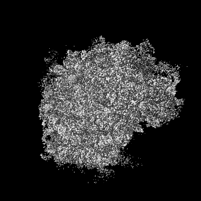
- EMDB-2875: Cryo electron microscopy of actively translating human polysomes ... -
+ Open data

- Basic information
| Entry | Database: EMDB / ID: EMD-2875 | |||||||||
|---|---|---|---|---|---|---|---|---|---|---|
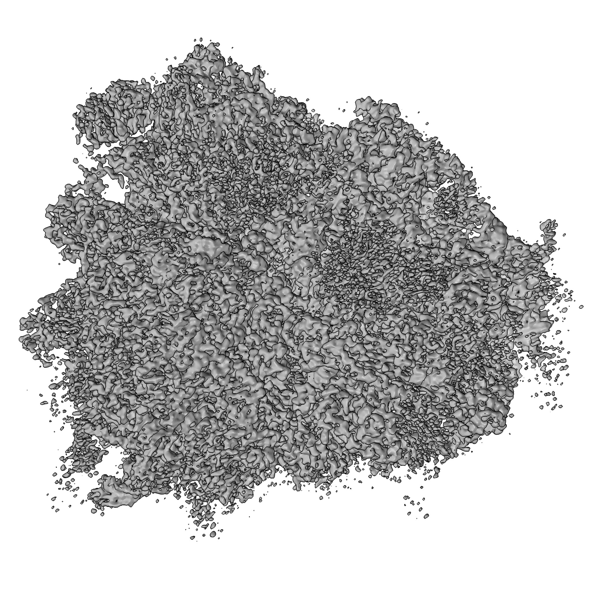
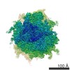
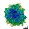
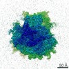
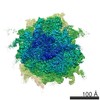
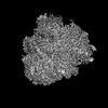
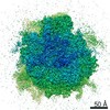
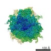
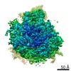
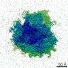
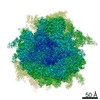
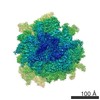
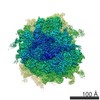
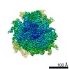
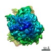
| Title | Cryo electron microscopy of actively translating human polysomes (POST state). | |||||||||
 Map data Map data | POST state of human polysomes | |||||||||
 Sample Sample |
| |||||||||
 Keywords Keywords | mammalian ribosome / translation / polysome / cryo electron microscopy / elongation cycle | |||||||||
| Function / homology |  Function and homology information Function and homology informationembryonic brain development / translation at presynapse / exit from mitosis / optic nerve development / response to insecticide / regulation of translation involved in cellular response to UV / eukaryotic 80S initiation complex / negative regulation of endoplasmic reticulum unfolded protein response / ribosomal protein import into nucleus / negative regulation of formation of translation preinitiation complex ...embryonic brain development / translation at presynapse / exit from mitosis / optic nerve development / response to insecticide / regulation of translation involved in cellular response to UV / eukaryotic 80S initiation complex / negative regulation of endoplasmic reticulum unfolded protein response / ribosomal protein import into nucleus / negative regulation of formation of translation preinitiation complex / axial mesoderm development / regulation of G1 to G0 transition / retinal ganglion cell axon guidance / oxidized pyrimidine DNA binding / response to TNF agonist / positive regulation of base-excision repair / positive regulation of ubiquitin-protein transferase activity / positive regulation of respiratory burst involved in inflammatory response / protein-DNA complex disassembly / positive regulation of intrinsic apoptotic signaling pathway in response to DNA damage by p53 class mediator / positive regulation of gastrulation / protein tyrosine kinase inhibitor activity / positive regulation of DNA-templated transcription initiation / positive regulation of intrinsic apoptotic signaling pathway in response to DNA damage / 90S preribosome assembly / IRE1-RACK1-PP2A complex / positive regulation of Golgi to plasma membrane protein transport / nucleolus organization / TNFR1-mediated ceramide production / alpha-beta T cell differentiation / positive regulation of DNA damage response, signal transduction by p53 class mediator / GAIT complex / negative regulation of RNA splicing / TORC2 complex binding / neural crest cell differentiation / supercoiled DNA binding / cytoplasmic translational initiation / NF-kappaB complex / negative regulation of DNA repair / G1 to G0 transition / oxidized purine DNA binding / cysteine-type endopeptidase activator activity involved in apoptotic process / middle ear morphogenesis / rRNA modification in the nucleus and cytosol / negative regulation of intrinsic apoptotic signaling pathway in response to hydrogen peroxide / negative regulation of bicellular tight junction assembly / ubiquitin-like protein conjugating enzyme binding / regulation of establishment of cell polarity / negative regulation of phagocytosis / erythrocyte homeostasis / cytoplasmic side of rough endoplasmic reticulum membrane / Formation of the ternary complex, and subsequently, the 43S complex / ion channel inhibitor activity / protein kinase A binding / laminin receptor activity / Ribosomal scanning and start codon recognition / pigmentation / homeostatic process / positive regulation of mitochondrial depolarization / Translation initiation complex formation / macrophage chemotaxis / lung morphogenesis / negative regulation of Wnt signaling pathway / positive regulation of natural killer cell proliferation / fibroblast growth factor binding / Protein hydroxylation / monocyte chemotaxis / BH3 domain binding / negative regulation of translational frameshifting / regulation of adenylate cyclase-activating G protein-coupled receptor signaling pathway / SARS-CoV-1 modulates host translation machinery / positive regulation of GTPase activity / TOR signaling / mTORC1-mediated signalling / iron-sulfur cluster binding / Peptide chain elongation / regulation of cell division / cellular response to ethanol / Selenocysteine synthesis / Formation of a pool of free 40S subunits / negative regulation of protein binding / positive regulation of intrinsic apoptotic signaling pathway by p53 class mediator / Eukaryotic Translation Termination / protein serine/threonine kinase inhibitor activity / blastocyst development / SRP-dependent cotranslational protein targeting to membrane / Response of EIF2AK4 (GCN2) to amino acid deficiency / ubiquitin ligase inhibitor activity / negative regulation of respiratory burst involved in inflammatory response / Viral mRNA Translation / endonucleolytic cleavage to generate mature 3'-end of SSU-rRNA from (SSU-rRNA, 5.8S rRNA, LSU-rRNA) / Nonsense Mediated Decay (NMD) independent of the Exon Junction Complex (EJC) / positive regulation of signal transduction by p53 class mediator / negative regulation of ubiquitin-dependent protein catabolic process / protein localization to nucleus / GTP hydrolysis and joining of the 60S ribosomal subunit / L13a-mediated translational silencing of Ceruloplasmin expression / Major pathway of rRNA processing in the nucleolus and cytosol / regulation of translational fidelity / protein targeting Similarity search - Function | |||||||||
| Biological species |  Homo sapiens (human) Homo sapiens (human) | |||||||||
| Method | single particle reconstruction / cryo EM / Resolution: 3.5 Å | |||||||||
 Authors Authors | Behrmann E / Loerke J / Budkevich TV / Yamamoto K / Schmidt A / Penczek PA / Vos MR / Burger J / Mielke T / Scheerer P / Spahn CMT | |||||||||
 Citation Citation | Journal: Cell / Year: 2015 Title: Structural snapshots of actively translating human ribosomes. Authors: Elmar Behrmann / Justus Loerke / Tatyana V Budkevich / Kaori Yamamoto / Andrea Schmidt / Pawel A Penczek / Matthijn R Vos / Jörg Bürger / Thorsten Mielke / Patrick Scheerer / Christian M T Spahn /    Abstract: Macromolecular machines, such as the ribosome, undergo large-scale conformational changes during their functional cycles. Although their mode of action is often compared to that of mechanical ...Macromolecular machines, such as the ribosome, undergo large-scale conformational changes during their functional cycles. Although their mode of action is often compared to that of mechanical machines, a crucial difference is that, at the molecular dimension, thermodynamic effects dominate functional cycles, with proteins fluctuating stochastically between functional states defined by energetic minima on an energy landscape. Here, we have used cryo-electron microscopy to image ex-vivo-derived human polysomes as a source of actively translating ribosomes. Multiparticle refinement and 3D variability analysis allowed us to visualize a variety of native translation intermediates. Significantly populated states include not only elongation cycle intermediates in pre- and post-translocational states, but also eEF1A-containing decoding and termination/recycling complexes. Focusing on the post-translocational state, we extended this assessment to the single-residue level, uncovering striking details of ribosome-ligand interactions and identifying both static and functionally important dynamic elements. | |||||||||
| History |
|
- Structure visualization
| Movie |
Movie viewer |
|---|---|
| Structure viewer | EM map: SurfViewMolmilJmol/JSmol |
| Supplemental images |
- Downloads & links
-EMDB archive
| Map data | emd_2875.map.gz | 152.8 MB | EMDB map data format | |
|---|---|---|---|---|
| Header (meta data) | emd-2875-v30.xmlemd-2875.xml | 10.9 KB 10.9 KB | Display Display | EMDB header |
| Images | post.tif | 3.1 MB | ||
| Archive directory |  http://ftp.pdbj.org/pub/emdb/structures/EMD-2875ftp://ftp.pdbj.org/pub/emdb/structures/EMD-2875 http://ftp.pdbj.org/pub/emdb/structures/EMD-2875ftp://ftp.pdbj.org/pub/emdb/structures/EMD-2875 | HTTPS FTP |
-Related structure data
| Related structure data | 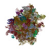 5aj0MC 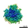 2902C  2903C 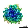 2904C 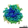 2905C 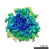 2906C 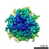 2907C 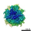 2908C  2909C 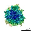 2910C 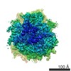 2911C M: atomic model generated by this map C: citing same article ( |
|---|---|
| Similar structure data |
-Links
| EMDB pages | EMDB (EBI/PDBe) / EMDataResource |
|---|---|
| Related items in Molecule of the Month |


-Map
| File | Download / File: emd_2875.map.gz / Format: CCP4 / Size: 238.4 MB / Type: IMAGE STORED AS FLOATING POINT NUMBER (4 BYTES) | ||||||||||||||||||||||||||||||||||||||||||||||||||||||||||||||||||||
|---|---|---|---|---|---|---|---|---|---|---|---|---|---|---|---|---|---|---|---|---|---|---|---|---|---|---|---|---|---|---|---|---|---|---|---|---|---|---|---|---|---|---|---|---|---|---|---|---|---|---|---|---|---|---|---|---|---|---|---|---|---|---|---|---|---|---|---|---|---|
| Annotation | POST state of human polysomes | ||||||||||||||||||||||||||||||||||||||||||||||||||||||||||||||||||||
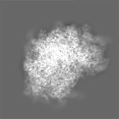
| Projections & slices | Image control
Images are generated by Spider. | ||||||||||||||||||||||||||||||||||||||||||||||||||||||||||||||||||||
| Voxel size | X=Y=Z: 0.945 Å | ||||||||||||||||||||||||||||||||||||||||||||||||||||||||||||||||||||
| Density |
| ||||||||||||||||||||||||||||||||||||||||||||||||||||||||||||||||||||
| Symmetry | Space group: 1 | ||||||||||||||||||||||||||||||||||||||||||||||||||||||||||||||||||||
| Details | EMDB XML:
CCP4 map header:
| ||||||||||||||||||||||||||||||||||||||||||||||||||||||||||||||||||||
 Z (Sec.)
 Y (Row.)
 X (Col.)


-Supplemental data
- Sample components
-Entire : Native ribosomal complex from polysomes - POST state
| Entire | Name: Native ribosomal complex from polysomes - POST state |
|---|---|
| Components |
|
-Supramolecule #1000: Native ribosomal complex from polysomes - POST state
| Supramolecule | Name: Native ribosomal complex from polysomes - POST state / type: sample / ID: 1000 / Details: The sample was monodisperse / Number unique components: 1 |
|---|---|
| Molecular weight | Experimental: 4.5 MDa / Theoretical: 4.5 MDa |
-Supramolecule #1: 80S ribosomal complex
| Supramolecule | Name: 80S ribosomal complex / type: complex / ID: 1 / Name.synonym: 80S ribosome Details: The sample was isolated as polysomes from the cell. Structural analysis shows that the complex contains 2 tRNAs and mRNA. Recombinant expression: No / Ribosome-details: ribosome-eukaryote: ALL |
|---|---|
| Source (natural) | Organism: Homo sapiens (human) / Strain: HEK 293T / synonym: Human / Tissue: Kidney / Cell: HEK 293T / Location in cell: Cytoplasm |
| Molecular weight | Experimental: 4.5 MDa / Theoretical: 4.5 MDa |
-Experimental details
-Structure determination
| Method | cryo EM |
|---|---|
 Processing Processing | single particle reconstruction |
| Aggregation state | particle |
-Sample preparation
| Concentration | 3.5 mg/mL |
|---|---|
| Buffer | pH: 7.5 Details: 20 mM Hepes-KOH, pH 7.5, 100 mM KCl, 1.5 mM MgCl2, 0.5 mM spermidine, 0.04 mM spermine, 1 mM DTT |
| Grid | Details: Quantifoil grids with additional continuous carbon support |
| Vitrification | Cryogen name: ETHANE / Chamber humidity: 100 % / Chamber temperature: 93 K / Instrument: FEI VITROBOT MARK II / Method: blot for 2-4 seconds before plunging |
- Electron microscopy #1
| Microscopy ID | 1 |
|---|---|
| Microscope | FEI POLARA 300 |
| Details | Data was collected automatically with Leginon |
| Date | Aug 20, 2012 |
| Image recording | Category: CCD / Film or detector model: TVIPS TEMCAM-F416 (4k x 4k) / Number real images: 51282 / Average electron dose: 20 e/Å2 |
| Electron beam | Acceleration voltage: 300 kV / Electron source:  FIELD EMISSION GUN FIELD EMISSION GUN |
| Electron optics | Calibrated magnification: 205000 / Illumination mode: FLOOD BEAM / Imaging mode: BRIGHT FIELD / Cs: 2.0 mm / Nominal defocus max: 4.5 µm / Nominal defocus min: 2.0 µm / Nominal magnification: 115000 |
| Sample stage | Specimen holder model: GATAN LIQUID NITROGEN |
| Experimental equipment |  Model: Tecnai Polara / Image courtesy: FEI Company |
-Electron microscopy #2
| Microscopy ID | 2 |
|---|---|
| Microscope | FEI TITAN KRIOS |
| Date | Nov 29, 2012 |
| Image recording | Category: CCD / Film or detector model: FEI FALCON II (4k x 4k) / Number real images: 51282 / Average electron dose: 20 e/Å2 |
| Electron beam | Acceleration voltage: 300 kV / Electron source: FIELD EMISSION GUN |
| Electron optics | Calibrated magnification: 172000 / Illumination mode: FLOOD BEAM / Imaging mode: BRIGHT FIELD / Cs: 2.7 mm / Nominal defocus max: 4.5 µm / Nominal defocus min: 1.5 µm / Nominal magnification: 96000 |
| Sample stage | Specimen holder model: FEI TITAN KRIOS AUTOGRID HOLDER |
| Experimental equipment |  Model: Titan Krios / Image courtesy: FEI Company |
-Image processing
| CTF correction | Details: Each micrograph |
|---|---|
| Final reconstruction | Applied symmetry - Point group: C1 (asymmetric) / Algorithm: OTHER / Resolution.type: BY AUTHOR / Resolution: 3.5 Å / Resolution method: OTHER / Software - Name: Signature, CTFFind3, Spider, SPARX Details: Maps were calculated from 2 datasets using SSNR-weighted combination. Number images used: 313321 |
