Movie
Movie Controller

[English] 日本語
 Yorodumi
- EMDB-28223: CryoEM structure of Resistance to Inhibitors of Cholinesterase-8B... -
+ Open data

- Basic information
| Entry |  | |||||||||
|---|---|---|---|---|---|---|---|---|---|---|
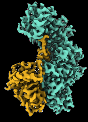
| Title | CryoEM structure of Resistance to Inhibitors of Cholinesterase-8B (Ric-8B) in complex with G alpha s | |||||||||
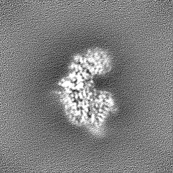
 Map data Map data | Sharpened cryoEM Map of Ric-8B/G alpha s complex | |||||||||
 Sample Sample |
| |||||||||
| Function / homology |  Function and homology information Function and homology informationPKA activation in glucagon signalling / hair follicle placode formation / developmental growth / G-protein alpha-subunit binding / D1 dopamine receptor binding / intracellular transport / Hedgehog 'off' state / adenylate cyclase-activating adrenergic receptor signaling pathway / activation of adenylate cyclase activity / adenylate cyclase activator activity ...PKA activation in glucagon signalling / hair follicle placode formation / developmental growth / G-protein alpha-subunit binding / D1 dopamine receptor binding / intracellular transport / Hedgehog 'off' state / adenylate cyclase-activating adrenergic receptor signaling pathway / activation of adenylate cyclase activity / adenylate cyclase activator activity / guanyl-nucleotide exchange factor activity / trans-Golgi network membrane / G-protein beta/gamma-subunit complex binding / bone development / adenylate cyclase-activating G protein-coupled receptor signaling pathway / Prostacyclin signalling through prostacyclin receptor / Glucagon signaling in metabolic regulation / cognition / platelet aggregation / Glucagon-type ligand receptors / Vasopressin regulates renal water homeostasis via Aquaporins / G alpha (z) signalling events / cellular response to catecholamine stimulus / Glucagon-like Peptide-1 (GLP1) regulates insulin secretion / ADORA2B mediated anti-inflammatory cytokines production / adenylate cyclase-activating dopamine receptor signaling pathway / cellular response to prostaglandin E stimulus / GPER1 signaling / heterotrimeric G-protein complex / sensory perception of smell / positive regulation of cold-induced thermogenesis / cell cortex / G alpha (i) signalling events / G alpha (s) signalling events / G protein-coupled receptor signaling pathway / GTPase activity / centrosome / GTP binding / extracellular exosome / membrane / metal ion binding / plasma membrane / cytoplasm / cytosol Similarity search - Function | |||||||||
| Biological species |  Homo sapiens (human) / Homo sapiens (human) /  | |||||||||
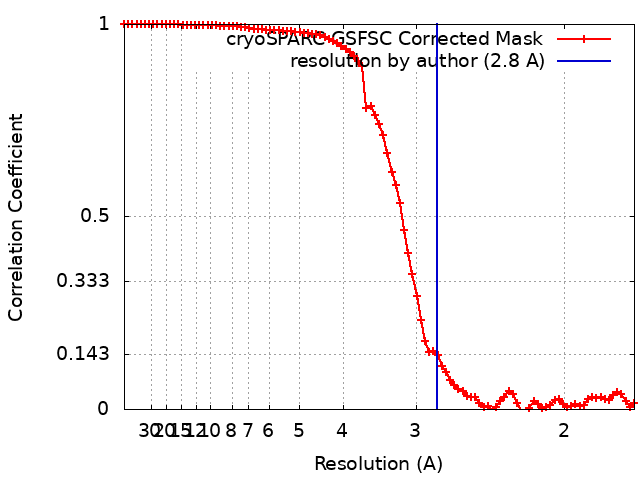
| Method | single particle reconstruction / cryo EM / Resolution: 2.8 Å | |||||||||
 Authors Authors | Papasergi-Scott MM / Skiniotis G | |||||||||
| Funding support |  United States, 1 items United States, 1 items
| |||||||||
 Citation Citation | Journal: Structure / Year: 2023 Title: Structures of Ric-8B in complex with Gα protein folding clients reveal isoform specificity mechanisms. Authors: Makaía M Papasergi-Scott / Frank E Kwarcinski / Maiya Yu / Ouliana Panova / Ann M Ovrutsky / Georgios Skiniotis / Gregory G Tall / Abstract: Mammalian Ric-8 proteins act as chaperones to regulate the cellular abundance of heterotrimeric G protein α subunits. The Ric-8A isoform chaperones Gαi/o, Gα12/13, and Gαq/11 subunits, while Ric- ...Mammalian Ric-8 proteins act as chaperones to regulate the cellular abundance of heterotrimeric G protein α subunits. The Ric-8A isoform chaperones Gαi/o, Gα12/13, and Gαq/11 subunits, while Ric-8B acts on Gαs/olf subunits. Here, we determined cryoelectron microscopy (cryo-EM) structures of Ric-8B in complex with Gαs and Gαolf, revealing isoform differences in the relative positioning and contacts between the C-terminal α5 helix of Gα within the concave pocket formed by Ric-8 α-helical repeat elements. Despite the overall architectural similarity with our earlier structures of Ric-8A complexed to Gαq and Gαi1, Ric-8B distinctly accommodates an extended loop found only in Gαs/olf proteins. The structures, along with results from Ric-8 protein thermal stability assays and cell-based Gαolf folding assays, support a requirement for the Gα C-terminal region for binding specificity, and highlight that multiple structural elements impart specificity for Ric-8/G protein binding. | |||||||||
| History |
|
- Structure visualization
| Supplemental images |
|---|
- Downloads & links
-EMDB archive
| Map data | emd_28223.map.gz | 54.7 MB | EMDB map data format | |
|---|---|---|---|---|
| Header (meta data) | emd-28223-v30.xmlemd-28223.xml | 18.9 KB 18.9 KB | Display Display | EMDB header |
| FSC (resolution estimation) | emd_28223_fsc.xml | 9.2 KB | Display | FSC data file |
| Images |  emd_28223.png emd_28223.png | 127.8 KB | ||
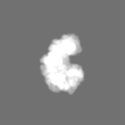
| Masks | emd_28223_msk_1.map | 58.2 MB | Mask map | |
| Others | emd_28223_half_map_1.map.gzemd_28223_half_map_2.map.gz | 54.1 MB 54.1 MB | ||
| Archive directory |  http://ftp.pdbj.org/pub/emdb/structures/EMD-28223ftp://ftp.pdbj.org/pub/emdb/structures/EMD-28223 http://ftp.pdbj.org/pub/emdb/structures/EMD-28223ftp://ftp.pdbj.org/pub/emdb/structures/EMD-28223 | HTTPS FTP |
-Validation report
| Summary document | emd_28223_validation.pdf.gz | 1 MB | Display | EMDB validaton report |
|---|---|---|---|---|
| Full document | emd_28223_full_validation.pdf.gz | 1 MB | Display | |
| Data in XML | emd_28223_validation.xml.gz | 16.4 KB | Display | |
| Data in CIF | emd_28223_validation.cif.gz | 20.9 KB | Display | |
| Arichive directory | https://ftp.pdbj.org/pub/emdb/validation_reports/EMD-28223ftp://ftp.pdbj.org/pub/emdb/validation_reports/EMD-28223 | HTTPS FTP |
-Related structure data
| Related structure data |  8el7MC  8el8C M: atomic model generated by this map C: citing same article ( |
|---|---|
| Similar structure data |

-Links
| EMDB pages | EMDB (EBI/PDBe) / EMDataResource |
|---|---|
| Related items in Molecule of the Month |


-Map
| File | Download / File: emd_28223.map.gz / Format: CCP4 / Size: 58.2 MB / Type: IMAGE STORED AS FLOATING POINT NUMBER (4 BYTES) | ||||||||||||||||||||
|---|---|---|---|---|---|---|---|---|---|---|---|---|---|---|---|---|---|---|---|---|---|
| Annotation | Sharpened cryoEM Map of Ric-8B/G alpha s complex | ||||||||||||||||||||
| Voxel size | X=Y=Z: 0.8521 Å | ||||||||||||||||||||
| Density |
| ||||||||||||||||||||
| Symmetry | Space group: 1 | ||||||||||||||||||||
| Details | EMDB XML:
|
-Supplemental data
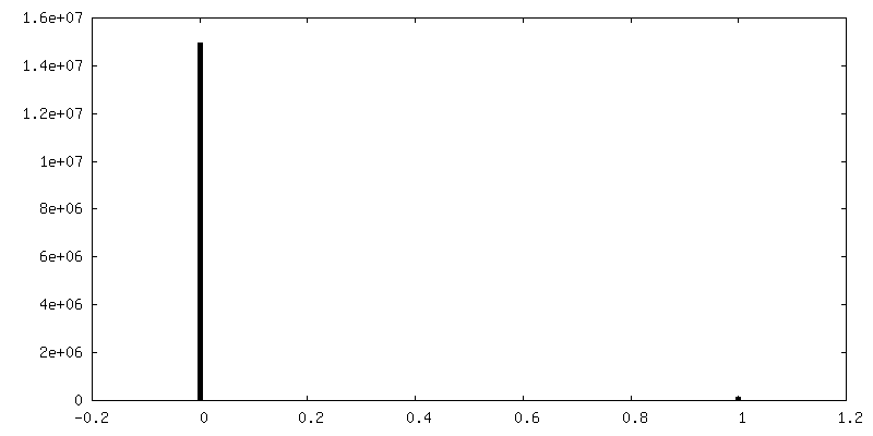
-Mask #1
| File | emd_28223_msk_1.map | ||||||||||||
|---|---|---|---|---|---|---|---|---|---|---|---|---|---|
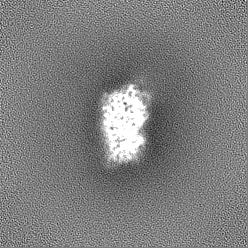
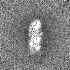
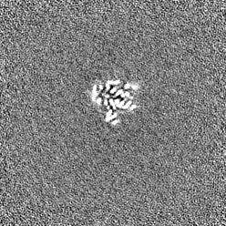
| Projections & Slices |
| ||||||||||||
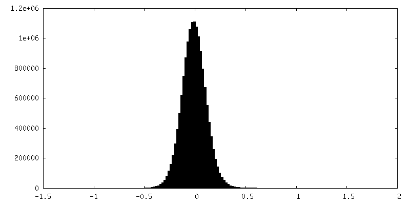
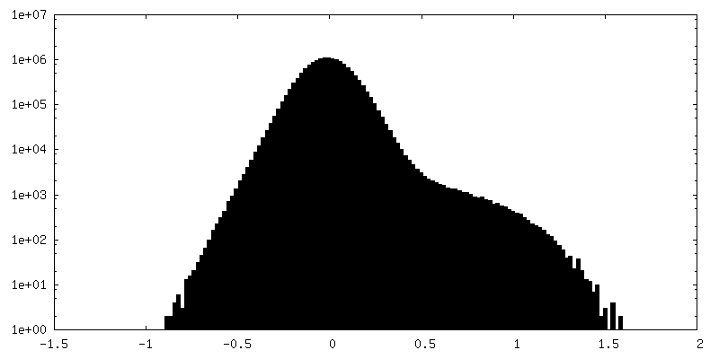
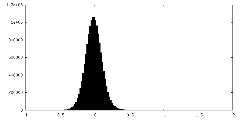
| Density Histograms |
 Z
Z Y
Y X
X


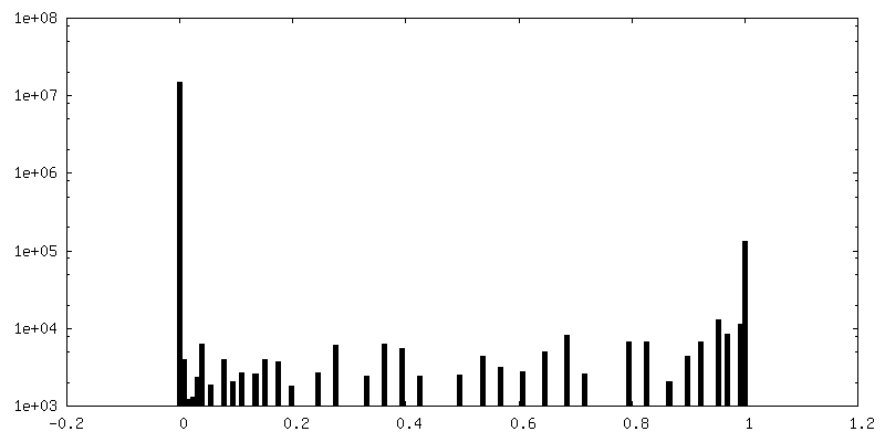
-Half map: Half Map 1 of Ric-8B/G alpha s complex
| File | emd_28223_half_map_1.map | ||||||||||||
|---|---|---|---|---|---|---|---|---|---|---|---|---|---|
| Annotation | Half Map 1 of Ric-8B/G alpha s complex | ||||||||||||
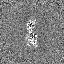
| Projections & Slices |
| ||||||||||||
| Density Histograms |


-Half map: Half Map 2 of Ric-8B/G alpha s complex
| File | emd_28223_half_map_2.map | ||||||||||||
|---|---|---|---|---|---|---|---|---|---|---|---|---|---|
| Annotation | Half Map 2 of Ric-8B/G alpha s complex | ||||||||||||
| Projections & Slices |
| ||||||||||||
| Density Histograms |

- Sample components
-Entire : Ric-8B complexed with G alpha s
| Entire | Name: Ric-8B complexed with G alpha s |
|---|---|
| Components |
|
-Supramolecule #1: Ric-8B complexed with G alpha s
| Supramolecule | Name: Ric-8B complexed with G alpha s / type: complex / ID: 1 / Chimera: Yes / Parent: 0 / Macromolecule list: #1-#2 |
|---|
-Supramolecule #2: Guanine nucleotide-binding protein G(s) subunit alpha isoforms short
| Supramolecule | Name: Guanine nucleotide-binding protein G(s) subunit alpha isoforms short type: complex / ID: 2 / Chimera: Yes / Parent: 1 / Macromolecule list: #1 |
|---|---|
| Source (natural) | Organism: Homo sapiens (human) |
-Supramolecule #3: Isoform 1 of Synembryn-B
| Supramolecule | Name: Isoform 1 of Synembryn-B / type: complex / ID: 3 / Chimera: Yes / Parent: 1 / Macromolecule list: #2 |
|---|---|
| Source (natural) | Organism: |
-Macromolecule #1: Guanine nucleotide-binding protein G(s) subunit alpha isoforms short
| Macromolecule | Name: Guanine nucleotide-binding protein G(s) subunit alpha isoforms short type: protein_or_peptide / ID: 1 / Number of copies: 1 / Enantiomer: LEVO |
|---|---|
| Source (natural) | Organism: Homo sapiens (human) |
| Molecular weight | Theoretical: 44.32616 KDa |
| Recombinant expression | Organism:  Trichoplusia ni (cabbage looper) Trichoplusia ni (cabbage looper) |
| Sequence | String: MGCLGNSKTE DQRNEEKAQR EANKKIEKQL QKDKQVYRAT HRLLLLGAGE SGKSTIVKQM RILHVNGFNG DSEKATKVQD IKNNLKEAI ETIVAAMSNL VPPVELANPE NQFRVDYILS VMNVPDFDFP PEFYEHAKAL WEDEGVRACY ERSNEYQLID C AQYFLDKI ...String: MGCLGNSKTE DQRNEEKAQR EANKKIEKQL QKDKQVYRAT HRLLLLGAGE SGKSTIVKQM RILHVNGFNG DSEKATKVQD IKNNLKEAI ETIVAAMSNL VPPVELANPE NQFRVDYILS VMNVPDFDFP PEFYEHAKAL WEDEGVRACY ERSNEYQLID C AQYFLDKI DVIKQADYVP SDQDLLRCRV LTSGIFETKF QVDKVNFHMF DVGGQRDERR KWIQCFNDVT AIIFVVASSS YN MVIREDN QTNRLQEALN LFKSIWNNRW LRTISVILFL NKQDLLAEKV LAGKSKIEDY FPEFARYTTP EDATPEPGED PRV TRAKYF IRDEFLRIST ASGDGRHYCY PHFTCAVDTE NIRRVFNDCR DIIQRMHLRQ YELL |
-Macromolecule #2: Isoform 1 of Synembryn-B
| Macromolecule | Name: Isoform 1 of Synembryn-B / type: protein_or_peptide / ID: 2 / Number of copies: 1 / Enantiomer: LEVO |
|---|---|
| Source (natural) | Organism: |
| Molecular weight | Theoretical: 63.928043 KDa |
| Recombinant expression | Organism: Trichoplusia ni (cabbage looper) |
| Sequence | String: GEFMDEERAL YIVRAGEAGA IERVLRDYSD KHRATFKFES ADEDKRKKLC EGIFKVLVKE VPTTCQVSCL EVLRILSRDK KILVPVTTK ENMQILLRLA KLHESDDSLE KVSEFPVIVE SLKCLCNIVF NSQMAQQLSL ELNLAAKLCN LLRKCKDRKF I NDIKCFDL ...String: GEFMDEERAL YIVRAGEAGA IERVLRDYSD KHRATFKFES ADEDKRKKLC EGIFKVLVKE VPTTCQVSCL EVLRILSRDK KILVPVTTK ENMQILLRLA KLHESDDSLE KVSEFPVIVE SLKCLCNIVF NSQMAQQLSL ELNLAAKLCN LLRKCKDRKF I NDIKCFDL RLLFVLSLLH TDIRSQLRYE LQGLPLLTQI LESAFSIKWT DEYESAIDHN GPPLSPQETD CAIEALKALF NV TVDSWKV HKESDSHQFR VMAAVLRHCL LIVGPTEDKT EELHSNAVNL LSNVPVSCLD VLICPLTHEE TAQEAATLDE LPS DKTTEK DTALKNSTMV YNGMNMEAIH VLLNFMEKRI DKGSSYREGL TPVLSLLTEC SRAHRNIRKF LKDQVLPPLR DVTN RPEVG STVRNKLVRL MTHVDLGVKQ IAAEFLFVLC KERVDSLLKY TGYGNAAGLL AARGLLAGGR GDNWY(SEP)EDED (TPO)DTEEYKNA KPNINLITGH LEEPMPNPID EMTEEQKEYE AMKLVNMLDK LSREELLKPM GLKPDGTITP LEEALSQ YS VIEETSSDTD |
-Macromolecule #3: water
| Macromolecule | Name: water / type: ligand / ID: 3 / Number of copies: 5 / Formula: HOH |
|---|---|
| Molecular weight | Theoretical: 18.015 Da |
| Chemical component information |  ChemComp-HOH: |
-Experimental details
-Structure determination
| Method | cryo EM |
|---|---|
 Processing Processing | single particle reconstruction |
| Aggregation state | particle |
-Sample preparation
| Concentration | 3.3 mg/mL |
|---|---|
| Buffer | pH: 8 |
| Grid | Model: UltrAuFoil R1.2/1.3 / Material: GOLD / Pretreatment - Type: GLOW DISCHARGE |
| Vitrification | Cryogen name: ETHANE / Instrument: FEI VITROBOT MARK IV |
- Electron microscopy
| Microscope | FEI TITAN KRIOS |
|---|---|
| Image recording | Film or detector model: GATAN K3 (6k x 4k) / Number real images: 9416 / Average exposure time: 2.497 sec. / Average electron dose: 60.43 e/Å2 |
| Electron beam | Acceleration voltage: 300 kV / Electron source:  FIELD EMISSION GUN FIELD EMISSION GUN |
| Electron optics | Calibrated magnification: 57050 / Illumination mode: FLOOD BEAM / Imaging mode: BRIGHT FIELD / Nominal defocus max: 1.8 µm / Nominal defocus min: 0.8 µm |
| Sample stage | Specimen holder model: FEI TITAN KRIOS AUTOGRID HOLDER |
| Experimental equipment |  Model: Titan Krios / Image courtesy: FEI Company |