[English] 日本語
 Yorodumi
- PDB-8el8: CryoEM structure of Resistance to Inhibitors of Cholinesterase-8B... -
+ Open data
- Basic information
| Entry | Database: PDB / ID: 8el8 | ||||||
|---|---|---|---|---|---|---|---|
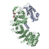
| Title | CryoEM structure of Resistance to Inhibitors of Cholinesterase-8B (Ric-8B) in complex with olfactory G protein alpha olf | ||||||
 Components Components |
| ||||||
 Keywords Keywords | SIGNALING PROTEIN / chaperone / armadillo repeat / GEF / Ras | ||||||
| Function / homology |  Function and homology information Function and homology informationAdenylate cyclase activating pathway / sensory perception of chemical stimulus / response to caffeine / G-protein alpha-subunit binding / Adenylate cyclase inhibitory pathway / protein folding chaperone / adenylate cyclase regulator activity / guanyl-nucleotide exchange factor activity / response to amphetamine / G protein-coupled receptor binding ...Adenylate cyclase activating pathway / sensory perception of chemical stimulus / response to caffeine / G-protein alpha-subunit binding / Adenylate cyclase inhibitory pathway / protein folding chaperone / adenylate cyclase regulator activity / guanyl-nucleotide exchange factor activity / response to amphetamine / G protein-coupled receptor binding / G-protein beta/gamma-subunit complex binding / Olfactory Signaling Pathway / adenylate cyclase-activating G protein-coupled receptor signaling pathway / sensory perception of smell / adenylate cyclase-activating dopamine receptor signaling pathway / heterotrimeric G-protein complex / G protein activity / cell cortex / Hydrolases; Acting on acid anhydrides; Acting on GTP to facilitate cellular and subcellular movement / G protein-coupled receptor signaling pathway / GTPase activity / centrosome / GTP binding / signal transduction / extracellular exosome / metal ion binding / plasma membrane / cytosol / cytoplasm Similarity search - Function | ||||||
| Biological species |  Homo sapiens (human) Homo sapiens (human) | ||||||
| Method | ELECTRON MICROSCOPY / single particle reconstruction / cryo EM / Resolution: 3.2 Å | ||||||
 Authors Authors | Papasergi-Scott, M.M. / Skiniotis, G. | ||||||
| Funding support |  United States, 1items United States, 1items
| ||||||
 Citation Citation |  Journal: Structure / Year: 2023 Journal: Structure / Year: 2023Title: Structures of Ric-8B in complex with Gα protein folding clients reveal isoform specificity mechanisms. Authors: Makaía M Papasergi-Scott / Frank E Kwarcinski / Maiya Yu / Ouliana Panova / Ann M Ovrutsky / Georgios Skiniotis / Gregory G Tall /  Abstract: Mammalian Ric-8 proteins act as chaperones to regulate the cellular abundance of heterotrimeric G protein α subunits. The Ric-8A isoform chaperones Gαi/o, Gα12/13, and Gαq/11 subunits, while Ric- ...Mammalian Ric-8 proteins act as chaperones to regulate the cellular abundance of heterotrimeric G protein α subunits. The Ric-8A isoform chaperones Gαi/o, Gα12/13, and Gαq/11 subunits, while Ric-8B acts on Gαs/olf subunits. Here, we determined cryoelectron microscopy (cryo-EM) structures of Ric-8B in complex with Gαs and Gαolf, revealing isoform differences in the relative positioning and contacts between the C-terminal α5 helix of Gα within the concave pocket formed by Ric-8 α-helical repeat elements. Despite the overall architectural similarity with our earlier structures of Ric-8A complexed to Gαq and Gαi1, Ric-8B distinctly accommodates an extended loop found only in Gαs/olf proteins. The structures, along with results from Ric-8 protein thermal stability assays and cell-based Gαolf folding assays, support a requirement for the Gα C-terminal region for binding specificity, and highlight that multiple structural elements impart specificity for Ric-8/G protein binding. | ||||||
| History |
|
- Structure visualization
| Structure viewer | Molecule:  Molmil Molmil Jmol/JSmol Jmol/JSmol |
|---|
- Downloads & links
- Download
| PDBx/mmCIF format |  8el8.cif.gz 8el8.cif.gz | 134.2 KB | Display |  PDBx/mmCIF format PDBx/mmCIF format |
|---|---|---|---|---|
| PDB format |  pdb8el8.ent.gz pdb8el8.ent.gz | 95.5 KB | Display |  PDB format PDB format |
| PDBx/mmJSON format |  8el8.json.gz 8el8.json.gz | Tree view |  PDBx/mmJSON format PDBx/mmJSON format | |
| Others |  Other downloads Other downloads |
-Validation report
| Arichive directory |  https://data.pdbj.org/pub/pdb/validation_reports/el/8el8 https://data.pdbj.org/pub/pdb/validation_reports/el/8el8 ftp://data.pdbj.org/pub/pdb/validation_reports/el/8el8 ftp://data.pdbj.org/pub/pdb/validation_reports/el/8el8 | HTTPS FTP |
|---|
-Related structure data
| Related structure data |  28224MC  8el7C M: map data used to model this data C: citing same article ( |
|---|---|
| Similar structure data | Similarity search - Function & homology  F&H Search F&H Search |
- Links
Links
- Assembly
| Deposited unit | 
|
|---|---|
| 1 |
|
- Components
| #1: Protein | Mass: 44370.430 Da / Num. of mol.: 1 Source method: isolated from a genetically manipulated source Source: (gene. exp.)  Homo sapiens (human) / Gene: GNAL / Production host: Homo sapiens (human) / Gene: GNAL / Production host:  Trichoplusia ni (cabbage looper) / References: UniProt: P38405 Trichoplusia ni (cabbage looper) / References: UniProt: P38405 |
|---|---|
| #2: Protein | Mass: 63928.043 Da / Num. of mol.: 1 Source method: isolated from a genetically manipulated source Source: (gene. exp.)   Trichoplusia ni (cabbage looper) / References: UniProt: Q80XE1 Trichoplusia ni (cabbage looper) / References: UniProt: Q80XE1 |
| Has ligand of interest | N |
| Has protein modification | Y |
-Experimental details
-Experiment
| Experiment | Method: ELECTRON MICROSCOPY |
|---|---|
| EM experiment | Aggregation state: PARTICLE / 3D reconstruction method: single particle reconstruction |
- Sample preparation
| Component | Name: Ric-8B in complex with G protein subunit alpha olf / Type: COMPLEX / Entity ID: all / Source: RECOMBINANT |
|---|---|
| Molecular weight | Experimental value: NO |
| Source (natural) | Organism:  Homo sapiens (human) Homo sapiens (human) |
| Source (recombinant) | Organism:  Trichoplusia ni (cabbage looper) Trichoplusia ni (cabbage looper) |
| Buffer solution | pH: 8 |
| Specimen | Conc.: 3.5 mg/ml / Embedding applied: NO / Shadowing applied: NO / Staining applied: NO / Vitrification applied: YES |
| Specimen support | Grid material: GOLD / Grid type: UltrAuFoil |
| Vitrification | Instrument: FEI VITROBOT MARK IV / Cryogen name: ETHANE |
- Electron microscopy imaging
| Experimental equipment |  Model: Titan Krios / Image courtesy: FEI Company |
|---|---|
| Microscopy | Model: FEI TITAN KRIOS |
| Electron gun | Electron source:  FIELD EMISSION GUN / Accelerating voltage: 300 kV / Illumination mode: FLOOD BEAM FIELD EMISSION GUN / Accelerating voltage: 300 kV / Illumination mode: FLOOD BEAM |
| Electron lens | Mode: BRIGHT FIELD / Calibrated magnification: 57050 X / Nominal defocus max: 1800 nm / Nominal defocus min: 800 nm |
| Specimen holder | Specimen holder model: FEI TITAN KRIOS AUTOGRID HOLDER |
| Image recording | Average exposure time: 2.49 sec. / Electron dose: 68.6 e/Å2 / Film or detector model: GATAN K3 (6k x 4k) / Num. of real images: 4670 |
- Processing
| EM software |
| ||||||||||||||||||||||||||||||||||||||||
|---|---|---|---|---|---|---|---|---|---|---|---|---|---|---|---|---|---|---|---|---|---|---|---|---|---|---|---|---|---|---|---|---|---|---|---|---|---|---|---|---|---|
| CTF correction | Type: NONE | ||||||||||||||||||||||||||||||||||||||||
| Particle selection | Num. of particles selected: 4695512 | ||||||||||||||||||||||||||||||||||||||||
| 3D reconstruction | Resolution: 3.2 Å / Resolution method: FSC 0.143 CUT-OFF / Num. of particles: 247416 / Symmetry type: POINT | ||||||||||||||||||||||||||||||||||||||||
| Atomic model building | B value: 171.1 / Protocol: AB INITIO MODEL / Space: REAL | ||||||||||||||||||||||||||||||||||||||||
| Atomic model building | PDB-ID: 8EL7 Accession code: 8EL7 / Source name: PDB / Type: experimental model |
 Movie
Movie Controller


 PDBj
PDBj


