 Movie
Movie Controller

[English] 日本語
 Yorodumi
- EMDB-27188: Subtomogram average of AP-1, Arf1 and Nef complexes on narrow mem... -
+ Open data

- Basic information
| Entry | 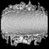 | ||||||||||||
|---|---|---|---|---|---|---|---|---|---|---|---|---|---|
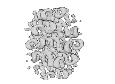
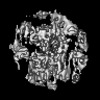
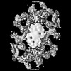
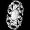
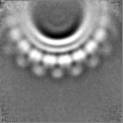
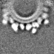
| Title | Subtomogram average of AP-1, Arf1 and Nef complexes on narrow membrane tubes centered on beta-Arf1 dimers | ||||||||||||
 Map data Map data | |||||||||||||
 Sample Sample |
| ||||||||||||
 Keywords Keywords | nef / AP / HIV / trafficking / VIRAL PROTEIN | ||||||||||||
| Function / homology |  Function and homology information Function and homology informationbasolateral protein secretion / synaptic vesicle budding from endosome / Lysosome Vesicle Biogenesis / positive regulation of natural killer cell degranulation / endosome to melanosome transport / protein trimerization / AP-1 adaptor complex / mitotic cleavage furrow ingression / trans-Golgi Network Vesicle Budding / negative regulation of glycoprotein biosynthetic process ...basolateral protein secretion / synaptic vesicle budding from endosome / Lysosome Vesicle Biogenesis / positive regulation of natural killer cell degranulation / endosome to melanosome transport / protein trimerization / AP-1 adaptor complex / mitotic cleavage furrow ingression / trans-Golgi Network Vesicle Budding / negative regulation of glycoprotein biosynthetic process / symbiont-mediated suppression of host antigen processing and presentation of peptide antigen via MHC class I / platelet dense granule organization / melanosome assembly / Glycosphingolipid transport / symbiont-mediated suppression of host antigen processing and presentation of peptide antigen via MHC class II / Golgi to lysosome transport / regulation of receptor internalization / Intra-Golgi traffic / Golgi to vacuole transport / Golgi Associated Vesicle Biogenesis / regulation of Arp2/3 complex-mediated actin nucleation / symbiont-mediated suppression of host autophagy / symbiont-mediated suppression of host apoptosis / Synthesis of PIPs at the Golgi membrane / clathrin-cargo adaptor activity / melanosome organization / MHC class II antigen presentation / GTP-dependent protein binding / thioesterase binding / Nef Mediated CD4 Down-regulation / positive regulation of natural killer cell mediated cytotoxicity / CD4 receptor binding / determination of left/right symmetry / dendritic spine organization / clathrin-coated vesicle / positive regulation of memory T cell activation / T cell mediated cytotoxicity directed against tumor cell target / long-term synaptic depression / positive regulation of CD8-positive, alpha-beta T cell activation / CD8-positive, alpha-beta T cell activation / positive regulation of CD8-positive, alpha-beta T cell proliferation / COPI-dependent Golgi-to-ER retrograde traffic / antigen processing and presentation of endogenous peptide antigen via MHC class I via ER pathway, TAP-dependent / TAP complex binding / Lysosome Vesicle Biogenesis / clathrin binding / antigen processing and presentation of exogenous peptide antigen via MHC class I / Golgi medial cisterna / Golgi Associated Vesicle Biogenesis / CD8 receptor binding / Synthesis of PIPs at the plasma membrane / cell leading edge / protection from natural killer cell mediated cytotoxicity / host cell Golgi membrane / beta-2-microglobulin binding / endoplasmic reticulum exit site / TAP binding / MHC class I protein binding / intracellular copper ion homeostasis / detection of bacterium / antigen processing and presentation of endogenous peptide antigen via MHC class Ib / antigen processing and presentation of endogenous peptide antigen via MHC class I via ER pathway, TAP-independent / kinesin binding / synaptic vesicle endocytosis / protein targeting / COPI-mediated anterograde transport / collagen binding / T cell receptor binding / viral life cycle / vesicle-mediated transport / Neutrophil degranulation / regulation of calcium-mediated signaling / clathrin-coated pit / MHC class II antigen presentation / Gene and protein expression by JAK-STAT signaling after Interleukin-12 stimulation / Nef mediated downregulation of MHC class I complex cell surface expression / cytoplasmic vesicle membrane / trans-Golgi network membrane / Endosomal/Vacuolar pathway / T cell mediated cytotoxicity / small monomeric GTPase / sarcomere / Antigen Presentation: Folding, assembly and peptide loading of class I MHC / lumenal side of endoplasmic reticulum membrane / kidney development / trans-Golgi network / peptide antigen assembly with MHC class I protein complex / ER to Golgi transport vesicle membrane / MHC class I peptide loading complex / positive regulation of T cell cytokine production / antigen processing and presentation of endogenous peptide antigen via MHC class I / MHC class I protein complex / intracellular protein transport / cellular response to virus / SH3 domain binding / positive regulation of T cell mediated cytotoxicity / recycling endosome / virion component / peptide antigen binding / positive regulation of type II interferon production Similarity search - Function | ||||||||||||
| Biological species |  Homo sapiens (human) Homo sapiens (human) | ||||||||||||
| Method | subtomogram averaging / cryo EM / Resolution: 20.0 Å | ||||||||||||
 Authors Authors | Hooy RM / Hurley JH | ||||||||||||
| Funding support |  United States, 3 items United States, 3 items
| ||||||||||||
 Citation Citation | Journal: Sci Adv / Year: 2022 Title: Self-assembly and structure of a clathrin-independent AP-1:Arf1 tubular membrane coat. Authors: Richard M Hooy / Yuichiro Iwamoto / Dan A Tudorica / Xuefeng Ren / James H Hurley / Abstract: The adaptor protein (AP) complexes not only form the inner layer of clathrin coats but also have clathrin-independent roles in membrane traffic whose mechanisms are unknown. HIV-1 Nef hijacks AP-1 to ...The adaptor protein (AP) complexes not only form the inner layer of clathrin coats but also have clathrin-independent roles in membrane traffic whose mechanisms are unknown. HIV-1 Nef hijacks AP-1 to sequester major histocompatibility complex class I (MHC-I), evading immune detection. We found that AP-1:Arf1:Nef:MHC-I forms a coat on tubulated membranes without clathrin and determined its structure. The coat assembles via Arf1 dimer interfaces. AP-1-positive tubules are enriched in cells upon clathrin knockdown. Nef localizes preferentially to AP-1 tubules in cells, explaining how Nef sequesters MHC-I. Coat contact residues are conserved across Arf isoforms and the Arf-dependent AP complexes AP-1, AP-3, and AP-4. Thus, AP complexes can self-assemble with Arf1 into tubular coats without clathrin or other scaffolding factors. The AP-1:Arf1 coat defines the structural basis of a broader class of tubulovesicular membrane coats as an intermediate in clathrin vesicle formation from internal membranes and as an MHC-I sequestration mechanism in HIV-1 infection. | ||||||||||||
| History |
|
- Structure visualization
| Supplemental images |
|---|
- Downloads & links
-EMDB archive
| Map data | emd_27188.map.gz | 3 MB | EMDB map data format | |
|---|---|---|---|---|
| Header (meta data) | emd-27188-v30.xmlemd-27188.xml | 20.3 KB 20.3 KB | Display Display | EMDB header |
| FSC (resolution estimation) | emd_27188_fsc.xml | 4.1 KB | Display | FSC data file |
| Images |  emd_27188.png emd_27188.png | 63.9 KB | ||
| Masks | emd_27188_msk_1.map | 5.4 MB | Mask map | |
| Filedesc metadata | emd-27188.cif.gz | 5.2 KB | ||
| Others | emd_27188_half_map_1.map.gzemd_27188_half_map_2.map.gz | 4.9 MB 4.9 MB | ||
| Archive directory |  http://ftp.pdbj.org/pub/emdb/structures/EMD-27188ftp://ftp.pdbj.org/pub/emdb/structures/EMD-27188 http://ftp.pdbj.org/pub/emdb/structures/EMD-27188ftp://ftp.pdbj.org/pub/emdb/structures/EMD-27188 | HTTPS FTP |
-Related structure data
| Related structure data |  8d9uMC 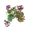 7ux3C  8d4cC  8d4dC  8d4eC  8d4fC  8d4gC 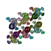 8d9rC  8d9sC  8d9tC  8d9vC  8d9wC C: citing same article ( M: atomic model generated by this map |
|---|---|
| Similar structure data |

-Links
| EMDB pages | EMDB (EBI/PDBe) / EMDataResource |
|---|---|
| Related items in Molecule of the Month |


-Map
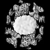
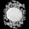
| File | Download / File: emd_27188.map.gz / Format: CCP4 / Size: 5.4 MB / Type: IMAGE STORED AS FLOATING POINT NUMBER (4 BYTES) | ||||||||||||||||||||||||||||||||||||
|---|---|---|---|---|---|---|---|---|---|---|---|---|---|---|---|---|---|---|---|---|---|---|---|---|---|---|---|---|---|---|---|---|---|---|---|---|---|
| Projections & slices | Image control
Images are generated by Spider. | ||||||||||||||||||||||||||||||||||||
| Voxel size | X=Y=Z: 4.2 Å | ||||||||||||||||||||||||||||||||||||
| Density |
| ||||||||||||||||||||||||||||||||||||
| Symmetry | Space group: 1 | ||||||||||||||||||||||||||||||||||||
| Details | EMDB XML:
|
 Z (Sec.)
 Y (Row.)
 X (Col.)


-Supplemental data
-Mask #1
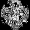
| File | emd_27188_msk_1.map | ||||||||||||
|---|---|---|---|---|---|---|---|---|---|---|---|---|---|
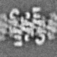
| Projections & Slices |
| ||||||||||||
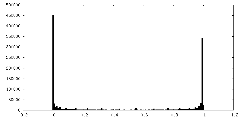
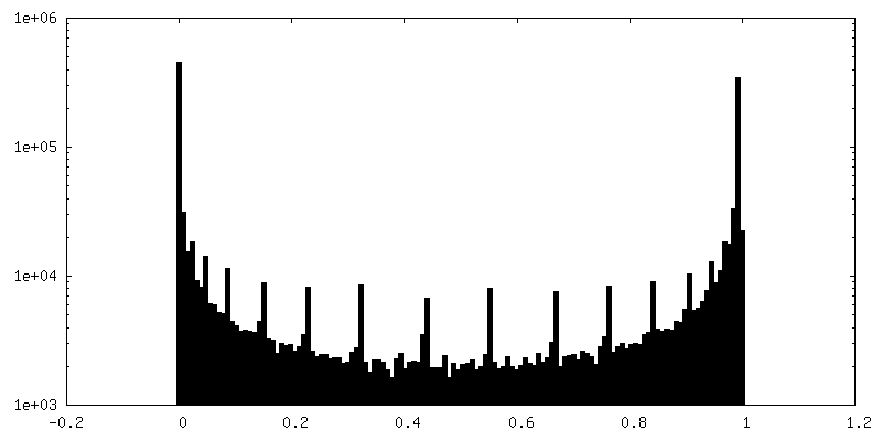
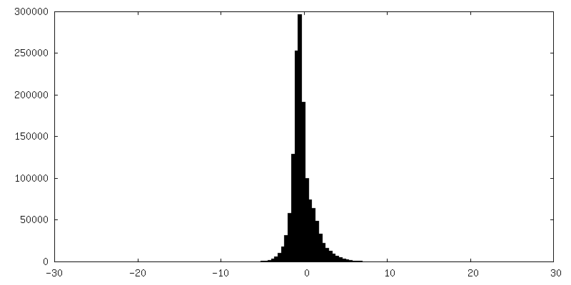
| Density Histograms |


-Half map: #1
| File | emd_27188_half_map_1.map | ||||||||||||
|---|---|---|---|---|---|---|---|---|---|---|---|---|---|
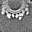
| Projections & Slices |
| ||||||||||||
| Density Histograms |


-Half map: #2
| File | emd_27188_half_map_2.map | ||||||||||||
|---|---|---|---|---|---|---|---|---|---|---|---|---|---|
| Projections & Slices |
| ||||||||||||
| Density Histograms |


- Sample components
-Entire : Complex of AP-1, Arf1, Nef and MHC-I cytosolic tail on a tubulate...
| Entire | Name: Complex of AP-1, Arf1, Nef and MHC-I cytosolic tail on a tubulated lipid bilayer |
|---|---|
| Components |
|
-Supramolecule #1: Complex of AP-1, Arf1, Nef and MHC-I cytosolic tail on a tubulate...
| Supramolecule | Name: Complex of AP-1, Arf1, Nef and MHC-I cytosolic tail on a tubulated lipid bilayer type: complex / ID: 1 / Parent: 0 / Macromolecule list: #1-#8 Details: Subtomogram average encompasses multiple AP-1 dimers |
|---|---|
| Source (natural) | Organism: Homo sapiens (human) |
-Supramolecule #2: AP-1 heterotetramer
| Supramolecule | Name: AP-1 heterotetramer / type: complex / ID: 2 / Parent: 1 / Macromolecule list: #4-#5, #7-#8 Details: All four subunits are co-expressed from the same plasmid. Assembly occurs in situ during expression. |
|---|---|
| Source (natural) | Organism: Homo sapiens (human) |
-Experimental details
-Structure determination
| Method | cryo EM |
|---|---|
 Processing Processing | subtomogram averaging |
| Aggregation state | particle |
-Sample preparation
| Concentration | 0.2 mg/mL | |||||||||||||||
|---|---|---|---|---|---|---|---|---|---|---|---|---|---|---|---|---|
| Buffer | pH: 7.2 Component:
Details: HEPES/KOAc concentrated stocks are diluted to their final concentrations then pH'd to 7.2 with KOH prior to use in experiments. | |||||||||||||||
| Grid | Model: EMS Lacey Carbon / Support film - Material: CARBON / Support film - topology: LACEY / Support film - Film thickness: 50 | |||||||||||||||
| Vitrification | Cryogen name: ETHANE / Chamber humidity: 100 % / Chamber temperature: 298 K / Instrument: FEI VITROBOT MARK IV Details: 60 second wait, 3-5 second blot, 597 filter paper, 0.5 second drain. Sample was supplemented with 10nm BSA-gold fiducials. 3.5ul of the mixture was double-side blotted.. |
- Electron microscopy
| Microscope | FEI TITAN KRIOS |
|---|---|
| Specialist optics | Energy filter - Slit width: 25 eV |
| Image recording | Film or detector model: GATAN K3 BIOQUANTUM (6k x 4k) / Digitization - Dimensions - Width: 5760 pixel / Digitization - Dimensions - Height: 4092 pixel / Number grids imaged: 1 / Average exposure time: 3.0 sec. / Average electron dose: 3.0 e/Å2 Details: Tilt images were collected in movie-mode. Each movie/tilt consisted of 3-4 frames each |
| Electron beam | Acceleration voltage: 300 kV / Electron source:  FIELD EMISSION GUN FIELD EMISSION GUN |
| Electron optics | Illumination mode: FLOOD BEAM / Imaging mode: BRIGHT FIELD / Cs: 2.7 mm / Nominal defocus max: 4.5 µm / Nominal defocus min: 1.5 µm / Nominal magnification: 42000 |
| Sample stage | Specimen holder model: FEI TITAN KRIOS AUTOGRID HOLDER / Cooling holder cryogen: NITROGEN |
| Experimental equipment |  Model: Titan Krios / Image courtesy: FEI Company |
+Image processing

-Atomic model buiding 1
| Refinement | Protocol: RIGID BODY FIT |
|---|---|
| Output model | PDB-8d9u: |
