Movie
Movie Controller

[English] 日本語
 Yorodumi
- EMDB-26960: Composite reconstruction of Class 1 of the erythrocyte ankyrin-1 ... -
+ Open data

- Basic information
| Entry |  | |||||||||
|---|---|---|---|---|---|---|---|---|---|---|
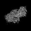
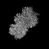
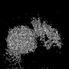
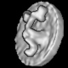
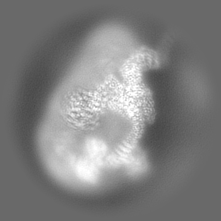
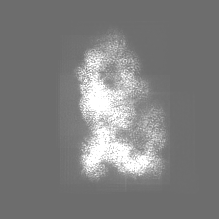
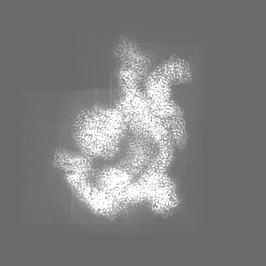
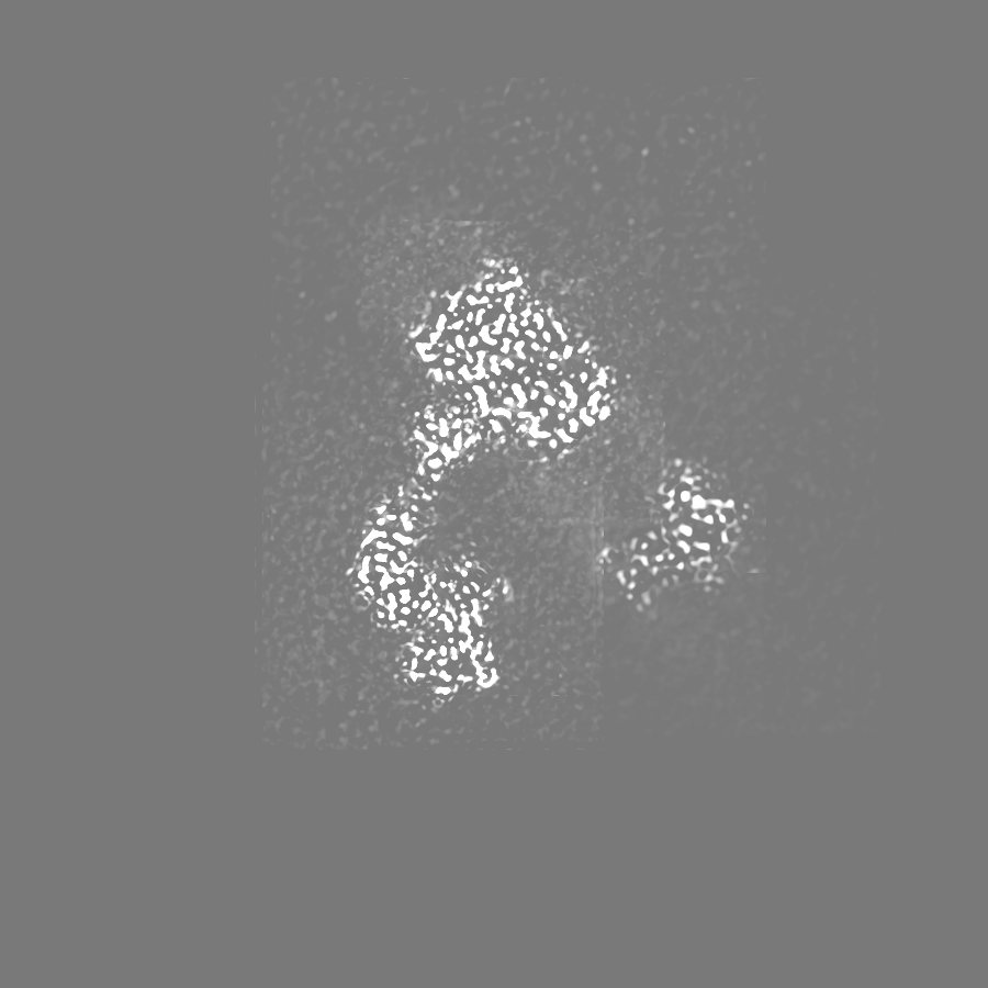
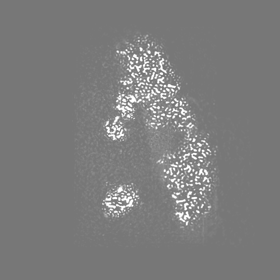
| Title | Composite reconstruction of Class 1 of the erythrocyte ankyrin-1 complex | |||||||||
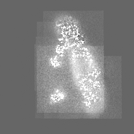
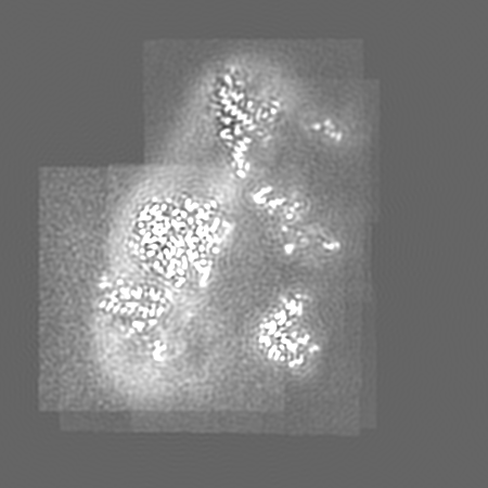
 Map data Map data | Composite reconstruction of EMDs: 26944, 26948, 26949, 26950, 26951, 26952, 26953, 26955, 26954, 26956, 26958 aligned to the consensus refinement. | |||||||||
 Sample Sample |
| |||||||||
 Keywords Keywords | Membrane Protein / Anion Exchange / Erythrocyte / Glycoprotein / TRANSPORT PROTEIN-STRUCTURAL PROTEIN complex | |||||||||
| Function / homology |  Function and homology information Function and homology informationmethylammonium transmembrane transport / Defective RHAG causes regulator type Rh-null hemolytic anemia (RHN) / Rhesus blood group biosynthesis / methylammonium transmembrane transporter activity / Rhesus glycoproteins mediate ammonium transport / carbon dioxide transmembrane transport / carbon dioxide transmembrane transporter activity / ammonium homeostasis / spectrin-associated cytoskeleton / hemoglobin metabolic process ...methylammonium transmembrane transport / Defective RHAG causes regulator type Rh-null hemolytic anemia (RHN) / Rhesus blood group biosynthesis / methylammonium transmembrane transporter activity / Rhesus glycoproteins mediate ammonium transport / carbon dioxide transmembrane transport / carbon dioxide transmembrane transporter activity / ammonium homeostasis / spectrin-associated cytoskeleton / hemoglobin metabolic process / maintenance of epithelial cell apical/basal polarity / protein-glutamine gamma-glutamyltransferase activity / response to increased oxygen levels / pH elevation / Defective SLC4A1 causes hereditary spherocytosis type 4 (HSP4), distal renal tubular acidosis (dRTA) and dRTA with hemolytic anemia (dRTA-HA) / negative regulation of urine volume / NrCAM interactions / Bicarbonate transporters / Neurofascin interactions / leak channel activity / ankyrin-1 complex / ammonium transmembrane transport / intracellular monoatomic ion homeostasis / ammonium channel activity / CHL1 interactions / monoatomic anion transmembrane transporter activity / plasma membrane phospholipid scrambling / chloride:bicarbonate antiporter activity / cytoskeletal adaptor activity / solute:inorganic anion antiporter activity / bicarbonate transmembrane transporter activity / bicarbonate transport / monoatomic anion transport / chloride transmembrane transporter activity / M band / : / chloride transport / Interaction between L1 and Ankyrins / ankyrin binding / hemoglobin binding / spectrin binding / erythrocyte maturation / negative regulation of glycolytic process through fructose-6-phosphate / exocytosis / cortical cytoskeleton / axolemma / endoplasmic reticulum to Golgi vesicle-mediated transport / spleen development / erythrocyte development / COPI-mediated anterograde transport / protein-membrane adaptor activity / cytoskeleton organization / chloride transmembrane transport / Cell surface interactions at the vascular wall / regulation of intracellular pH / sarcoplasmic reticulum / protein localization to plasma membrane / carbon dioxide transport / Erythrocytes take up oxygen and release carbon dioxide / Erythrocytes take up carbon dioxide and release oxygen / sarcolemma / multicellular organismal-level iron ion homeostasis / structural constituent of cytoskeleton / transmembrane transport / cell morphogenesis / cytoplasmic side of plasma membrane / Z disc / blood coagulation / regulation of cell shape / ATPase binding / virus receptor activity / protein phosphatase binding / blood microparticle / basolateral plasma membrane / cytoskeleton / transmembrane transporter binding / postsynaptic membrane / neuron projection / structural molecule activity / enzyme binding / signal transduction / protein homodimerization activity / extracellular exosome / nucleoplasm / ATP binding / membrane / identical protein binding / plasma membrane / cytosol Similarity search - Function | |||||||||
| Biological species |  Homo sapiens (human) Homo sapiens (human) | |||||||||
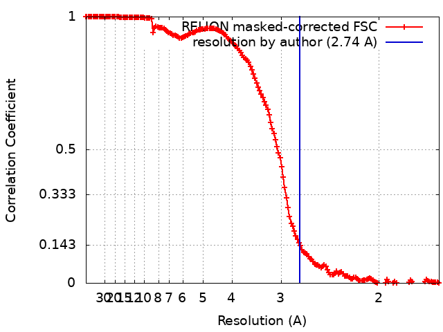
| Method | single particle reconstruction / cryo EM / Resolution: 2.74 Å | |||||||||
 Authors Authors | Vallese F / Kim K / Yen LY / Johnston JD / Noble AJ / Cali T / Clarke OB | |||||||||
| Funding support | 1 items
| |||||||||
 Citation Citation | Journal: Nat Struct Mol Biol / Year: 2022 Title: Architecture of the human erythrocyte ankyrin-1 complex. Authors: Francesca Vallese / Kookjoo Kim / Laura Y Yen / Jake D Johnston / Alex J Noble / Tito Calì / Oliver Biggs Clarke /   Abstract: The stability and shape of the erythrocyte membrane is provided by the ankyrin-1 complex, but how it tethers the spectrin-actin cytoskeleton to the lipid bilayer and the nature of its association ...The stability and shape of the erythrocyte membrane is provided by the ankyrin-1 complex, but how it tethers the spectrin-actin cytoskeleton to the lipid bilayer and the nature of its association with the band 3 anion exchanger and the Rhesus glycoproteins remains unknown. Here we present structures of ankyrin-1 complexes purified from human erythrocytes. We reveal the architecture of a core complex of ankyrin-1, the Rhesus proteins RhAG and RhCE, the band 3 anion exchanger, protein 4.2, glycophorin A and glycophorin B. The distinct T-shaped conformation of membrane-bound ankyrin-1 facilitates recognition of RhCE and, unexpectedly, the water channel aquaporin-1. Together, our results uncover the molecular details of ankyrin-1 association with the erythrocyte membrane, and illustrate the mechanism of ankyrin-mediated membrane protein clustering. | |||||||||
| History |
|
- Structure visualization
| Supplemental images |
|---|

- Downloads & links
-EMDB archive
| Map data | emd_26960.map.gz | 57.3 MB | EMDB map data format | |
|---|---|---|---|---|
| Header (meta data) | emd-26960-v30.xmlemd-26960.xml | 50.1 KB 50.1 KB | Display Display | EMDB header |
| FSC (resolution estimation) | emd_26960_fsc.xml | 15.9 KB | Display | FSC data file |
| Images | 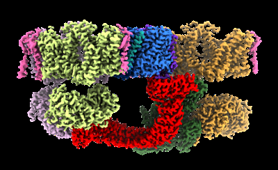 emd_26960.png emd_26960.png | 119.5 KB | ||
| Filedesc metadata | emd-26960.cif.gz | 10.4 KB | ||
| Others | emd_26960_additional_1.map.gzemd_26960_additional_2.map.gzemd_26960_additional_3.map.gzemd_26960_additional_4.map.gzemd_26960_additional_5.map.gzemd_26960_additional_6.map.gzemd_26960_additional_7.map.gzemd_26960_additional_8.map.gz | 328.1 MB 322.9 MB 173.3 MB 71.3 MB 322.9 MB 324.4 MB 444.7 MB 1 MB | ||
| Archive directory |  http://ftp.pdbj.org/pub/emdb/structures/EMD-26960ftp://ftp.pdbj.org/pub/emdb/structures/EMD-26960 http://ftp.pdbj.org/pub/emdb/structures/EMD-26960ftp://ftp.pdbj.org/pub/emdb/structures/EMD-26960 | HTTPS FTP |
-Related structure data
| Related structure data |  8cs9MC  7uz3C  7uzeC  7uzqC  7uzsC  7uzuC  7uzvC  7v07C  7v0kC  7v0mC  7v0qC  7v0sC  7v0tC  7v0uC 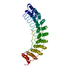 7v0xC  7v0yC  7v19C  8crqC  8crrC  8crtC  8cslC 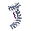 8csvC  8cswC  8csxC  8csyC  8ct2C  8ct3C  8cteC C: citing same article ( M: atomic model generated by this map |
|---|---|
| Similar structure data |


-Links
| EMDB pages | EMDB (EBI/PDBe) / EMDataResource |
|---|---|
| Related items in Molecule of the Month |


-Map
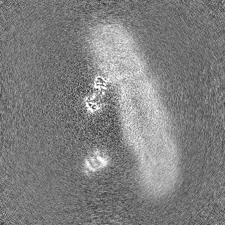
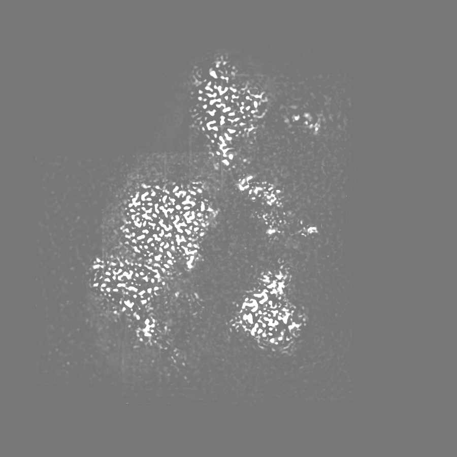
| File | Download / File: emd_26960.map.gz / Format: CCP4 / Size: 347.6 MB / Type: IMAGE STORED AS FLOATING POINT NUMBER (4 BYTES) | ||||||||||||||||||||||||||||||||||||
|---|---|---|---|---|---|---|---|---|---|---|---|---|---|---|---|---|---|---|---|---|---|---|---|---|---|---|---|---|---|---|---|---|---|---|---|---|---|
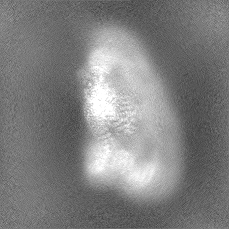
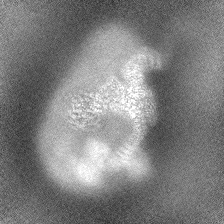
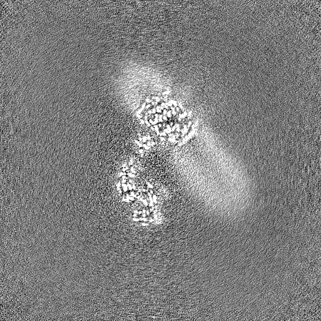
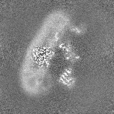
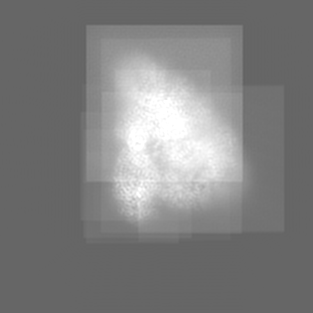
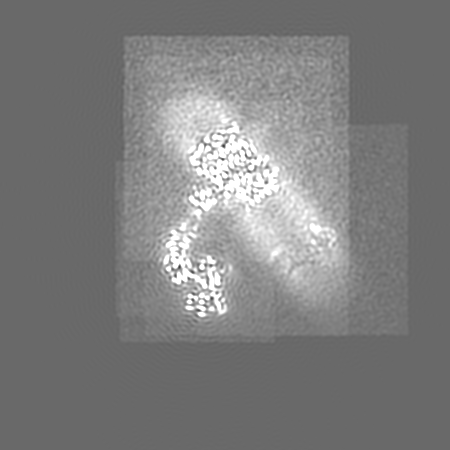
| Annotation | Composite reconstruction of EMDs: 26944, 26948, 26949, 26950, 26951, 26952, 26953, 26955, 26954, 26956, 26958 aligned to the consensus refinement. | ||||||||||||||||||||||||||||||||||||
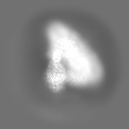
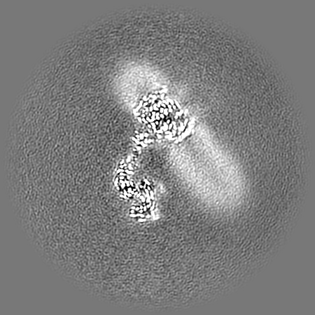
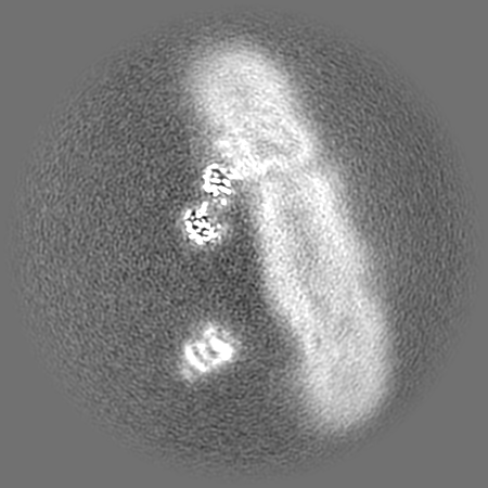
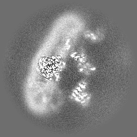
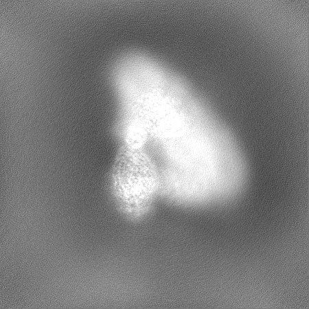
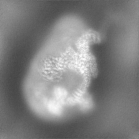
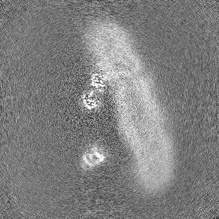
| Projections & slices | Image control
Images are generated by Spider. | ||||||||||||||||||||||||||||||||||||
| Voxel size | X=Y=Z: 0.83 Å | ||||||||||||||||||||||||||||||||||||
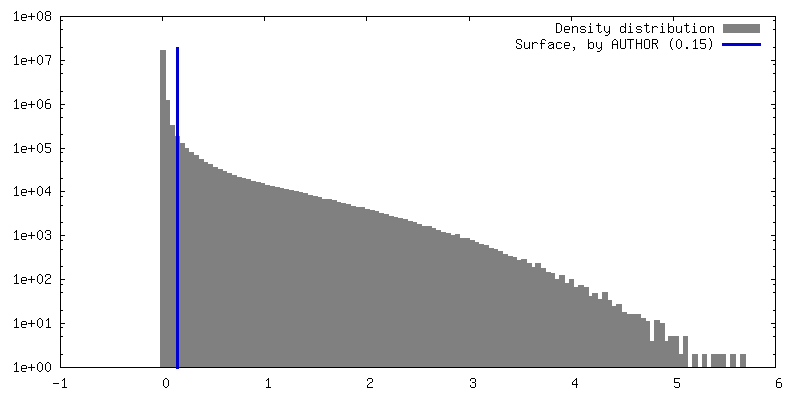
| Density |
| ||||||||||||||||||||||||||||||||||||
| Symmetry | Space group: 1 | ||||||||||||||||||||||||||||||||||||
| Details | EMDB XML:
|
 Z (Sec.)
 Y (Row.)
 X (Col.)


-Supplemental data
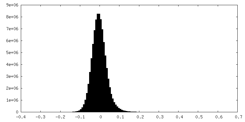
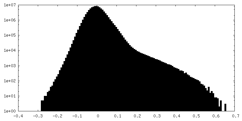
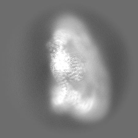
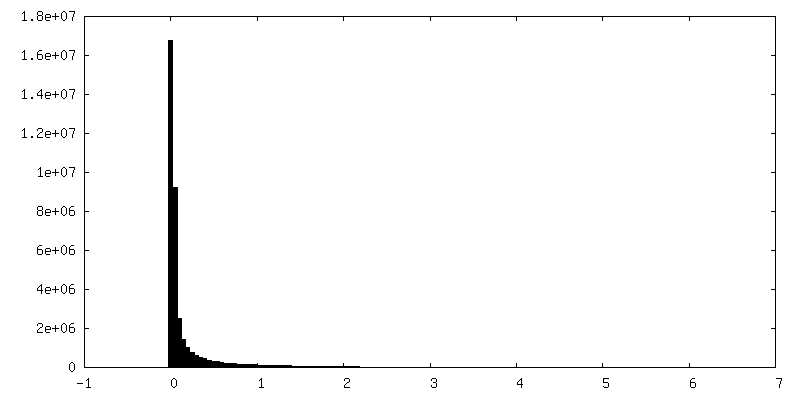
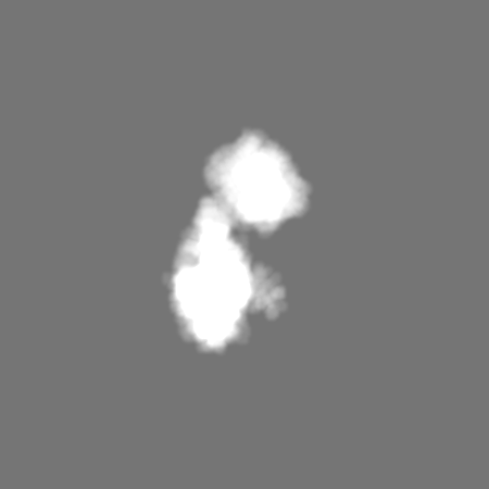
-Additional map: Consensus refinement (sharpened).
| File | emd_26960_additional_1.map | ||||||||||||
|---|---|---|---|---|---|---|---|---|---|---|---|---|---|
| Annotation | Consensus refinement (sharpened). | ||||||||||||
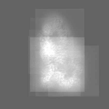
| Projections & Slices |
| ||||||||||||
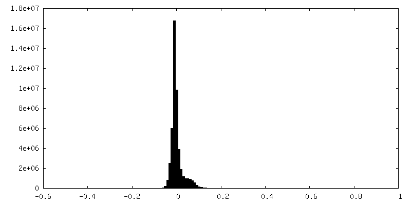
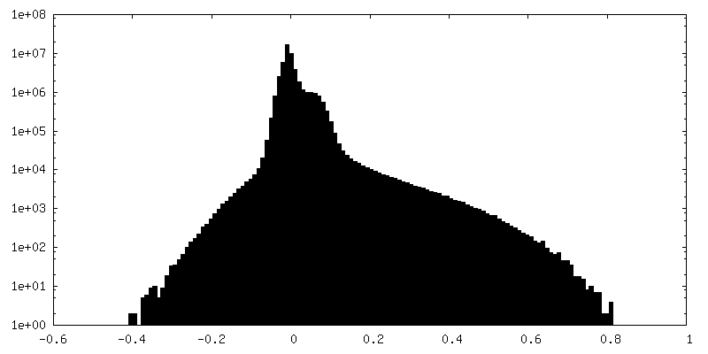
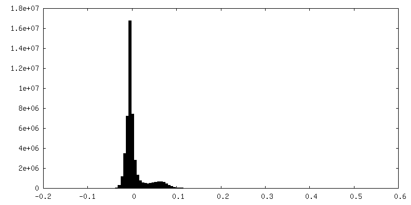
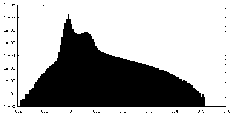
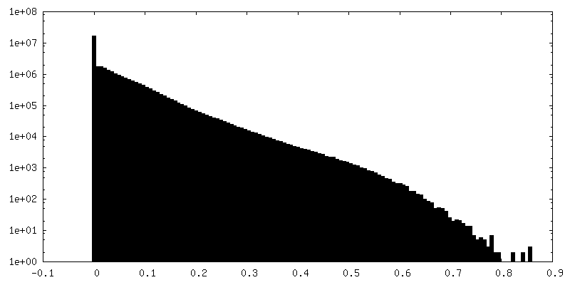
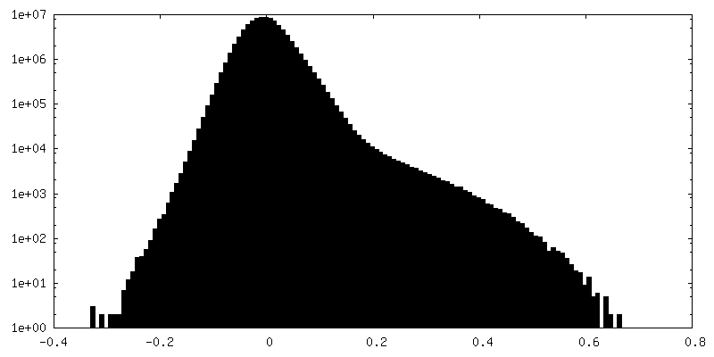
| Density Histograms |


-Additional map: Consensus refinement (half map 1)
| File | emd_26960_additional_2.map | ||||||||||||
|---|---|---|---|---|---|---|---|---|---|---|---|---|---|
| Annotation | Consensus refinement (half map 1) | ||||||||||||
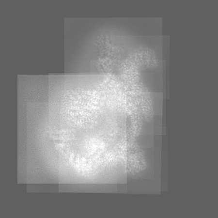
| Projections & Slices |
| ||||||||||||
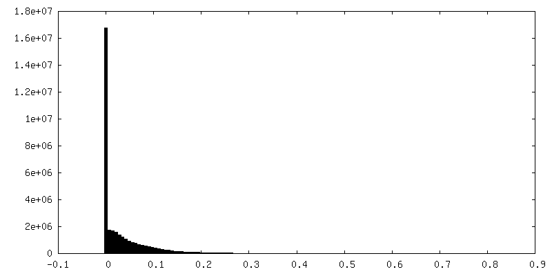
| Density Histograms |


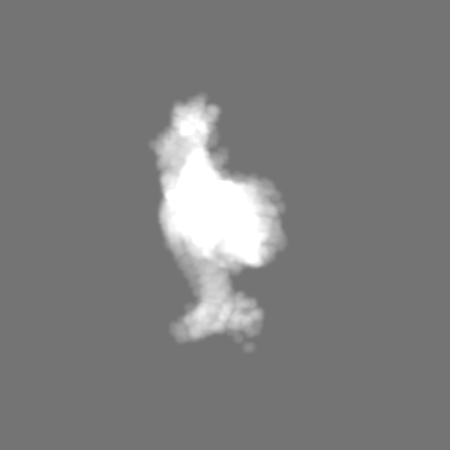
-Additional map: Consensus refinement (unsharpened).
| File | emd_26960_additional_3.map | ||||||||||||
|---|---|---|---|---|---|---|---|---|---|---|---|---|---|
| Annotation | Consensus refinement (unsharpened). | ||||||||||||
| Projections & Slices |
| ||||||||||||
| Density Histograms |


-Additional map: Composite reconstruction of EMDs: 26944, 26948, 26949, 26950,...
| File | emd_26960_additional_4.map | ||||||||||||
|---|---|---|---|---|---|---|---|---|---|---|---|---|---|
| Annotation | Composite reconstruction of EMDs: 26944, 26948, 26949, 26950, 26951, 26952, 26953, 26955, 26954, 26956, 26958 aligned to the consensus refinement. Unsharpened. | ||||||||||||
| Projections & Slices |
| ||||||||||||
| Density Histograms |


-Additional map: Consensus refinement (half map 1)
| File | emd_26960_additional_5.map | ||||||||||||
|---|---|---|---|---|---|---|---|---|---|---|---|---|---|
| Annotation | Consensus refinement (half map 1) | ||||||||||||
| Projections & Slices |
| ||||||||||||
| Density Histograms |


-Additional map: Composite reconstruction of EMDs: 26944, 26948, 26949, 26950,...
| File | emd_26960_additional_6.map | ||||||||||||
|---|---|---|---|---|---|---|---|---|---|---|---|---|---|
| Annotation | Composite reconstruction of EMDs: 26944, 26948, 26949, 26950, 26951, 26952, 26953, 26955, 26954, 26956, 26958 aligned to the consensus refinement. Unsharpened, filtered to 4A to aid visualization. | ||||||||||||
| Projections & Slices |
| ||||||||||||
| Density Histograms |


-Additional map: Composite reconstruction of EMDs: 26944, 26948, 26949, 26950,...
| File | emd_26960_additional_7.map | ||||||||||||
|---|---|---|---|---|---|---|---|---|---|---|---|---|---|
| Annotation | Composite reconstruction of EMDs: 26944, 26948, 26949, 26950, 26951, 26952, 26953, 26955, 26954, 26956, 26958 aligned to the consensus refinement. Resampled on 2x finer grid to aid visualization. | ||||||||||||
| Projections & Slices |
| ||||||||||||
| Density Histograms |


-Additional map: Mask used for FSC calculation of consensus refinement
| File | emd_26960_additional_8.map | ||||||||||||
|---|---|---|---|---|---|---|---|---|---|---|---|---|---|
| Annotation | Mask used for FSC calculation of consensus refinement | ||||||||||||
| Projections & Slices |
| ||||||||||||
| Density Histograms |


- Sample components
+Entire : Class 1 of erythrocyte ankyrin complex (composite map)
+Supramolecule #1: Class 1 of erythrocyte ankyrin complex (composite map)
+Macromolecule #1: Ankyrin-1
+Macromolecule #2: Blood group Rh(CE) polypeptide
+Macromolecule #3: Ammonium transporter Rh type A
+Macromolecule #4: Protein 4.2
+Macromolecule #5: Glycophorin-B
+Macromolecule #6: Glycophorin-A
+Macromolecule #7: Band 3 anion transport protein
+Macromolecule #9: CHOLESTEROL

+Macromolecule #10: Digitonin
+Macromolecule #11: 2-acetamido-2-deoxy-beta-D-glucopyranose

+Macromolecule #12: [(2R)-2-octanoyloxy-3-[oxidanyl-[(1R,2R,3S,4R,5R,6S)-2,3,6-tris(o...

+Macromolecule #13: water

-Experimental details
-Structure determination
| Method | cryo EM |
|---|---|
 Processing Processing | single particle reconstruction |
| Aggregation state | particle |
-Sample preparation
| Concentration | 8 mg/mL |
|---|---|
| Buffer | pH: 7.4 Details: Final gel filtration buffer contained 0.05% w/v digitonin, 130 mM KCl, 20 mM HEPES, pH 7.4, 1 mM ATP, 1 mM MgCl2, 1 mM PMSF. Peak fractions were concentrated to 8 mg/mL, and 0.01% w/v ...Details: Final gel filtration buffer contained 0.05% w/v digitonin, 130 mM KCl, 20 mM HEPES, pH 7.4, 1 mM ATP, 1 mM MgCl2, 1 mM PMSF. Peak fractions were concentrated to 8 mg/mL, and 0.01% w/v glycyrrhizic acid was added immediately prior to vitrification. |
| Vitrification | Cryogen name: ETHANE / Chamber humidity: 100 % / Chamber temperature: 277 K / Instrument: FEI VITROBOT MARK IV / Details: 4-6 seconds, wait time 30 seconds. |
| Details | Ankyrin complex mixture purified from digitonin-solubilized erythrocyte ghost membranes |
- Electron microscopy
| Microscope | FEI TITAN KRIOS |
|---|---|
| Specialist optics | Energy filter - Name: GIF Bioquantum / Energy filter - Slit width: 20 eV |
| Image recording | Film or detector model: GATAN K3 (6k x 4k) / Number grids imaged: 2 / Number real images: 14464 / Average exposure time: 2.5 sec. / Average electron dose: 58.0 e/Å2 / Details: Two grids were imaged in a single session. |
| Electron beam | Acceleration voltage: 300 kV / Electron source:  FIELD EMISSION GUN FIELD EMISSION GUN |
| Electron optics | Illumination mode: FLOOD BEAM / Imaging mode: BRIGHT FIELD / Cs: 2.7 mm / Nominal defocus max: 1.5 µm / Nominal defocus min: 0.5 µm |
| Sample stage | Specimen holder model: FEI TITAN KRIOS AUTOGRID HOLDER / Cooling holder cryogen: NITROGEN |
| Experimental equipment |  Model: Titan Krios / Image courtesy: FEI Company |