Movie
Movie Controller


+ Open data

- Basic information
| Entry |  | |||||||||
|---|---|---|---|---|---|---|---|---|---|---|
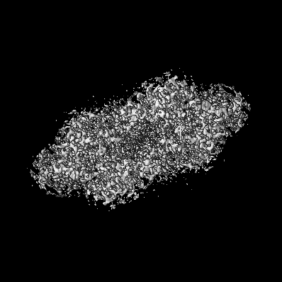
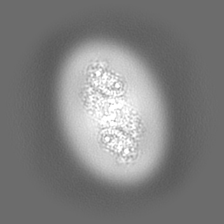
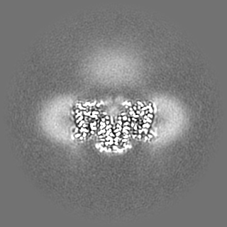
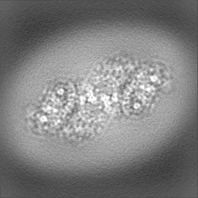
| Title | Band 3-Glycophorin A complex, outward facing | |||||||||
 Map data Map data | Main map used for building/refinement. Density modified using phenix.resolve_cryo_em and cropped to minimal box, then resampled to 2x finer spacing to aid visualization (original pixel size was 0.83) | |||||||||
 Sample Sample |
| |||||||||
 Keywords Keywords | Membrane Protein / Anion Exchange / Erythrocyte / Glycoprotein / TRANSPORT PROTEIN | |||||||||
| Function / homology |  Function and homology information Function and homology informationresponse to increased oxygen levels / pH elevation / Defective SLC4A1 causes hereditary spherocytosis type 4 (HSP4), distal renal tubular acidosis (dRTA) and dRTA with hemolytic anemia (dRTA-HA) / negative regulation of urine volume / Bicarbonate transporters / ankyrin-1 complex / intracellular monoatomic ion homeostasis / monoatomic anion transmembrane transporter activity / plasma membrane phospholipid scrambling / chloride:bicarbonate antiporter activity ...response to increased oxygen levels / pH elevation / Defective SLC4A1 causes hereditary spherocytosis type 4 (HSP4), distal renal tubular acidosis (dRTA) and dRTA with hemolytic anemia (dRTA-HA) / negative regulation of urine volume / Bicarbonate transporters / ankyrin-1 complex / intracellular monoatomic ion homeostasis / monoatomic anion transmembrane transporter activity / plasma membrane phospholipid scrambling / chloride:bicarbonate antiporter activity / solute:inorganic anion antiporter activity / bicarbonate transmembrane transporter activity / bicarbonate transport / monoatomic anion transport / chloride transmembrane transporter activity / chloride transport / ankyrin binding / hemoglobin binding / negative regulation of glycolytic process through fructose-6-phosphate / cortical cytoskeleton / erythrocyte development / protein-membrane adaptor activity / chloride transmembrane transport / Cell surface interactions at the vascular wall / regulation of intracellular pH / protein localization to plasma membrane / Erythrocytes take up oxygen and release carbon dioxide / Erythrocytes take up carbon dioxide and release oxygen / transmembrane transport / cytoplasmic side of plasma membrane / Z disc / blood coagulation / virus receptor activity / blood microparticle / basolateral plasma membrane / protein homodimerization activity / extracellular exosome / nucleoplasm / membrane / identical protein binding / plasma membrane / cytosol Similarity search - Function | |||||||||
| Biological species |  Homo sapiens (human) Homo sapiens (human) | |||||||||
| Method | single particle reconstruction / cryo EM / Resolution: 2.35 Å | |||||||||
 Authors Authors | Vallese F / Kim K / Yen LY / Johnston JD / Noble AJ / Cali T / Clarke OB | |||||||||
| Funding support | 1 items
| |||||||||
 Citation Citation | Journal: Nat Struct Mol Biol / Year: 2022 Title: Architecture of the human erythrocyte ankyrin-1 complex. Authors: Francesca Vallese / Kookjoo Kim / Laura Y Yen / Jake D Johnston / Alex J Noble / Tito Calì / Oliver Biggs Clarke /   Abstract: The stability and shape of the erythrocyte membrane is provided by the ankyrin-1 complex, but how it tethers the spectrin-actin cytoskeleton to the lipid bilayer and the nature of its association ...The stability and shape of the erythrocyte membrane is provided by the ankyrin-1 complex, but how it tethers the spectrin-actin cytoskeleton to the lipid bilayer and the nature of its association with the band 3 anion exchanger and the Rhesus glycoproteins remains unknown. Here we present structures of ankyrin-1 complexes purified from human erythrocytes. We reveal the architecture of a core complex of ankyrin-1, the Rhesus proteins RhAG and RhCE, the band 3 anion exchanger, protein 4.2, glycophorin A and glycophorin B. The distinct T-shaped conformation of membrane-bound ankyrin-1 facilitates recognition of RhCE and, unexpectedly, the water channel aquaporin-1. Together, our results uncover the molecular details of ankyrin-1 association with the erythrocyte membrane, and illustrate the mechanism of ankyrin-mediated membrane protein clustering. | |||||||||
| History |
|
- Structure visualization
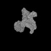
| Supplemental images |
|---|

- Downloads & links
-EMDB archive
| Map data | emd_26874.map.gz | 224.3 MB | EMDB map data format | |
|---|---|---|---|---|
| Header (meta data) | emd-26874-v30.xmlemd-26874.xml | 37.5 KB 37.5 KB | Display Display | EMDB header |
| FSC (resolution estimation) | emd_26874_fsc.xml | 11.3 KB | Display | FSC data file |
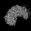
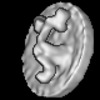
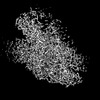
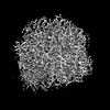
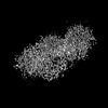
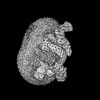
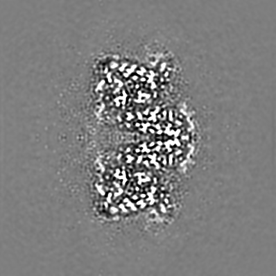
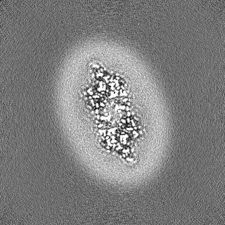
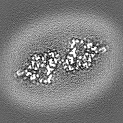
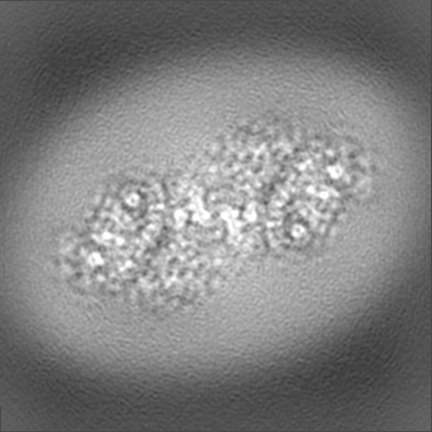
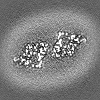
| Images |  emd_26874.png emd_26874.png | 108.3 KB | ||
| Filedesc metadata | emd-26874.cif.gz | 7.7 KB | ||
| Others | emd_26874_additional_1.map.gzemd_26874_additional_2.map.gzemd_26874_additional_3.map.gzemd_26874_additional_4.map.gzemd_26874_additional_5.map.gzemd_26874_half_map_1.map.gzemd_26874_half_map_2.map.gz | 115.7 MB 118 MB 115.7 MB 395.3 KB 62.5 MB 223 MB 223 MB | ||
| Archive directory |  http://ftp.pdbj.org/pub/emdb/structures/EMD-26874ftp://ftp.pdbj.org/pub/emdb/structures/EMD-26874 http://ftp.pdbj.org/pub/emdb/structures/EMD-26874ftp://ftp.pdbj.org/pub/emdb/structures/EMD-26874 | HTTPS FTP |
-Related structure data
| Related structure data |  7uz3MC  7uzeC  7uzqC  7uzsC  7uzuC  7uzvC  7v07C  7v0kC  7v0mC  7v0qC  7v0sC  7v0tC  7v0uC 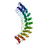 7v0xC  7v0yC  7v19C  8crqC  8crrC  8crtC  8cs9C  8cslC 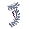 8csvC  8cswC  8csxC  8csyC  8ct2C  8ct3C  8cteC C: citing same article ( M: atomic model generated by this map |
|---|---|
| Similar structure data |


-Links
| EMDB pages | EMDB (EBI/PDBe) / EMDataResource |
|---|
-Map
| File | Download / File: emd_26874.map.gz / Format: CCP4 / Size: 240.5 MB / Type: IMAGE STORED AS FLOATING POINT NUMBER (4 BYTES) | ||||||||||||||||||||||||||||||||||||
|---|---|---|---|---|---|---|---|---|---|---|---|---|---|---|---|---|---|---|---|---|---|---|---|---|---|---|---|---|---|---|---|---|---|---|---|---|---|
| Annotation | Main map used for building/refinement. Density modified using phenix.resolve_cryo_em and cropped to minimal box, then resampled to 2x finer spacing to aid visualization (original pixel size was 0.83) | ||||||||||||||||||||||||||||||||||||
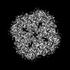
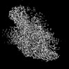
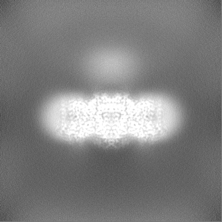
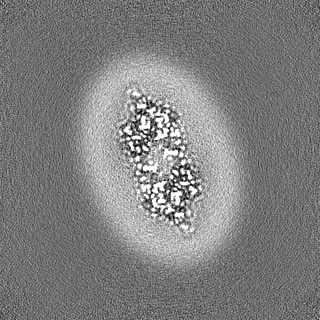
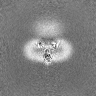
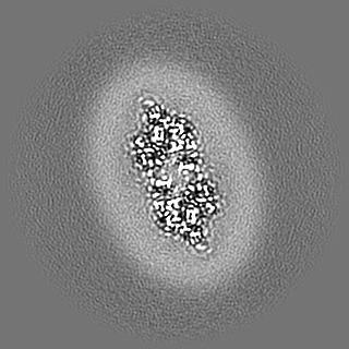
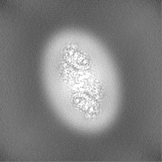
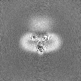
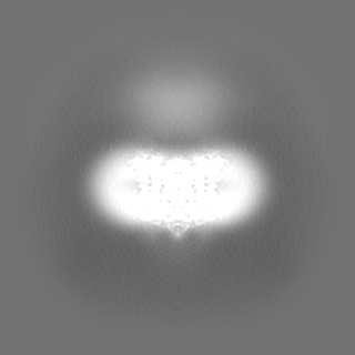
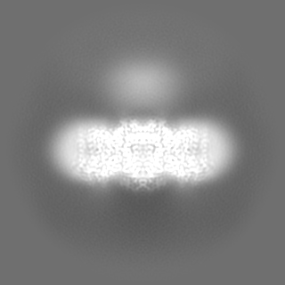
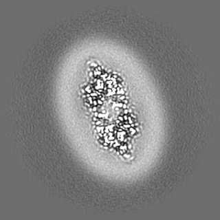
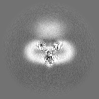
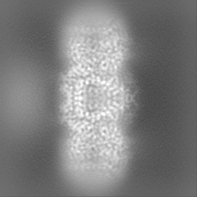
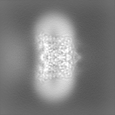
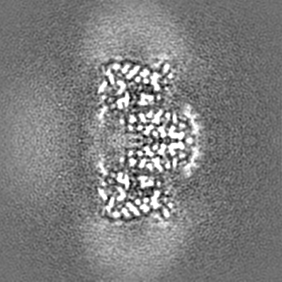
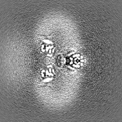
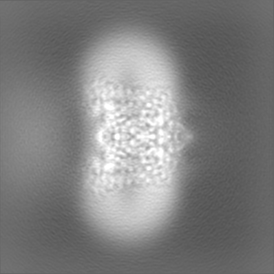
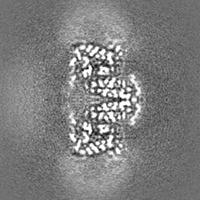
| Projections & slices | Image control
Images are generated by Spider. | ||||||||||||||||||||||||||||||||||||
| Voxel size | X=Y=Z: 0.415 Å | ||||||||||||||||||||||||||||||||||||
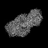
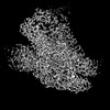
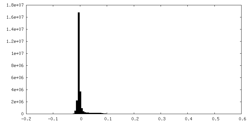
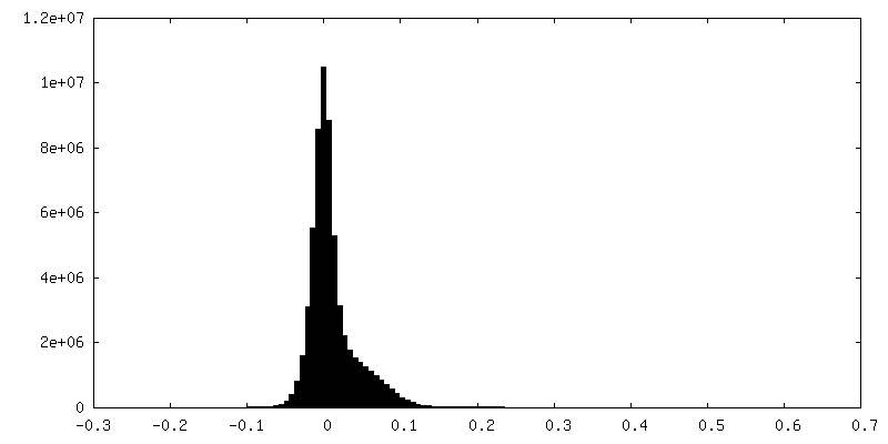
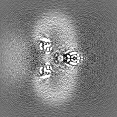
| Density |
| ||||||||||||||||||||||||||||||||||||
| Symmetry | Space group: 1 | ||||||||||||||||||||||||||||||||||||
| Details | EMDB XML:
|
 Z (Sec.)
 Y (Row.)
 X (Col.)


-Supplemental data
-Additional map: Unfiltered half map 1. Used for FSC calculation.
| File | emd_26874_additional_1.map | ||||||||||||
|---|---|---|---|---|---|---|---|---|---|---|---|---|---|
| Annotation | Unfiltered half map 1. Used for FSC calculation. | ||||||||||||
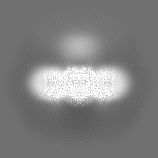
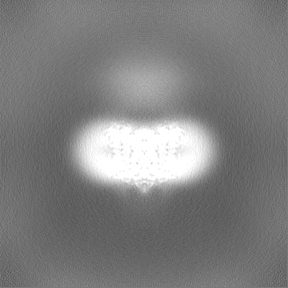
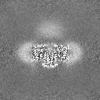
| Projections & Slices |
| ||||||||||||
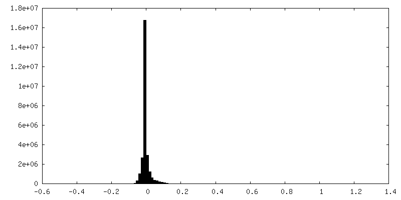
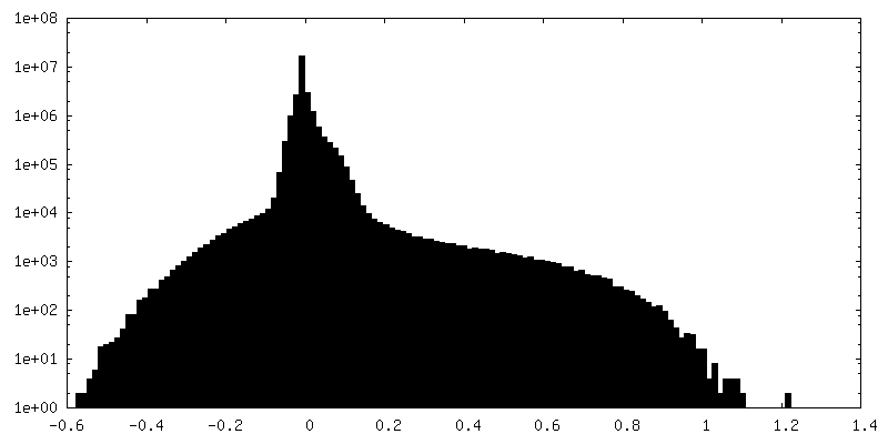
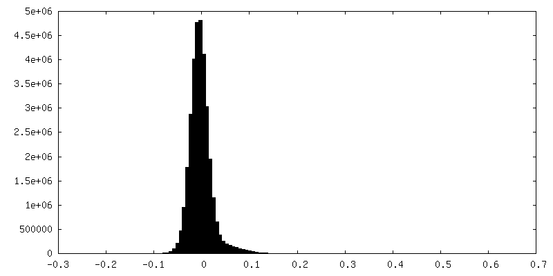
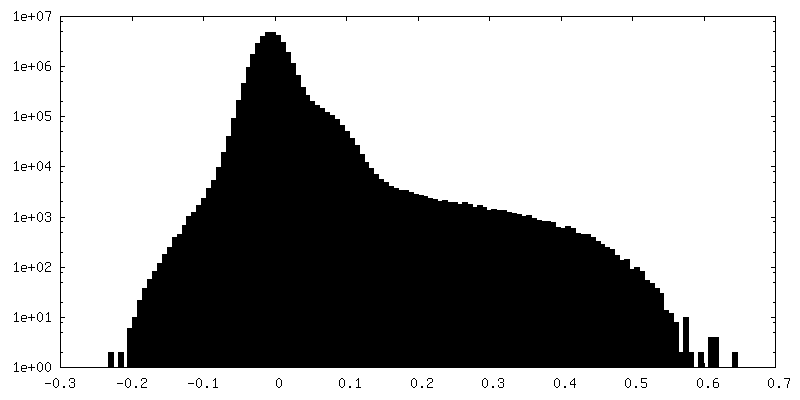
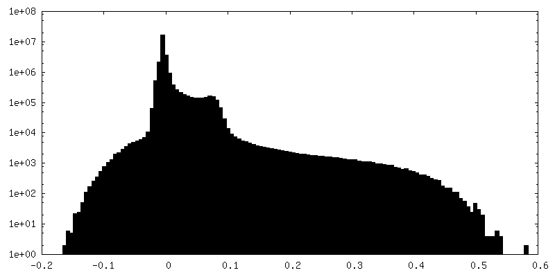
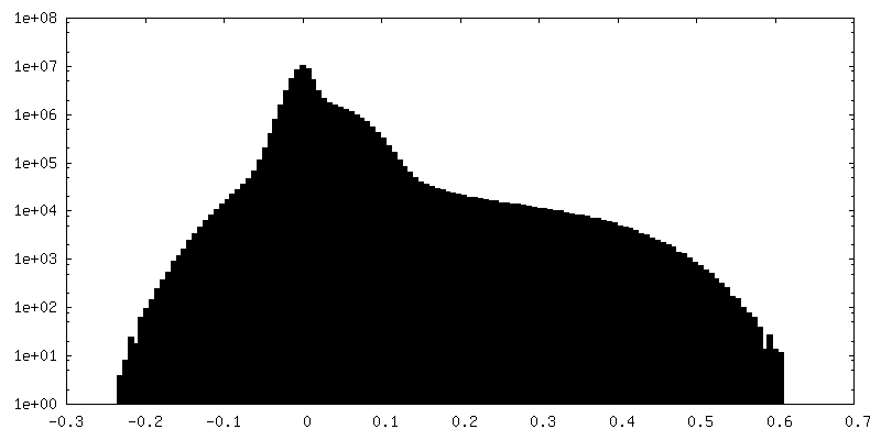
| Density Histograms |


-Additional map: Map auto-sharpened with B=48 (not cropped).
| File | emd_26874_additional_2.map | ||||||||||||
|---|---|---|---|---|---|---|---|---|---|---|---|---|---|
| Annotation | Map auto-sharpened with B=48 (not cropped). | ||||||||||||
| Projections & Slices |
| ||||||||||||
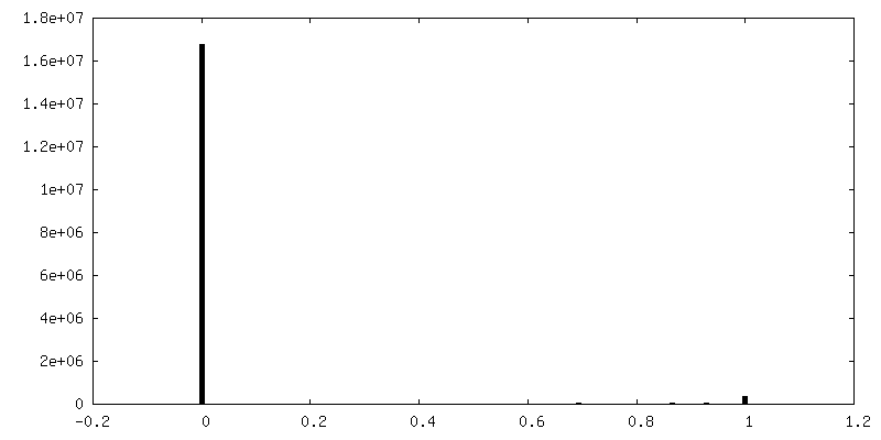
| Density Histograms |


-Additional map: Unfiltered half map 2. Used for FSC calculation.
| File | emd_26874_additional_3.map | ||||||||||||
|---|---|---|---|---|---|---|---|---|---|---|---|---|---|
| Annotation | Unfiltered half map 2. Used for FSC calculation. | ||||||||||||
| Projections & Slices |
| ||||||||||||
| Density Histograms |
-Additional map: Mask used for FSC calculation.
| File | emd_26874_additional_4.map | ||||||||||||
|---|---|---|---|---|---|---|---|---|---|---|---|---|---|
| Annotation | Mask used for FSC calculation. | ||||||||||||
| Projections & Slices |
| ||||||||||||
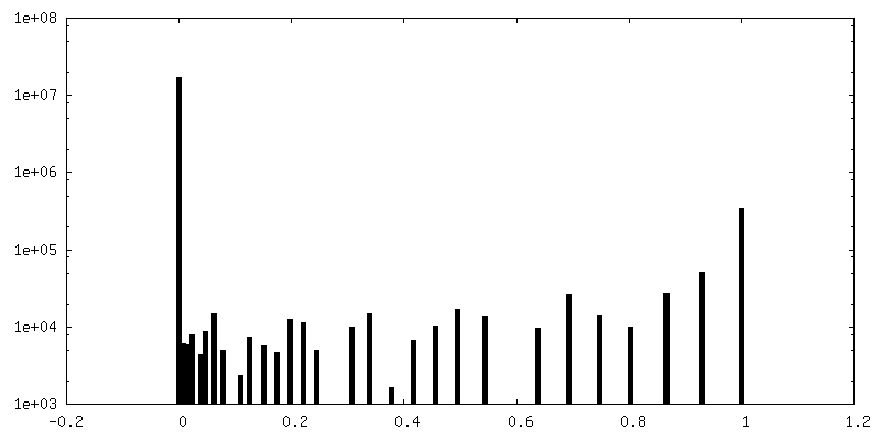
| Density Histograms |


-Additional map: Unsharpened map (not cropped).
| File | emd_26874_additional_5.map | ||||||||||||
|---|---|---|---|---|---|---|---|---|---|---|---|---|---|
| Annotation | Unsharpened map (not cropped). | ||||||||||||
| Projections & Slices |
| ||||||||||||
| Density Histograms |
-Half map: Unfiltered half map 2, cropped and resampled to...
| File | emd_26874_half_map_1.map | ||||||||||||
|---|---|---|---|---|---|---|---|---|---|---|---|---|---|
| Annotation | Unfiltered half map 2, cropped and resampled to match the main map and model, but otherwise unaltered. Original half maps and FSC mask are provided as additional maps. | ||||||||||||
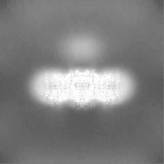
| Projections & Slices |
| ||||||||||||
| Density Histograms |
-Half map: Unfiltered half map 1, cropped and resampled to...
| File | emd_26874_half_map_2.map | ||||||||||||
|---|---|---|---|---|---|---|---|---|---|---|---|---|---|
| Annotation | Unfiltered half map 1, cropped and resampled to match the main map and model, but otherwise unaltered. Original half maps are provided as additional maps. | ||||||||||||
| Projections & Slices |
| ||||||||||||
| Density Histograms |


- Sample components
-Entire : Band 3 anion exchanger complexed with glycophorin A, in outward f...
| Entire | Name: Band 3 anion exchanger complexed with glycophorin A, in outward facing state. |
|---|---|
| Components |
|
-Supramolecule #1: Band 3 anion exchanger complexed with glycophorin A, in outward f...
| Supramolecule | Name: Band 3 anion exchanger complexed with glycophorin A, in outward facing state. type: complex / ID: 1 / Parent: 0 / Macromolecule list: #1-#2 Details: Particle set isolated by 3D classification from mixture mostly containing ankyrin complexes. |
|---|---|
| Source (natural) | Organism: Homo sapiens (human) / Organ: Blood / Tissue: Erythrocytes / Location in cell: Plasma membrane |
-Macromolecule #1: Glycophorin-A
| Macromolecule | Name: Glycophorin-A / type: protein_or_peptide / ID: 1 / Number of copies: 2 / Enantiomer: LEVO |
|---|---|
| Source (natural) | Organism: Homo sapiens (human) / Organ: Blood / Tissue: Erythrocytes |
| Molecular weight | Theoretical: 16.348433 KDa |
| Sequence | String: MYGKIIFVLL LSEIVSISAS STTGVAMHTS TSSSVTKSYI SSQTNDTHKR DTYAATPRAH EVSEISVRTV YPPEEETGER VQLAHHFSE PEITLIIFGV MAGVIGTILL ISYGIRRLIK KSPSDVKPLP SPDTDVPLSS VEIENPETSD Q UniProtKB: Glycophorin-A |
-Macromolecule #2: Band 3 anion transport protein
| Macromolecule | Name: Band 3 anion transport protein / type: protein_or_peptide / ID: 2 / Number of copies: 2 / Enantiomer: LEVO |
|---|---|
| Source (natural) | Organism: Homo sapiens (human) / Organ: Blood / Tissue: Erythrocytes |
| Molecular weight | Theoretical: 101.883859 KDa |
| Sequence | String: MEELQDDYED MMEENLEQEE YEDPDIPESQ MEEPAAHDTE ATATDYHTTS HPGTHKVYVE LQELVMDEKN QELRWMEAAR WVQLEENLG ENGAWGRPHL SHLTFWSLLE LRRVFTKGTV LLDLQETSLA GVANQLLDRF IFEDQIRPQD REELLRALLL K HSHAGELE ...String: MEELQDDYED MMEENLEQEE YEDPDIPESQ MEEPAAHDTE ATATDYHTTS HPGTHKVYVE LQELVMDEKN QELRWMEAAR WVQLEENLG ENGAWGRPHL SHLTFWSLLE LRRVFTKGTV LLDLQETSLA GVANQLLDRF IFEDQIRPQD REELLRALLL K HSHAGELE ALGGVKPAVL TRSGDPSQPL LPQHSSLETQ LFCEQGDGGT EGHSPSGILE KIPPDSEATL VLVGRADFLE QP VLGFVRL QEAAELEAVE LPVPIRFLFV LLGPEAPHID YTQLGRAAAT LMSERVFRID AYMAQSRGEL LHSLEGFLDC SLV LPPTDA PSEQALLSLV PVQRELLRRR YQSSPAKPDS SFYKGLDLNG GPDDPLQQTG QLFGGLVRDI RRRYPYYLSD ITDA FSPQV LAAVIFIYFA ALSPAITFGG LLGEKTRNQM GVSELLISTA VQGILFALLG AQPLLVVGFS GPLLVFEEAF FSFCE TNGL EYIVGRVWIG FWLILLVVLV VAFEGSFLVR FISRYTQEIF SFLISLIFIY ETFSKLIKIF QDHPLQKTYN YNVLMV PKP QGPLPNTALL SLVLMAGTFF FAMMLRKFKN SSYFPGKLRR VIGDFGVPIS ILIMVLVDFF IQDTYTQKLS VPDGFKV SN SSARGWVIHP LGLRSEFPIW MMFASALPAL LVFILIFLES QITTLIVSKP ERKMVKGSGF HLDLLLVVGM GGVAALFG M PWLSATTVRS VTHANALTVM GKASTPGAAA QIQEVKEQRI SGLLVAVLVG LSILMEPILS RIPLAVLFGI FLYMGVTSL SGIQLFDRIL LLFKPPKYHP DVPYVKRVKT WRMHLFTGIQ IICLAVLWVV KSTPASLALP FVLILTVPLR RVLLPLIFRN VELQCLDAD DAKATFDEEE GRDEYDEVAM PV UniProtKB: Band 3 anion transport protein |
-Macromolecule #4: CHOLESTEROL
| Macromolecule | Name: CHOLESTEROL / type: ligand / ID: 4 / Number of copies: 4 / Formula: CLR |
|---|---|
| Molecular weight | Theoretical: 386.654 Da |
| Chemical component information |  ChemComp-CLR: |
-Macromolecule #5: DIUNDECYL PHOSPHATIDYL CHOLINE
| Macromolecule | Name: DIUNDECYL PHOSPHATIDYL CHOLINE / type: ligand / ID: 5 / Number of copies: 2 / Formula: PLC |
|---|---|
| Molecular weight | Theoretical: 622.834 Da |
| Chemical component information |  ChemComp-PLC: |
-Macromolecule #6: [(2R)-2-octanoyloxy-3-[oxidanyl-[(1R,2R,3S,4R,5R,6S)-2,3,6-tris(o...
| Macromolecule | Name: [(2R)-2-octanoyloxy-3-[oxidanyl-[(1R,2R,3S,4R,5R,6S)-2,3,6-tris(oxidanyl)-4,5-diphosphonooxy-cyclohexyl]oxy-phosphoryl]oxy-propyl] octanoate type: ligand / ID: 6 / Number of copies: 2 / Formula: PIO |
|---|---|
| Molecular weight | Theoretical: 746.566 Da |
| Chemical component information |  ChemComp-PIO: |
-Macromolecule #7: water
| Macromolecule | Name: water / type: ligand / ID: 7 / Number of copies: 153 / Formula: HOH |
|---|---|
| Molecular weight | Theoretical: 18.015 Da |
| Chemical component information |  ChemComp-HOH: |
-Experimental details
-Structure determination
| Method | cryo EM |
|---|---|
 Processing Processing | single particle reconstruction |
| Aggregation state | particle |
-Sample preparation
| Concentration | 8 mg/mL |
|---|---|
| Buffer | pH: 7.4 Details: Final gel filtration buffer contained 0.05 % (w/v) digitonin, 130mM KCl, 20mM HEPES pH 7.4, 1mM ATP, 1mM MgCl2, 1mM PMSF. Peak fractions were concentrated to 8mg/mL, and 0.01% (w/v) of ...Details: Final gel filtration buffer contained 0.05 % (w/v) digitonin, 130mM KCl, 20mM HEPES pH 7.4, 1mM ATP, 1mM MgCl2, 1mM PMSF. Peak fractions were concentrated to 8mg/mL, and 0.01% (w/v) of glycyrrhizic acid was added immediately prior to vitrification. |
| Grid | Model: UltrAuFoil R0.6/1 / Material: GOLD / Mesh: 300 / Pretreatment - Type: GLOW DISCHARGE |
| Vitrification | Cryogen name: ETHANE / Chamber humidity: 100 % / Chamber temperature: 277 K / Instrument: FEI VITROBOT MARK IV / Details: 4-6 seconds, wait time 30 seconds.. |
| Details | Ankyrin complex mixture, purified from digitonin-solubilized erythrocyte ghost membranes. |
- Electron microscopy
| Microscope | FEI TITAN KRIOS |
|---|---|
| Specialist optics | Energy filter - Name: GIF Bioquantum / Energy filter - Slit width: 20 eV |
| Image recording | Film or detector model: GATAN K3 (6k x 4k) / Number grids imaged: 2 / Number real images: 14464 / Average exposure time: 2.5 sec. / Average electron dose: 58.0 e/Å2 / Details: Two grids were imaged in a single session. |
| Electron beam | Acceleration voltage: 300 kV / Electron source:  FIELD EMISSION GUN FIELD EMISSION GUN |
| Electron optics | Illumination mode: FLOOD BEAM / Imaging mode: BRIGHT FIELD / Cs: 2.7 mm / Nominal defocus max: 1.5 µm / Nominal defocus min: 0.5 µm |
| Sample stage | Specimen holder model: FEI TITAN KRIOS AUTOGRID HOLDER / Cooling holder cryogen: NITROGEN |
| Experimental equipment |  Model: Titan Krios / Image courtesy: FEI Company |