Movie
Movie Controller


+ Open data

- Basic information
| Entry |  | |||||||||
|---|---|---|---|---|---|---|---|---|---|---|
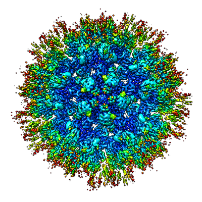
| Title | Structure of HIV-1 capsid declination | |||||||||
 Map data Map data | Declination of in vitro HIV-1 capsids | |||||||||
 Sample Sample |
| |||||||||
 Keywords Keywords | HIV-1 / capsid / declination / pentamer / hexamer / VIRUS LIKE PARTICLE | |||||||||
| Function / homology |  Function and homology information Function and homology informationviral budding via host ESCRT complex / host multivesicular body / ISG15 antiviral mechanism / viral nucleocapsid / viral translational frameshifting / host cell nucleus / host cell plasma membrane / virion membrane / structural molecule activity / RNA binding / zinc ion binding Similarity search - Function | |||||||||
| Biological species |   Human immunodeficiency virus 1 Human immunodeficiency virus 1 | |||||||||
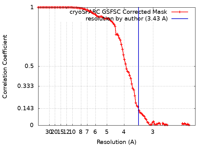
| Method | single particle reconstruction / cryo EM / Resolution: 3.43 Å | |||||||||
 Authors Authors | Pornillos O / Ganser-Pornillos BK / Schirra RT | |||||||||
| Funding support |  United States, 2 items United States, 2 items
| |||||||||
 Citation Citation | Journal: Nat Struct Mol Biol / Year: 2023 Title: A molecular switch modulates assembly and host factor binding of the HIV-1 capsid. Authors: Randall T Schirra / Nayara F B Dos Santos / Kaneil K Zadrozny / Iga Kucharska / Barbie K Ganser-Pornillos / Owen Pornillos /  Abstract: The HIV-1 capsid is a fullerene cone made of quasi-equivalent hexamers and pentamers of the viral CA protein. Typically, quasi-equivalent assembly of viral capsid subunits is controlled by a ...The HIV-1 capsid is a fullerene cone made of quasi-equivalent hexamers and pentamers of the viral CA protein. Typically, quasi-equivalent assembly of viral capsid subunits is controlled by a molecular switch. Here, we identify a Thr-Val-Gly-Gly motif that modulates CA hexamer/pentamer switching by folding into a 3 helix in the pentamer and random coil in the hexamer. Manipulating the coil/helix configuration of the motif allowed us to control pentamer and hexamer formation in a predictable manner, thus proving its function as a molecular switch. Importantly, the switch also remodels the common binding site for host factors that are critical for viral replication and the new ultra-potent HIV-1 inhibitor lenacapavir. This study reveals that a critical assembly element also modulates the post-assembly and viral replication functions of the HIV-1 capsid and provides new insights on capsid function and inhibition. | |||||||||
| History |
|
- Structure visualization
| Supplemental images |
|---|
- Downloads & links
-EMDB archive
| Map data | emd_26715.map.gz | 104.9 MB | EMDB map data format | |
|---|---|---|---|---|
| Header (meta data) | emd-26715-v30.xmlemd-26715.xml | 15.4 KB 15.4 KB | Display Display | EMDB header |
| FSC (resolution estimation) | emd_26715_fsc.xml | 11.4 KB | Display | FSC data file |
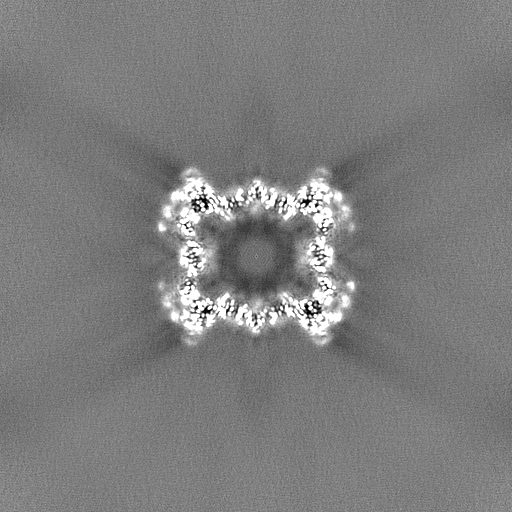
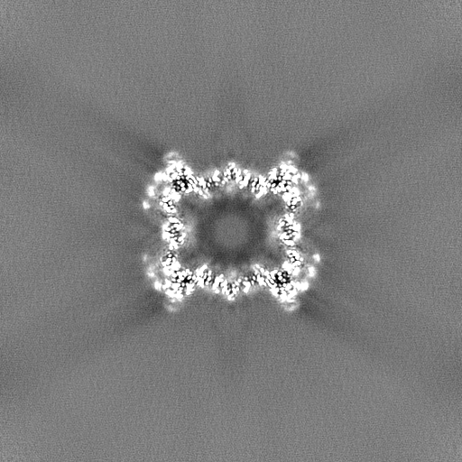
| Images |  emd_26715.png emd_26715.png | 240.2 KB | ||
| Filedesc metadata | emd-26715.cif.gz | 5.7 KB | ||
| Others | emd_26715_half_map_1.map.gzemd_26715_half_map_2.map.gz | 474.7 MB 474.7 MB | ||
| Archive directory |  http://ftp.pdbj.org/pub/emdb/structures/EMD-26715ftp://ftp.pdbj.org/pub/emdb/structures/EMD-26715 http://ftp.pdbj.org/pub/emdb/structures/EMD-26715ftp://ftp.pdbj.org/pub/emdb/structures/EMD-26715 | HTTPS FTP |
-Related structure data
| Related structure data |  7urnMC 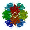 7urtC  8eepC 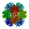 8eetC  8ejlC M: atomic model generated by this map C: citing same article ( |
|---|---|
| Similar structure data |


-Links
| EMDB pages | EMDB (EBI/PDBe) / EMDataResource |
|---|---|
| Related items in Molecule of the Month |


-Map
| File | Download / File: emd_26715.map.gz / Format: CCP4 / Size: 111.5 MB / Type: IMAGE STORED AS FLOATING POINT NUMBER (4 BYTES) | ||||||||||||||||||||||||||||||||||||
|---|---|---|---|---|---|---|---|---|---|---|---|---|---|---|---|---|---|---|---|---|---|---|---|---|---|---|---|---|---|---|---|---|---|---|---|---|---|
| Annotation | Declination of in vitro HIV-1 capsids | ||||||||||||||||||||||||||||||||||||
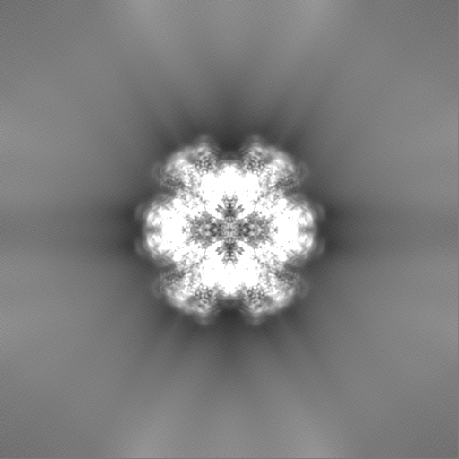
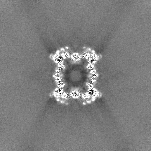
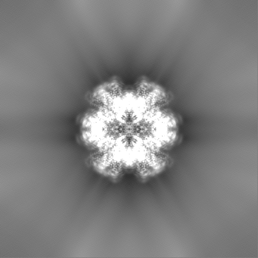
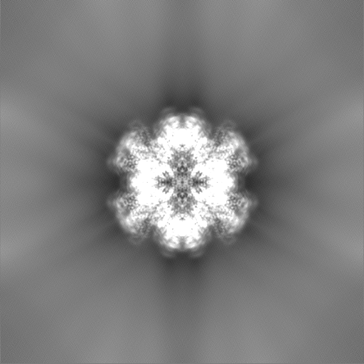
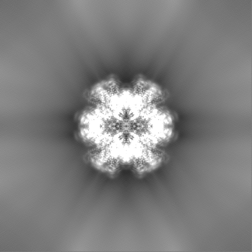
| Projections & slices | Image control
Images are generated by Spider. | ||||||||||||||||||||||||||||||||||||
| Voxel size | X=Y=Z: 1.08 Å | ||||||||||||||||||||||||||||||||||||
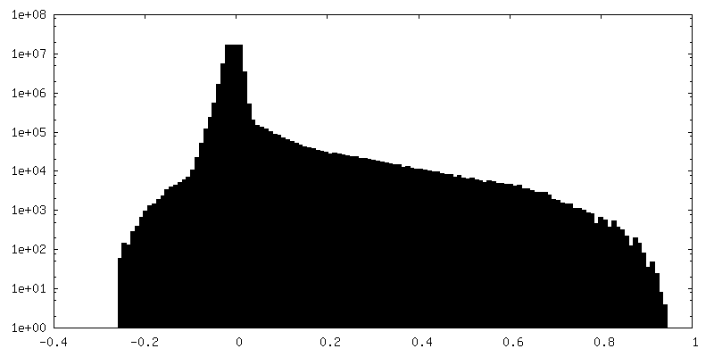
| Density |
| ||||||||||||||||||||||||||||||||||||
| Symmetry | Space group: 1 | ||||||||||||||||||||||||||||||||||||
| Details | EMDB XML:
|
 Z (Sec.)
 Y (Row.)
 X (Col.)


-Supplemental data
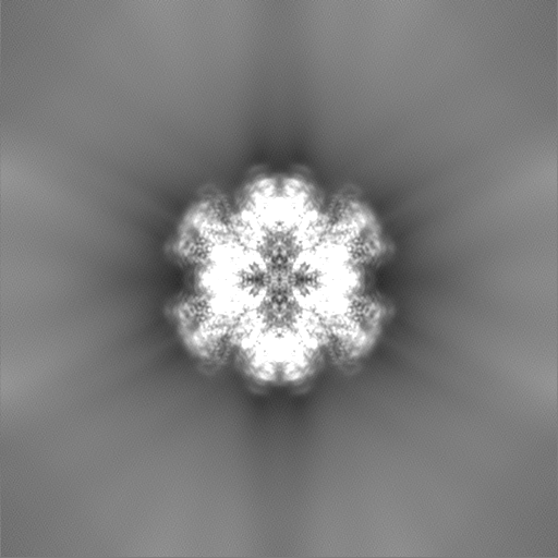
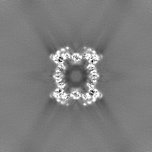
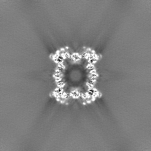
-Half map: Half map B
| File | emd_26715_half_map_1.map | ||||||||||||
|---|---|---|---|---|---|---|---|---|---|---|---|---|---|
| Annotation | Half map B | ||||||||||||
| Projections & Slices |
| ||||||||||||
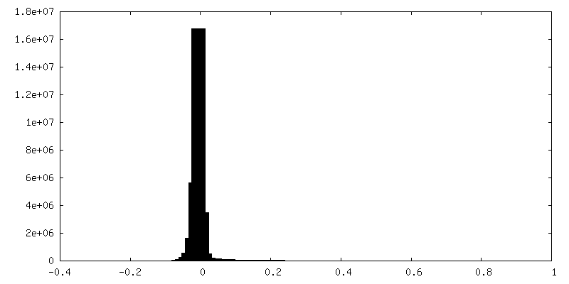
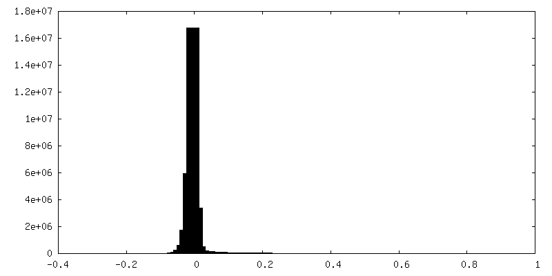
| Density Histograms |
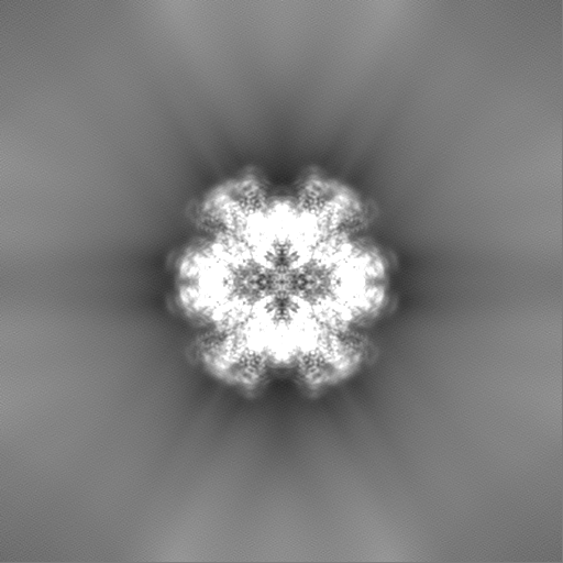
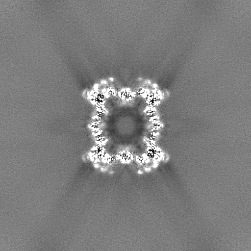
-Half map: Half map A
| File | emd_26715_half_map_2.map | ||||||||||||
|---|---|---|---|---|---|---|---|---|---|---|---|---|---|
| Annotation | Half map A | ||||||||||||
| Projections & Slices |
| ||||||||||||
| Density Histograms |

- Sample components
-Entire : Capsid-like particles of in vitro assembled HIV-1 CA protein
| Entire | Name: Capsid-like particles of in vitro assembled HIV-1 CA protein |
|---|---|
| Components |
|
-Supramolecule #1: Capsid-like particles of in vitro assembled HIV-1 CA protein
| Supramolecule | Name: Capsid-like particles of in vitro assembled HIV-1 CA protein type: complex / ID: 1 / Parent: 0 / Macromolecule list: #1 |
|---|---|
| Source (natural) | Organism: Human immunodeficiency virus 1 |
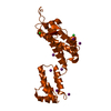
-Macromolecule #1: HIV-1 capsid protein
| Macromolecule | Name: HIV-1 capsid protein / type: protein_or_peptide / ID: 1 / Number of copies: 7 / Enantiomer: LEVO |
|---|---|
| Source (natural) | Organism: Human immunodeficiency virus 1 / Strain: NL4-3 |
| Molecular weight | Theoretical: 25.630426 KDa |
| Recombinant expression | Organism:  |
| Sequence | String: PIVQNLQGQM VHQAISPRTL NAWVKVVEEK AFSPEVIPMF SALSEGATPQ DLNTMLNTVG GHQAAMQMLK ETINEEAAEW DRLHPVHAG PIAPGQMREP RGSDIAGTTS TLQEQIGWMT HNPPIPVGEI YKRWIILGLN KIVRMYSPTS ILDIRQGPKE P FRDYVDRF ...String: PIVQNLQGQM VHQAISPRTL NAWVKVVEEK AFSPEVIPMF SALSEGATPQ DLNTMLNTVG GHQAAMQMLK ETINEEAAEW DRLHPVHAG PIAPGQMREP RGSDIAGTTS TLQEQIGWMT HNPPIPVGEI YKRWIILGLN KIVRMYSPTS ILDIRQGPKE P FRDYVDRF YKTLRAEQAS QEVKNWMTET LLVQNANPDC KTILKALGPG ATLEEMMTAC QGVGGPGHKA RVL UniProtKB: Gag polyprotein |
-Macromolecule #2: INOSITOL HEXAKISPHOSPHATE
| Macromolecule | Name: INOSITOL HEXAKISPHOSPHATE / type: ligand / ID: 2 / Number of copies: 2 / Formula: IHP |
|---|---|
| Molecular weight | Theoretical: 660.035 Da |
| Chemical component information |  ChemComp-IHP: |
-Experimental details
-Structure determination
| Method | cryo EM |
|---|---|
 Processing Processing | single particle reconstruction |
| Aggregation state | particle |
-Sample preparation
| Buffer | pH: 6 |
|---|---|
| Vitrification | Cryogen name: ETHANE / Instrument: HOMEMADE PLUNGER / Details: Manual plunge-freezing. |
- Electron microscopy
| Microscope | FEI TITAN KRIOS |
|---|---|
| Image recording | Film or detector model: GATAN K3 (6k x 4k) / Average electron dose: 50.0 e/Å2 |
| Electron beam | Acceleration voltage: 300 kV / Electron source:  FIELD EMISSION GUN FIELD EMISSION GUN |
| Electron optics | Illumination mode: FLOOD BEAM / Imaging mode: BRIGHT FIELD / Nominal defocus max: 2.5 µm / Nominal defocus min: 0.5 µm |
| Experimental equipment |  Model: Titan Krios / Image courtesy: FEI Company |
+Image processing
-Atomic model buiding 1
| Initial model | PDB ID: Chain - Source name: PDB / Chain - Initial model type: experimental model |
|---|---|
| Details | The following protein chains were refined separately in real space using Phenix: A (pentamer); L, M and N (half-hexamer). Chain M was then rigid-body fit to generate chains O, P and Q to complete the hexamer. Chains O, P and Q are deposited as polyALA models. |
| Refinement | Space: REAL / Protocol: OTHER |
| Output model | PDB-7urn: |