National Institutes of Health/National Institute of General Medical Sciences (NIH/NIGMS)
5R35GM130393
United States
Citation
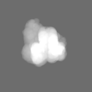
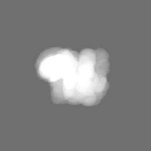
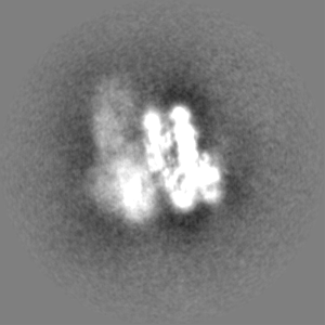
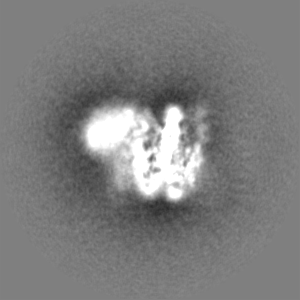
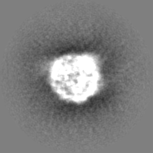
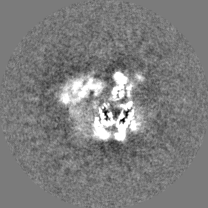
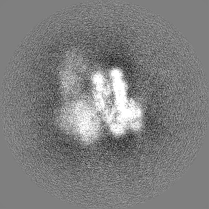
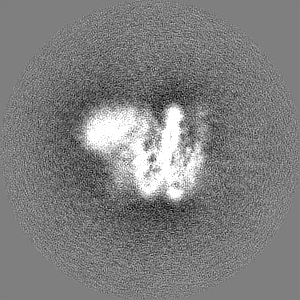
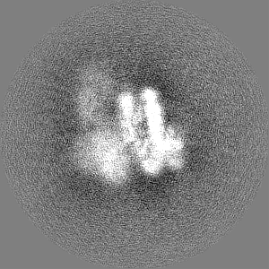
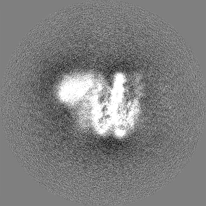
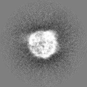
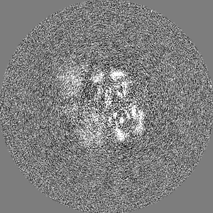
Journal: Proc Natl Acad Sci U S A / Year: 2022 Title: Multistate structures of the MLL1-WRAD complex bound to H2B-ubiquitinated nucleosome. Authors: Sanim Rahman / Niklas A Hoffmann / Evan J Worden / Marissa L Smith / Kevin E W Namitz / Bruce A Knutson / Michael S Cosgrove / Cynthia Wolberger / Abstract: The human Mixed Lineage Leukemia-1 (MLL1) complex methylates histone H3K4 to promote transcription and is stimulated by monoubiquitination of histone H2B. Recent structures of the MLL1-WRAD core ...The human Mixed Lineage Leukemia-1 (MLL1) complex methylates histone H3K4 to promote transcription and is stimulated by monoubiquitination of histone H2B. Recent structures of the MLL1-WRAD core complex, which comprises the MLL1 methyltransferase, DR5, bBp5, sh2L, and PY-30, have revealed variability in the docking of MLL1-WRAD on nucleosomes. In addition, portions of the Ash2L structure and the position of DPY30 remain ambiguous. We used an integrated approach combining cryoelectron microscopy (cryo-EM) and mass spectrometry cross-linking to determine a structure of the MLL1-WRAD complex bound to ubiquitinated nucleosomes. The resulting model contains the Ash2L intrinsically disordered region (IDR), SPRY insertion region, Sdc1-DPY30 interacting region (SDI-motif), and the DPY30 dimer. We also resolved three additional states of MLL1-WRAD lacking one or more subunits, which may reflect different steps in the assembly of MLL1-WRAD. The docking of subunits in all four states differs from structures of MLL1-WRAD bound to unmodified nucleosomes, suggesting that H2B-ubiquitin favors assembly of the active complex. Our results provide a more complete picture of MLL1-WRAD and the role of ubiquitin in promoting formation of the active methyltransferase complex.
In the structure databanks used in Yorodumi, some data are registered as the other names, "COVID-19 virus" and "2019-nCoV". Here are the details of the virus and the list of structure data.
Jan 31, 2019. EMDB accession codes are about to change! (news from PDBe EMDB page)
EMDB accession codes are about to change! (news from PDBe EMDB page)
The allocation of 4 digits for EMDB accession codes will soon come to an end. Whilst these codes will remain in use, new EMDB accession codes will include an additional digit and will expand incrementally as the available range of codes is exhausted. The current 4-digit format prefixed with “EMD-” (i.e. EMD-XXXX) will advance to a 5-digit format (i.e. EMD-XXXXX), and so on. It is currently estimated that the 4-digit codes will be depleted around Spring 2019, at which point the 5-digit format will come into force.
The EM Navigator/Yorodumi systems omit the EMD- prefix.
Related info.:Q: What is EMD? / ID/Accession-code notation in Yorodumi/EM Navigator
Yorodumi is a browser for structure data from EMDB, PDB, SASBDB, etc.
This page is also the successor to EM Navigator detail page, and also detail information page/front-end page for Omokage search.
The word "yorodu" (or yorozu) is an old Japanese word meaning "ten thousand". "mi" (miru) is to see.
Related info.:EMDB / PDB / SASBDB / Comparison of 3 databanks / Yorodumi Search / Aug 31, 2016. New EM Navigator & Yorodumi / Yorodumi Papers / Jmol/JSmol / Function and homology information / Changes in new EM Navigator and Yorodumi
 Movie
Movie Controller


 Open data
 Basic information
 Map data
 Sample
 Function and homology information
 Homo sapiens (human) /
  Authors
 United States, 1 items
  Citation
 Structure visualization
 Downloads & links
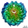
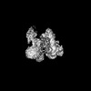
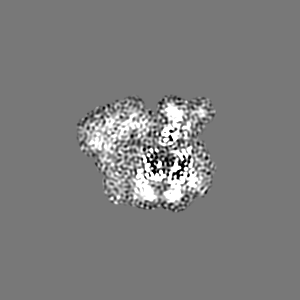
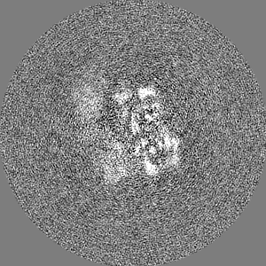
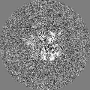
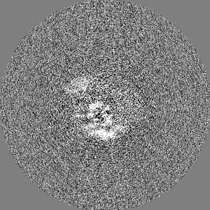
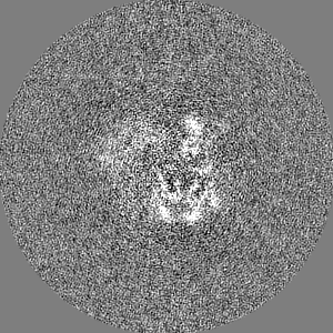
 emd_26454.png
 http://ftp.pdbj.org/pub/emdb/structures/EMD-26454
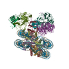


 Z (Sec.)
 Y (Row.)
 X (Col.)


 Sample components

 Processing
 Electron microscopy
 FIELD EMISSION GUN