 Movie
Movie Controller


+ Open data

- Basic information
| Entry | Database: PDB / ID: 8du4 | ||||||
|---|---|---|---|---|---|---|---|
| Title | Complex between RbBP5-WDR5 and an H2B-ubiquitinated nucleosome | ||||||
 Components Components |
| ||||||
 Keywords Keywords | DNA BINDING PROTEIN / ubiquitin / chromatin / MLL / methylation | ||||||
| Function / homology |  Function and homology information Function and homology informationhistone H3Q5ser reader activity / histone H3K4me1 reader activity / Loss of Function of KMT2D in MLL4 Complex Formation in Kabuki Syndrome / Epigenetic regulation of gene expression by MLL3 and MLL4 complexes / MLL3/4 complex / Set1C/COMPASS complex / ATAC complex / MLL1/2 complex / NSL complex / histone H3K4 methyltransferase activity ...histone H3Q5ser reader activity / histone H3K4me1 reader activity / Loss of Function of KMT2D in MLL4 Complex Formation in Kabuki Syndrome / Epigenetic regulation of gene expression by MLL3 and MLL4 complexes / MLL3/4 complex / Set1C/COMPASS complex / ATAC complex / MLL1/2 complex / NSL complex / histone H3K4 methyltransferase activity / Cardiogenesis / Formation of WDR5-containing histone-modifying complexes / histone methyltransferase complex / MLL1 complex / histone acetyltransferase complex / regulation of embryonic development / regulation of cell division / positive regulation of gluconeogenesis / transcription initiation-coupled chromatin remodeling / gluconeogenesis / skeletal system development / Deactivation of the beta-catenin transactivating complex / Formation of the beta-catenin:TCF transactivating complex / RUNX1 regulates genes involved in megakaryocyte differentiation and platelet function / response to estrogen / PKMTs methylate histone lysines / Activation of anterior HOX genes in hindbrain development during early embryogenesis / RMTs methylate histone arginines / structural constituent of chromatin / mitotic spindle / nucleosome / nucleosome assembly / heterochromatin formation / HATs acetylate histones / MLL4 and MLL3 complexes regulate expression of PPARG target genes in adipogenesis and hepatic steatosis / sperm principal piece / Neddylation / histone binding / mitochondrial outer membrane / regulation of cell cycle / transcription cis-regulatory region binding / protein heterodimerization activity / DNA damage response / regulation of transcription by RNA polymerase II / regulation of DNA-templated transcription / positive regulation of DNA-templated transcription / nucleolus / negative regulation of transcription by RNA polymerase II / DNA binding / nucleoplasm / nucleus Similarity search - Function | ||||||
| Biological species | synthetic construct (others)  Homo sapiens (human) Homo sapiens (human) | ||||||
| Method | ELECTRON MICROSCOPY / single particle reconstruction / cryo EM / Resolution: 3.55 Å | ||||||
 Authors Authors | Niklas, H.A. / Rahman, S. / Worden, E.J. / Wolberger, C. | ||||||
| Funding support |  United States, 1items United States, 1items
| ||||||
 Citation Citation | Journal: Proc Natl Acad Sci U S A / Year: 2022 Title: Multistate structures of the MLL1-WRAD complex bound to H2B-ubiquitinated nucleosome. Authors: Sanim Rahman / Niklas A Hoffmann / Evan J Worden / Marissa L Smith / Kevin E W Namitz / Bruce A Knutson / Michael S Cosgrove / Cynthia Wolberger / Abstract: The human Mixed Lineage Leukemia-1 (MLL1) complex methylates histone H3K4 to promote transcription and is stimulated by monoubiquitination of histone H2B. Recent structures of the MLL1-WRAD core ...The human Mixed Lineage Leukemia-1 (MLL1) complex methylates histone H3K4 to promote transcription and is stimulated by monoubiquitination of histone H2B. Recent structures of the MLL1-WRAD core complex, which comprises the MLL1 methyltransferase, DR5, bBp5, sh2L, and PY-30, have revealed variability in the docking of MLL1-WRAD on nucleosomes. In addition, portions of the Ash2L structure and the position of DPY30 remain ambiguous. We used an integrated approach combining cryoelectron microscopy (cryo-EM) and mass spectrometry cross-linking to determine a structure of the MLL1-WRAD complex bound to ubiquitinated nucleosomes. The resulting model contains the Ash2L intrinsically disordered region (IDR), SPRY insertion region, Sdc1-DPY30 interacting region (SDI-motif), and the DPY30 dimer. We also resolved three additional states of MLL1-WRAD lacking one or more subunits, which may reflect different steps in the assembly of MLL1-WRAD. The docking of subunits in all four states differs from structures of MLL1-WRAD bound to unmodified nucleosomes, suggesting that H2B-ubiquitin favors assembly of the active complex. Our results provide a more complete picture of MLL1-WRAD and the role of ubiquitin in promoting formation of the active methyltransferase complex. | ||||||
| History |
|
- Structure visualization
| Structure viewer | Molecule: MolmilJmol/JSmol |
|---|
- Downloads & links
-Download
| PDBx/mmCIF format | 8du4.cif.gz | 462.2 KB | Display | PDBx/mmCIF format |
|---|---|---|---|---|
| PDB format | pdb8du4.ent.gz | 349.4 KB | Display | PDB format |
| PDBx/mmJSON format | 8du4.json.gz | Tree view | PDBx/mmJSON format | |
| Others |  Other downloads Other downloads |
-Validation report
| Arichive directory | https://data.pdbj.org/pub/pdb/validation_reports/du/8du4ftp://data.pdbj.org/pub/pdb/validation_reports/du/8du4 | HTTPS FTP |
|---|
-Related structure data
| Related structure data |  27715MC 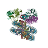 7ud5C C: citing same article ( M: map data used to model this data |
|---|---|
| Similar structure data |
-Links
 PDBj
PDBj


- Assembly
| Deposited unit | 
|
|---|---|
| 1 |
|
-Components
-Protein , 7 types, 11 molecules AEBFCGDHLNO
| #1: Protein | Mass: 15383.028 Da / Num. of mol.: 2 / Mutation: K4Nle, M90Nle, M120Nle Source method: isolated from a genetically manipulated source Source: (gene. exp.)  #2: Protein | Mass: 11394.426 Da / Num. of mol.: 2 Source method: isolated from a genetically manipulated source Source: (gene. exp.) #3: Protein | Mass: 14109.436 Da / Num. of mol.: 2 Source method: isolated from a genetically manipulated source Source: (gene. exp.) #4: Protein | Mass: 13629.911 Da / Num. of mol.: 2 / Mutation: S32T, K120C Source method: isolated from a genetically manipulated source Source: (gene. exp.) #7: Protein | | Mass: 36648.371 Da / Num. of mol.: 1 Source method: isolated from a genetically manipulated source Source: (gene. exp.) Homo sapiens (human) / Gene: WDR5, BIG3 / Production host: #8: Protein | | Mass: 59223.477 Da / Num. of mol.: 1 Source method: isolated from a genetically manipulated source Source: (gene. exp.) Homo sapiens (human) / Gene: RBBP5, RBQ3 / Production host: #9: Protein | | Mass: 9164.521 Da / Num. of mol.: 1 / Mutation: G76C Source method: isolated from a genetically manipulated source Source: (gene. exp.) Homo sapiens (human) / Gene: UBB / Production host: |
|---|
-601 DNA (146- ... , 2 types, 2 molecules IJ
| #5: DNA chain | Mass: 44825.559 Da / Num. of mol.: 1 Source method: isolated from a genetically manipulated source Source: (gene. exp.) synthetic construct (others) / Production host: |
|---|---|
| #6: DNA chain | Mass: 45305.852 Da / Num. of mol.: 1 Source method: isolated from a genetically manipulated source Source: (gene. exp.) synthetic construct (others) / Production host: |
-Details
| Has ligand of interest | N |
|---|---|
| Has protein modification | Y |
-Experimental details
-Experiment
| Experiment | Method: ELECTRON MICROSCOPY |
|---|---|
| EM experiment | Aggregation state: PARTICLE / 3D reconstruction method: single particle reconstruction |
- Sample preparation
| Component |
| ||||||||||||||||||||||||
|---|---|---|---|---|---|---|---|---|---|---|---|---|---|---|---|---|---|---|---|---|---|---|---|---|---|
| Source (natural) |
| ||||||||||||||||||||||||
| Source (recombinant) |
| ||||||||||||||||||||||||
| Buffer solution | pH: 8 | ||||||||||||||||||||||||
| Specimen | Embedding applied: NO / Shadowing applied: NO / Staining applied: NO / Vitrification applied: YES | ||||||||||||||||||||||||
| Vitrification | Cryogen name: ETHANE |
- Electron microscopy imaging
| Experimental equipment |  Model: Titan Krios / Image courtesy: FEI Company |
|---|---|
| Microscopy | Model: FEI TITAN KRIOS |
| Electron gun | Electron source:  FIELD EMISSION GUN / Accelerating voltage: 300 kV / Illumination mode: FLOOD BEAM FIELD EMISSION GUN / Accelerating voltage: 300 kV / Illumination mode: FLOOD BEAM |
| Electron lens | Mode: BRIGHT FIELD / Nominal defocus max: 2800 nm / Nominal defocus min: 1000 nm |
| Image recording | Electron dose: 60 e/Å2 / Film or detector model: GATAN K3 (6k x 4k) / Num. of real images: 5091 |
- Processing
| Software | Name: PHENIX / Version: 1.20.1_4487: / Classification: refinement | ||||||||||||||||||||||||
|---|---|---|---|---|---|---|---|---|---|---|---|---|---|---|---|---|---|---|---|---|---|---|---|---|---|
| EM software | Name: PHENIX / Category: model refinement | ||||||||||||||||||||||||
| CTF correction | Type: PHASE FLIPPING AND AMPLITUDE CORRECTION | ||||||||||||||||||||||||
| 3D reconstruction | Resolution: 3.55 Å / Resolution method: FSC 0.143 CUT-OFF / Num. of particles: 134528 / Symmetry type: POINT | ||||||||||||||||||||||||
| Refine LS restraints |
|
