 ムービー
ムービー コントローラー


+ データを開く

- 基本情報
基本情報
| 登録情報 | データベース: EMDB / ID: EMD-2638 | |||||||||
|---|---|---|---|---|---|---|---|---|---|---|
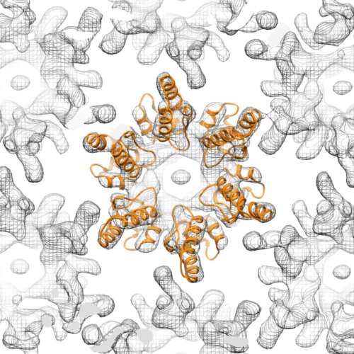
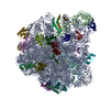
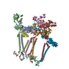
| タイトル | Cryo-electron microscopy of tubular arrays of HIV-1 Gag resolves structures essential for immature virus assembly. | |||||||||
 マップデータ マップデータ | Reconstruction of HIV-1 CANC immature-like tubular particles | |||||||||
 試料 試料 |
| |||||||||
 キーワード キーワード | HIV-1 / capsid / SP1 / helical reconstruction | |||||||||
| 機能・相同性 | : / gag protein p24 N-terminal domain / viral process / Retroviral nucleocapsid Gag protein p24, C-terminal domain / Gag protein p24 C-terminal domain / Retrovirus capsid, C-terminal / Retrovirus capsid, N-terminal / viral capsid / Gag protein 機能・相同性情報 機能・相同性情報 | |||||||||
| 生物種 |   Human immunodeficiency virus 1 (ヒト免疫不全ウイルス) Human immunodeficiency virus 1 (ヒト免疫不全ウイルス) | |||||||||
| 手法 | らせん対称体再構成法 / クライオ電子顕微鏡法 / 解像度: 9.4 Å | |||||||||
 データ登録者 データ登録者 | Bharat TAM / Castillo-Menendez LR / Hagen WH / Lux V / Igonet S / Schorb M / Schur FKM / Krauesslich HG / Briggs JAG | |||||||||
 引用 引用 | ジャーナル: Proc Natl Acad Sci U S A / 年: 2014 タイトル: Cryo-electron microscopy of tubular arrays of HIV-1 Gag resolves structures essential for immature virus assembly. 著者: Tanmay A M Bharat / Luis R Castillo Menendez / Wim J H Hagen / Vanda Lux / Sebastien Igonet / Martin Schorb / Florian K M Schur / Hans-Georg Kräusslich / John A G Briggs /    要旨: The assembly of HIV-1 is mediated by oligomerization of the major structural polyprotein, Gag, into a hexameric protein lattice at the plasma membrane of the infected cell. This leads to budding and ...The assembly of HIV-1 is mediated by oligomerization of the major structural polyprotein, Gag, into a hexameric protein lattice at the plasma membrane of the infected cell. This leads to budding and release of progeny immature virus particles. Subsequent proteolytic cleavage of Gag triggers rearrangement of the particles to form mature infectious virions. Obtaining a structural model of the assembled lattice of Gag within immature virus particles is necessary to understand the interactions that mediate assembly of HIV-1 particles in the infected cell, and to describe the substrate that is subsequently cleaved by the viral protease. An 8-Å resolution structure of an immature virus-like tubular array assembled from a Gag-derived protein of the related retrovirus Mason-Pfizer monkey virus (M-PMV) has previously been reported, and a model for the arrangement of the HIV-1 capsid (CA) domains has been generated based on homology to this structure. Here we have assembled tubular arrays of a HIV-1 Gag-derived protein with an immature-like arrangement of the C-terminal CA domains and have solved their structure by using hybrid cryo-EM and tomography analysis. The structure reveals the arrangement of the C-terminal domain of CA within an immature-like HIV-1 Gag lattice, and provides, to our knowledge, the first high-resolution view of the region immediately downstream of CA, which is essential for assembly, and is significantly different from the respective region in M-PMV. Our results reveal a hollow column of density for this region in HIV-1 that is compatible with the presence of a six-helix bundle at this position. | |||||||||
| 履歴 |
|
- 構造の表示
構造の表示
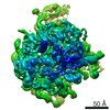
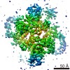
| ムービー |
ムービービューア |
|---|---|
| 構造ビューア | EMマップ: SurfViewMolmilJmol/JSmol |
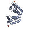
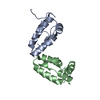
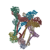
| 添付画像 |
- ダウンロードとリンク
-EMDBアーカイブ
| マップデータ | emd_2638.map.gz | 3.5 MB | EMDBマップデータ形式 | |
|---|---|---|---|---|
| ヘッダ (付随情報) | emd-2638-v30.xmlemd-2638.xml | 12.7 KB 12.7 KB | 表示 表示 | EMDBヘッダ |
| 画像 |  EMD-2638-image_for_EMDB1_500x500.jpg EMD-2638-image_for_EMDB1_500x500.jpg | 77.7 KB | ||
| アーカイブディレクトリ |  http://ftp.pdbj.org/pub/emdb/structures/EMD-2638ftp://ftp.pdbj.org/pub/emdb/structures/EMD-2638 http://ftp.pdbj.org/pub/emdb/structures/EMD-2638ftp://ftp.pdbj.org/pub/emdb/structures/EMD-2638 | HTTPS FTP |
-検証レポート
| 文書・要旨 | emd_2638_validation.pdf.gz | 301.1 KB | 表示 | EMDB検証レポート |
|---|---|---|---|---|
| 文書・詳細版 | emd_2638_full_validation.pdf.gz | 300.2 KB | 表示 | |
| XML形式データ | emd_2638_validation.xml.gz | 5.6 KB | 表示 | |
| アーカイブディレクトリ | https://ftp.pdbj.org/pub/emdb/validation_reports/EMD-2638ftp://ftp.pdbj.org/pub/emdb/validation_reports/EMD-2638 | HTTPS FTP |
-関連構造データ
-リンク
| EMDBのページ | EMDB (EBI/PDBe) / EMDataResource |
|---|---|
| 「今月の分子」の関連する項目 |

-マップ
| ファイル | ダウンロード / ファイル: emd_2638.map.gz / 形式: CCP4 / 大きさ: 3.7 MB / タイプ: IMAGE STORED AS FLOATING POINT NUMBER (4 BYTES) | ||||||||||||||||||||||||||||||||||||||||||||||||||||||||||||||||||||
|---|---|---|---|---|---|---|---|---|---|---|---|---|---|---|---|---|---|---|---|---|---|---|---|---|---|---|---|---|---|---|---|---|---|---|---|---|---|---|---|---|---|---|---|---|---|---|---|---|---|---|---|---|---|---|---|---|---|---|---|---|---|---|---|---|---|---|---|---|---|
| 注釈 | Reconstruction of HIV-1 CANC immature-like tubular particles | ||||||||||||||||||||||||||||||||||||||||||||||||||||||||||||||||||||
| 投影像・断面図 | 画像のコントロール
画像は Spider により作成 | ||||||||||||||||||||||||||||||||||||||||||||||||||||||||||||||||||||
| ボクセルのサイズ | X=Y=Z: 1.53 Å | ||||||||||||||||||||||||||||||||||||||||||||||||||||||||||||||||||||
| 密度 |
| ||||||||||||||||||||||||||||||||||||||||||||||||||||||||||||||||||||
| 対称性 | 空間群: 1 | ||||||||||||||||||||||||||||||||||||||||||||||||||||||||||||||||||||
| 詳細 | EMDB XML:
CCP4マップ ヘッダ情報:
| ||||||||||||||||||||||||||||||||||||||||||||||||||||||||||||||||||||
 Z (Sec.)
 Y (Row.)
 X (Col.)


-添付データ
- 試料の構成要素
-全体 : HIV-1 CANC Y169L/S protein assembled with 73mer DNA into tubular ...
| 全体 | 名称: HIV-1 CANC Y169L/S protein assembled with 73mer DNA into tubular particles |
|---|---|
| 要素 |
|
-超分子 #1000: HIV-1 CANC Y169L/S protein assembled with 73mer DNA into tubular ...
| 超分子 | 名称: HIV-1 CANC Y169L/S protein assembled with 73mer DNA into tubular particles タイプ: sample / ID: 1000 / 詳細: Helical tubular crystals / 集合状態: helical / Number unique components: 1 |
|---|
-分子 #1: Human immunodeficiency virus 1 Gag
| 分子 | 名称: Human immunodeficiency virus 1 Gag / タイプ: protein_or_peptide / ID: 1 / Name.synonym: HIV-1 Gag 詳細: 10nm protein-A conjugated gold particles were added to the sample. Protein construct consists of CA to NC domains with mutation in position Y169. 集合状態: Helical / 組換発現: Yes |
|---|---|
| 由来(天然) | 生物種: Human immunodeficiency virus 1 (ヒト免疫不全ウイルス) 別称: HIV-1 |
| 組換発現 | 生物種:  |
| 配列 | UniProtKB: Gag protein |
-実験情報
-構造解析
| 手法 | クライオ電子顕微鏡法 |
|---|---|
 解析 解析 | らせん対称体再構成法 |
| 試料の集合状態 | helical array |
-試料調製
| 濃度 | 2 mg/mL |
|---|---|
| 緩衝液 | pH: 6 / 詳細: 30 mM MES, 1 mM EDTA, 1 mM DTT, 0.5 M NaCl |
| グリッド | 詳細: 300 mesh C-Flat copper grids were glow discharged for 20 seconds |
| 凍結 | 凍結剤: ETHANE / チャンバー内湿度: 100 % / 装置: HOMEMADE PLUNGER |
| 詳細 | Protein stock solutions were dialyzed for 2 h at 4 degree Celsius against the assembly buffer (50 mM Tris HCl pH 7.5) in the presence of nucleic acid |
- 電子顕微鏡法
| 顕微鏡 | FEI TITAN KRIOS |
|---|---|
| 日付 | 2012年12月25日 |
| 撮影 | カテゴリ: FILM / フィルム・検出器のモデル: KODAK SO-163 FILM / デジタル化 - スキャナー: ZEISS SCAI / 実像数: 82 / 平均電子線量: 23 e/Å2 |
| 電子線 | 加速電圧: 300 kV / 電子線源:  FIELD EMISSION GUN FIELD EMISSION GUN |
| 電子光学系 | 照射モード: FLOOD BEAM / 撮影モード: BRIGHT FIELD / Cs: 2.7 mm / 最大 デフォーカス(公称値): -0.0045 µm / 最小 デフォーカス(公称値): -0.001 µm |
| 試料ステージ | 試料ホルダーモデル: FEI TITAN KRIOS AUTOGRID HOLDER |
| 実験機器 |  モデル: Titan Krios / 画像提供: FEI Company |
-画像解析
| 詳細 | Helical reconstruction was carried out as described in Sachse et al, J. Mol. Biol. (2007), based on symmetries determined by sub-tomogram averaging as described in Bharat et al Nature (2012). Averaging of the asymmetric unit was carried out in 3D using methods described in Briggs et al PNAS (2009). |
|---|---|
| 最終 再構成 | 想定した対称性 - らせんパラメータ - Δz: 1.92 Å 想定した対称性 - らせんパラメータ - ΔΦ: 11.07 ° 想定した対称性 - らせんパラメータ - 軸対称性: C1 (非対称) 解像度のタイプ: BY AUTHOR / 解像度: 9.4 Å / 解像度の算出法: OTHER / ソフトウェア - 名称: Spider, AV3 詳細: Resolution is calculated using the independently aligned and averaged data sets (gold standard technique) |
-原子モデル構築 1
| 初期モデル | PDB ID: |
|---|---|
| ソフトウェア | 名称: Chimera, MDFF |
| 詳細 | For generating flexible fits of HIV-1 CA, six copies each of the PDB files corresponding to the CA-NTD (PDB 2JPR), and CA-CTD (CA-CTD Y169S, PDB 4COP) were rigid-body fitted into the cryoEM map using UCSF Chimera). The rigid-body fitted structures of the CA-NTD and CA-CTD that corresponded to the same Gag molecule were joined together manually in Coot. This rigid body fit of all proteins was refined using the molecular dynamics flexible fitting (MDFF) package. Secondary structure elements in the input PDB file were constrained during the simulation. The simulation was conducted in an explicit solvent model with periodic boundary conditions. |
| 精密化 | 空間: REAL / プロトコル: FLEXIBLE FIT |
| 得られたモデル |  PDB-4d1k: |
-原子モデル構築 2
| 初期モデル | PDB ID: |
|---|---|
| ソフトウェア | 名称: Chimera, MDFF |
| 詳細 | For generating flexible fits of HIV-1 CA, six copies each of the PDB files corresponding to the CA-NTD (PDB 2JPR), and CA-CTD (CA-CTD Y169S, PDB 4COP) were rigid-body fitted into the cryoEM map using UCSF Chimera). The rigid-body fitted structures of the CA-NTD and CA-CTD that corresponded to the same Gag molecule were joined together manually in Coot. This rigid body fit of all proteins was refined using the molecular dynamics flexible fitting (MDFF) package. Secondary structure elements in the input PDB file were constrained during the simulation. The simulation was conducted in an explicit solvent model with periodic boundary conditions. |
| 精密化 | 空間: REAL / プロトコル: FLEXIBLE FIT |
| 得られたモデル | PDB-4d1k: |

