+ Open data
- Basic information
| Entry |  | |||||||||
|---|---|---|---|---|---|---|---|---|---|---|
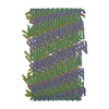
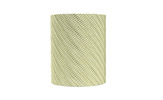
| Title | Structure of the smaller diameter PSMalpha3 nanotubes | |||||||||
 Map data Map data | ||||||||||
 Sample Sample |
| |||||||||
 Keywords Keywords | nanotubes / amyloid / cross-alpha / PROTEIN FIBRIL | |||||||||
| Function / homology | : / Phenol-soluble modulin alpha peptide / Phenol-soluble modulin alpha peptide family / killing of cells of another organism / Phenol-soluble modulin alpha 3 peptide Function and homology information Function and homology information | |||||||||
| Biological species |  | |||||||||
| Method | helical reconstruction / cryo EM / Resolution: 3.9 Å | |||||||||
 Authors Authors | Beltran LC / Kreutzberger MA | |||||||||
| Funding support |  United States, 2 items United States, 2 items
| |||||||||
 Citation Citation |  Journal: Proc Natl Acad Sci U S A / Year: 2022 Journal: Proc Natl Acad Sci U S A / Year: 2022Title: Phenol-soluble modulins PSMα3 and PSMβ2 form nanotubes that are cross-α amyloids. Authors: Mark A B Kreutzberger / Shengyuan Wang / Leticia C Beltran / Abraham Tuachi / Xiaobing Zuo / Edward H Egelman / Vincent P Conticello /  Abstract: Phenol-soluble modulins (PSMs) are peptide-based virulence factors that play significant roles in the pathogenesis of staphylococcal strains in community-associated and hospital-associated infections. ...Phenol-soluble modulins (PSMs) are peptide-based virulence factors that play significant roles in the pathogenesis of staphylococcal strains in community-associated and hospital-associated infections. In addition to cytotoxicity, PSMs display the propensity to self-assemble into fibrillar species, which may be mediated through the formation of amphipathic conformations. Here, we analyze the self-assembly behavior of two PSMs, PSMα3 and PSMβ2, which are derived from peptides expressed by methicillin-resistant Staphylococcus aureus (MRSA), a significant human pathogen. In both cases, we observed the formation of a mixture of self-assembled species including twisted filaments, helical ribbons, and nanotubes, which can reversibly interconvert in vitro. Cryo–electron microscopy structural analysis of three PSM nanotubes, two derived from PSMα3 and one from PSMβ2, revealed that the assemblies displayed remarkably similar structures based on lateral association of cross-α amyloid protofilaments. The amphipathic helical conformations of PSMα3 and PSMβ2 enforced a bilayer arrangement within the protofilaments that defined the structures of the respective PSMα3 and PSMβ2 nanotubes. We demonstrate that, similar to amyloids based on cross-β protofilaments, cross-α amyloids derived from these PSMs display polymorphism, not only in terms of the global morphology (e.g., twisted filament, helical ribbon, and nanotube) but also with respect to the number of protofilaments within a given peptide assembly. These results suggest that the folding landscape of PSM derivatives may be more complex than originally anticipated and that the assemblies are able to sample a wide range of supramolecular structural space. | |||||||||
| History |
|
- Structure visualization
| Supplemental images |
|---|
- Downloads & links
-EMDB archive
| Map data |  emd_25573.map.gz emd_25573.map.gz | 86.7 MB |  EMDB map data format EMDB map data format | |
|---|---|---|---|---|
| Header (meta data) |  emd-25573-v30.xml emd-25573-v30.xml emd-25573.xml emd-25573.xml | 9.2 KB 9.2 KB | Display Display |  EMDB header EMDB header |
| Images |  emd_25573.png emd_25573.png | 66.6 KB | ||
| Filedesc metadata |  emd-25573.cif.gz emd-25573.cif.gz | 4.3 KB | ||
| Archive directory |  http://ftp.pdbj.org/pub/emdb/structures/EMD-25573 http://ftp.pdbj.org/pub/emdb/structures/EMD-25573 ftp://ftp.pdbj.org/pub/emdb/structures/EMD-25573 ftp://ftp.pdbj.org/pub/emdb/structures/EMD-25573 | HTTPS FTP |
-Validation report
| Summary document |  emd_25573_validation.pdf.gz emd_25573_validation.pdf.gz | 663.9 KB | Display |  EMDB validaton report EMDB validaton report |
|---|---|---|---|---|
| Full document |  emd_25573_full_validation.pdf.gz emd_25573_full_validation.pdf.gz | 663.4 KB | Display | |
| Data in XML |  emd_25573_validation.xml.gz emd_25573_validation.xml.gz | 7.4 KB | Display | |
| Data in CIF |  emd_25573_validation.cif.gz emd_25573_validation.cif.gz | 8.4 KB | Display | |
| Arichive directory |  https://ftp.pdbj.org/pub/emdb/validation_reports/EMD-25573 https://ftp.pdbj.org/pub/emdb/validation_reports/EMD-25573 ftp://ftp.pdbj.org/pub/emdb/validation_reports/EMD-25573 ftp://ftp.pdbj.org/pub/emdb/validation_reports/EMD-25573 | HTTPS FTP |
-Related structure data
| Related structure data |  7szzMC  7t0xC  7t8uC M: atomic model generated by this map C: citing same article ( |
|---|---|
| Similar structure data | Similarity search - Function & homology  F&H Search F&H Search |
- Links
Links
| EMDB pages |  EMDB (EBI/PDBe) / EMDB (EBI/PDBe) /  EMDataResource EMDataResource |
|---|
- Map
Map
| File |  Download / File: emd_25573.map.gz / Format: CCP4 / Size: 282.6 MB / Type: IMAGE STORED AS FLOATING POINT NUMBER (4 BYTES) Download / File: emd_25573.map.gz / Format: CCP4 / Size: 282.6 MB / Type: IMAGE STORED AS FLOATING POINT NUMBER (4 BYTES) | ||||||||||||||||||||||||||||||||||||
|---|---|---|---|---|---|---|---|---|---|---|---|---|---|---|---|---|---|---|---|---|---|---|---|---|---|---|---|---|---|---|---|---|---|---|---|---|---|
| Projections & slices | Image control
Images are generated by Spider. | ||||||||||||||||||||||||||||||||||||
| Voxel size | X=Y=Z: 1.08 Å | ||||||||||||||||||||||||||||||||||||
| Density |
| ||||||||||||||||||||||||||||||||||||
| Symmetry | Space group: 1 | ||||||||||||||||||||||||||||||||||||
| Details | EMDB XML:
|
-Supplemental data
- Sample components
-Entire : Nanotube assembly of PSMalpha3
| Entire | Name: Nanotube assembly of PSMalpha3 |
|---|---|
| Components |
|
-Supramolecule #1: Nanotube assembly of PSMalpha3
| Supramolecule | Name: Nanotube assembly of PSMalpha3 / type: complex / ID: 1 / Parent: 0 / Macromolecule list: all |
|---|---|
| Source (natural) | Organism:  |
-Macromolecule #1: Phenol-soluble modulin PSM-alpha-3
| Macromolecule | Name: Phenol-soluble modulin PSM-alpha-3 / type: protein_or_peptide / ID: 1 / Number of copies: 4 / Enantiomer: LEVO |
|---|---|
| Source (natural) | Organism:  |
| Molecular weight | Theoretical: 2.611149 KDa |
| Sequence | String: MEFVAKLFKF FKDLLGKFLG NN UniProtKB: Phenol-soluble modulin alpha 3 peptide |
-Experimental details
-Structure determination
| Method | cryo EM |
|---|---|
 Processing Processing | helical reconstruction |
| Aggregation state | filament |
- Sample preparation
| Buffer | pH: 2 |
|---|---|
| Vitrification | Cryogen name: ETHANE |
- Electron microscopy
| Microscope | TFS KRIOS |
|---|---|
| Image recording | Film or detector model: GATAN K3 (6k x 4k) / Average electron dose: 50.0 e/Å2 |
| Electron beam | Acceleration voltage: 300 kV / Electron source:  FIELD EMISSION GUN FIELD EMISSION GUN |
| Electron optics | Illumination mode: FLOOD BEAM / Imaging mode: BRIGHT FIELD |
| Experimental equipment |  Model: Titan Krios / Image courtesy: FEI Company |
- Image processing
| Final reconstruction | Applied symmetry - Helical parameters - Δz: 0.63 Å Applied symmetry - Helical parameters - Δ&Phi: 138.3 ° Applied symmetry - Helical parameters - Axial symmetry: C1 (asymmetric) Resolution.type: BY AUTHOR / Resolution: 3.9 Å / Resolution method: FSC 0.143 CUT-OFF / Number images used: 48684 |
|---|---|
| Startup model | Type of model: PDB ENTRY PDB model - PDB ID: |
| Final angle assignment | Type: NOT APPLICABLE |
 Movie
Movie Controller


 Z (Sec.)
 Y (Row.)
 X (Col.)


