 Movie
Movie Controller

[English] 日本語
 Yorodumi
- PDB-5i55: Crystal Structure of the Virulent PSM-alpha3 Peptide Forming a Cr... -
+ Open data

- Basic information
| Entry | Database: PDB / ID: 5i55 | ||||||||||||
|---|---|---|---|---|---|---|---|---|---|---|---|---|---|
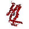
| Title | Crystal Structure of the Virulent PSM-alpha3 Peptide Forming a Cross-alpha amyloid-like Fibril | ||||||||||||
 Components Components | Psm alpha-3 | ||||||||||||
 Keywords Keywords | PROTEIN FIBRIL / THE CROSS-ALPHA AMYLOID-LIKE FOLD IS COMPOSED OF MATING ALPHA-HELICAL SHEETS | ||||||||||||
| Function / homology | : / Phenol-soluble modulin alpha peptide / Phenol-soluble modulin alpha peptide family / killing of cells of another organism / ACETATE ION / Phenol-soluble modulin alpha 3 peptide / Phenol-soluble modulin alpha 3 peptide Function and homology information Function and homology information | ||||||||||||
| Biological species |   Staphylococcus aureus (bacteria) Staphylococcus aureus (bacteria) | ||||||||||||
| Method |  X-RAY DIFFRACTION / SYNCHROTRON / AB INITIO PHASING / Resolution: 1.45 Å X-RAY DIFFRACTION / SYNCHROTRON / AB INITIO PHASING / Resolution: 1.45 Å | ||||||||||||
| Model details | Phenol Soluble Modulin | ||||||||||||
 Authors Authors | Landau, M. / Moshe, A. / Tayeb-Fligelman, E. / Sawaya, M.R. / Coquelle, N. / Colletier, J.-P. | ||||||||||||
| Funding support |  United States, United States,  Israel, 3items Israel, 3items
| ||||||||||||
 Citation Citation | Journal: Science / Year: 2017 Title: The cytotoxic Staphylococcus aureus PSM alpha 3 reveals a cross-alpha amyloid-like fibril. Authors: Tayeb-Fligelman, E. / Tabachnikov, O. / Moshe, A. / Goldshmidt-Tran, O. / Sawaya, M.R. / Coquelle, N. / Colletier, J.P. / Landau, M. | ||||||||||||
| History |
|
- Structure visualization
| Structure viewer | Molecule: MolmilJmol/JSmol |
|---|
- Downloads & links
-Download
| PDBx/mmCIF format | 5i55.cif.gz | 17.6 KB | Display | PDBx/mmCIF format |
|---|---|---|---|---|
| PDB format | pdb5i55.ent.gz | 9.1 KB | Display | PDB format |
| PDBx/mmJSON format | 5i55.json.gz | Tree view | PDBx/mmJSON format | |
| Others |  Other downloads Other downloads |
-Validation report
| Arichive directory | https://data.pdbj.org/pub/pdb/validation_reports/i5/5i55ftp://data.pdbj.org/pub/pdb/validation_reports/i5/5i55 | HTTPS FTP |
|---|
-Related structure data
| Similar structure data |
|---|


-Links
 PDBj
PDBj- Assembly
| Deposited unit | 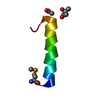
| ||||||||
|---|---|---|---|---|---|---|---|---|---|
| 1 | x 20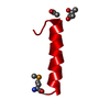
| ||||||||
| Unit cell |
|
-Components
| #1: Protein/peptide | Mass: 2658.044 Da / Num. of mol.: 1 / Source method: obtained synthetically / Details: PSM-alpha3 from S. aureus, synthesized / Source: (synth.) Staphylococcus aureus (bacteria) / References: UniProt: H9BRQ7, UniProt: P0C805*PLUS |
|---|---|
| #2: Chemical | ChemComp-MPD / (  Mass: 118.174 Da / Num. of mol.: 1 / Source method: obtained synthetically / Formula: C6H14O2 / Comment: precipitant*YM Mass: 118.174 Da / Num. of mol.: 1 / Source method: obtained synthetically / Formula: C6H14O2 / Comment: precipitant*YM |
| #3: Chemical | ChemComp-ACT /   Mass: 59.044 Da / Num. of mol.: 1 / Source method: obtained synthetically / Formula: C2H3O2 Mass: 59.044 Da / Num. of mol.: 1 / Source method: obtained synthetically / Formula: C2H3O2 |
| #4: Water | ChemComp-HOH /  Mass: 18.015 Da / Num. of mol.: 12 / Source method: isolated from a natural source / Formula: H2O Mass: 18.015 Da / Num. of mol.: 12 / Source method: isolated from a natural source / Formula: H2O |
| Has protein modification | Y |
-Experimental details
-Experiment
| Experiment | Method: X-RAY DIFFRACTION / Number of used crystals: 1 |
|---|
- Sample preparation
| Crystal | Density Matthews: 1.61 Å3/Da / Density % sol: 23.48 % / Description: Needle-shaped microcrystal |
|---|---|
| Crystal grow | Temperature: 293 K / Method: vapor diffusion, hanging drop Details: reservoir contained 0.2 M ammonium acetate, 0.1M BisTris pH 5.5, 45% MPD |
-Data collection
| Diffraction | Mean temperature: 100 K | |||||||||||||||||||||||||||||||||||||||||||||||||||||||||||||||||||||||||||||||||||||||||||||||||||||||||||||||||||||||||||||||||||||||||||||||||||||||||||||||||||||||||||||||||||||||||||||||||||||||||||||||||||||||||||||||||||||||
|---|---|---|---|---|---|---|---|---|---|---|---|---|---|---|---|---|---|---|---|---|---|---|---|---|---|---|---|---|---|---|---|---|---|---|---|---|---|---|---|---|---|---|---|---|---|---|---|---|---|---|---|---|---|---|---|---|---|---|---|---|---|---|---|---|---|---|---|---|---|---|---|---|---|---|---|---|---|---|---|---|---|---|---|---|---|---|---|---|---|---|---|---|---|---|---|---|---|---|---|---|---|---|---|---|---|---|---|---|---|---|---|---|---|---|---|---|---|---|---|---|---|---|---|---|---|---|---|---|---|---|---|---|---|---|---|---|---|---|---|---|---|---|---|---|---|---|---|---|---|---|---|---|---|---|---|---|---|---|---|---|---|---|---|---|---|---|---|---|---|---|---|---|---|---|---|---|---|---|---|---|---|---|---|---|---|---|---|---|---|---|---|---|---|---|---|---|---|---|---|---|---|---|---|---|---|---|---|---|---|---|---|---|---|---|---|---|---|---|---|---|---|---|---|---|---|---|---|---|---|---|---|---|
| Diffraction source | Source: SYNCHROTRON / Site: APS / Beamline: 24-ID-C / Wavelength: 0.9792,0.9794,0.9796 | |||||||||||||||||||||||||||||||||||||||||||||||||||||||||||||||||||||||||||||||||||||||||||||||||||||||||||||||||||||||||||||||||||||||||||||||||||||||||||||||||||||||||||||||||||||||||||||||||||||||||||||||||||||||||||||||||||||||
| Detector | Type: DECTRIS PILATUS 6M-F / Detector: PIXEL / Date: Apr 18, 2015 | |||||||||||||||||||||||||||||||||||||||||||||||||||||||||||||||||||||||||||||||||||||||||||||||||||||||||||||||||||||||||||||||||||||||||||||||||||||||||||||||||||||||||||||||||||||||||||||||||||||||||||||||||||||||||||||||||||||||
| Radiation | Protocol: MAD / Monochromatic (M) / Laue (L): M / Scattering type: x-ray | |||||||||||||||||||||||||||||||||||||||||||||||||||||||||||||||||||||||||||||||||||||||||||||||||||||||||||||||||||||||||||||||||||||||||||||||||||||||||||||||||||||||||||||||||||||||||||||||||||||||||||||||||||||||||||||||||||||||
| Radiation wavelength |
| |||||||||||||||||||||||||||||||||||||||||||||||||||||||||||||||||||||||||||||||||||||||||||||||||||||||||||||||||||||||||||||||||||||||||||||||||||||||||||||||||||||||||||||||||||||||||||||||||||||||||||||||||||||||||||||||||||||||
| Reflection | Resolution: 1.45→16.55 Å / Num. obs: 3126 / % possible obs: 96.2 % / Observed criterion σ(I): -3 / Redundancy: 18.97 % / Biso Wilson estimate: 18.998 Å2 / CC1/2: 0.998 / Rmerge(I) obs: 0.124 / Rrim(I) all: 0.13 / Χ2: 1.019 / Net I/σ(I): 10.01 / Num. measured all: 59309 | |||||||||||||||||||||||||||||||||||||||||||||||||||||||||||||||||||||||||||||||||||||||||||||||||||||||||||||||||||||||||||||||||||||||||||||||||||||||||||||||||||||||||||||||||||||||||||||||||||||||||||||||||||||||||||||||||||||||
| Reflection shell | Diffraction-ID: 1 / Rejects: _
|
- Processing
| Software |
| |||||||||||||||||||||||||||||||||||||||||||||||||||||||
|---|---|---|---|---|---|---|---|---|---|---|---|---|---|---|---|---|---|---|---|---|---|---|---|---|---|---|---|---|---|---|---|---|---|---|---|---|---|---|---|---|---|---|---|---|---|---|---|---|---|---|---|---|---|---|---|---|
| Refinement | Method to determine structure: AB INITIO PHASING / Resolution: 1.45→16.55 Å / Cor.coef. Fo:Fc: 0.983 / Cor.coef. Fo:Fc free: 0.967 / WRfactor Rfree: 0.1785 / WRfactor Rwork: 0.1412 / FOM work R set: 0.8277 / SU B: 1.744 / SU ML: 0.059 / SU R Cruickshank DPI: 0.0727 / SU Rfree: 0.0777 / Cross valid method: THROUGHOUT / σ(F): 0 / ESU R: 0.073 / ESU R Free: 0.078 / Stereochemistry target values: MAXIMUM LIKELIHOOD Details: HYDROGENS HAVE BEEN ADDED IN THE RIDING POSITIONS U VALUES : REFINED INDIVIDUALLY
| |||||||||||||||||||||||||||||||||||||||||||||||||||||||
| Solvent computation | Ion probe radii: 0.8 Å / Shrinkage radii: 0.8 Å / VDW probe radii: 1.2 Å / Solvent model: MASK | |||||||||||||||||||||||||||||||||||||||||||||||||||||||
| Displacement parameters | Biso max: 53.03 Å2 / Biso mean: 16.958 Å2 / Biso min: 10.85 Å2
| |||||||||||||||||||||||||||||||||||||||||||||||||||||||
| Refinement step | Cycle: final / Resolution: 1.45→16.55 Å
| |||||||||||||||||||||||||||||||||||||||||||||||||||||||
| Refine LS restraints |
| |||||||||||||||||||||||||||||||||||||||||||||||||||||||
| LS refinement shell | Resolution: 1.45→1.487 Å / Total num. of bins used: 20
|
