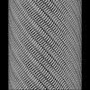
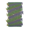
PROTEIN FIBRIL / nanotubes / amyloid / cross-alpha
Function / homology
Phenol-soluble modulin beta protein / Phenol-soluble modulin beta protein / toxin activity / killing of cells of another organism / extracellular region / Antibacterial protein
Function and homology information
Biological species
Staphylococcus aureus (bacteria)
Method
ELECTRON MICROSCOPY / helical reconstruction / cryo EM / Resolution: 4.3 Å
National Institutes of Health/National Institute of General Medical Sciences (NIH/NIGMS)
GM122510
United States
Citation
Journal: Proc Natl Acad Sci U S A / Year: 2022 Title: Phenol-soluble modulins PSMα3 and PSMβ2 form nanotubes that are cross-α amyloids. Authors: Mark A B Kreutzberger / Shengyuan Wang / Leticia C Beltran / Abraham Tuachi / Xiaobing Zuo / Edward H Egelman / Vincent P Conticello / Abstract: Phenol-soluble modulins (PSMs) are peptide-based virulence factors that play significant roles in the pathogenesis of staphylococcal strains in community-associated and hospital-associated infections. ...Phenol-soluble modulins (PSMs) are peptide-based virulence factors that play significant roles in the pathogenesis of staphylococcal strains in community-associated and hospital-associated infections. In addition to cytotoxicity, PSMs display the propensity to self-assemble into fibrillar species, which may be mediated through the formation of amphipathic conformations. Here, we analyze the self-assembly behavior of two PSMs, PSMα3 and PSMβ2, which are derived from peptides expressed by methicillin-resistant Staphylococcus aureus (MRSA), a significant human pathogen. In both cases, we observed the formation of a mixture of self-assembled species including twisted filaments, helical ribbons, and nanotubes, which can reversibly interconvert in vitro. Cryo–electron microscopy structural analysis of three PSM nanotubes, two derived from PSMα3 and one from PSMβ2, revealed that the assemblies displayed remarkably similar structures based on lateral association of cross-α amyloid protofilaments. The amphipathic helical conformations of PSMα3 and PSMβ2 enforced a bilayer arrangement within the protofilaments that defined the structures of the respective PSMα3 and PSMβ2 nanotubes. We demonstrate that, similar to amyloids based on cross-β protofilaments, cross-α amyloids derived from these PSMs display polymorphism, not only in terms of the global morphology (e.g., twisted filament, helical ribbon, and nanotube) but also with respect to the number of protofilaments within a given peptide assembly. These results suggest that the folding landscape of PSM derivatives may be more complex than originally anticipated and that the assemblies are able to sample a wide range of supramolecular structural space.
Helical symmetry: (Circular symmetry: 6 / Dyad axis: no / N subunits divisor: 1 / Num. of operations: 120 / Rise per n subunits: 8.3 Å / Rotation per n subunits: 28.6 °)
-
Components
#1: Protein/peptide
Antibacterialprotein / Antibacterial protein 3 / Antibacterial protein 3 homolog / Psm beta2 / Uncharacterized protein
In the structure databanks used in Yorodumi, some data are registered as the other names, "COVID-19 virus" and "2019-nCoV". Here are the details of the virus and the list of structure data.
Jan 31, 2019. EMDB accession codes are about to change! (news from PDBe EMDB page)
EMDB accession codes are about to change! (news from PDBe EMDB page)
The allocation of 4 digits for EMDB accession codes will soon come to an end. Whilst these codes will remain in use, new EMDB accession codes will include an additional digit and will expand incrementally as the available range of codes is exhausted. The current 4-digit format prefixed with “EMD-” (i.e. EMD-XXXX) will advance to a 5-digit format (i.e. EMD-XXXXX), and so on. It is currently estimated that the 4-digit codes will be depleted around Spring 2019, at which point the 5-digit format will come into force.
The EM Navigator/Yorodumi systems omit the EMD- prefix.
Related info.:Q: What is EMD? / ID/Accession-code notation in Yorodumi/EM Navigator
Yorodumi is a browser for structure data from EMDB, PDB, SASBDB, etc.
This page is also the successor to EM Navigator detail page, and also detail information page/front-end page for Omokage search.
The word "yorodu" (or yorozu) is an old Japanese word meaning "ten thousand". "mi" (miru) is to see.
Related info.:EMDB / PDB / SASBDB / Comparison of 3 databanks / Yorodumi Search / Aug 31, 2016. New EM Navigator & Yorodumi / Yorodumi Papers / Jmol/JSmol / Function and homology information / Changes in new EM Navigator and Yorodumi
 Movie
Movie Controller


 Open data
 Basic information
 Components
 Keywords
 Function and homology information
 Staphylococcus aureus (bacteria)
 Authors
 United States, 2items
  Citation
 Structure visualization
 Downloads & links
 Other downloads


 PDBj
PDBj
 Assembly
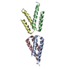
 Sample preparation
 Electron microscopy imaging
 FIELD EMISSION GUN / Accelerating voltage: 300 kV / Illumination mode: FLOOD BEAM
 Processing