National Institutes of Health/National Institute Of Allergy and Infectious Diseases (NIH/NIAID)
R01 AI136680
United States
National Institutes of Health/National Institute Of Allergy and Infectious Diseases (NIH/NIAID)
AI27690
United States
National Institutes of Health/National Institute Of Allergy and Infectious Diseases (NIH/NIAID)
U54AI150472
United States
Citation
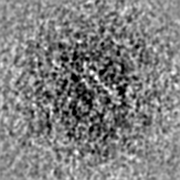
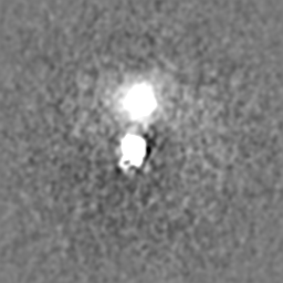
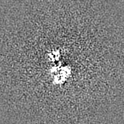
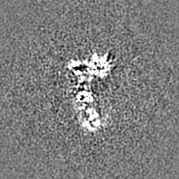
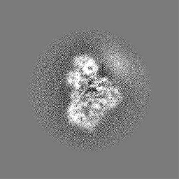
Journal: Sci Adv / Year: 2022 Title: Cryo-EM structure of the HIV-1 Pol polyprotein provides insights into virion maturation. Authors: Jerry Joe E K Harrison / Dario Oliveira Passos / Jessica F Bruhn / Joseph D Bauman / Lynda Tuberty / Jeffrey J DeStefano / Francesc Xavier Ruiz / Dmitry Lyumkis / Eddy Arnold / Abstract: Key proteins of retroviruses and other RNA viruses are translated and subsequently processed from polyprotein precursors by the viral protease (PR). Processing of the HIV Gag-Pol polyprotein yields ...Key proteins of retroviruses and other RNA viruses are translated and subsequently processed from polyprotein precursors by the viral protease (PR). Processing of the HIV Gag-Pol polyprotein yields the HIV structural proteins and enzymes. Structures of the mature enzymes PR, reverse transcriptase (RT), and integrase (IN) aided understanding of catalysis and design of antiretrovirals, but knowledge of the Pol precursor architecture and function before PR cleavage is limited. We developed a system to produce stable HIV-1 Pol and determined its cryo-electron microscopy structure. RT in Pol has a similar arrangement to the mature RT heterodimer, and its dimerization may draw together two PR monomers to activate proteolytic processing. HIV-1 thus may leverage the dimerization interfaces in Pol to regulate assembly and maturation of polyprotein precursors.
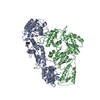
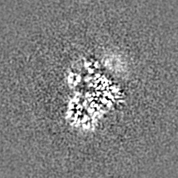
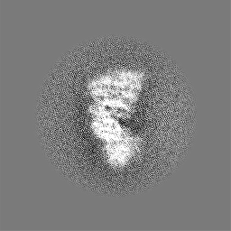
Name: PR-RT portion of HIV-1 Pol / type: complex / ID: 1 / Parent: 0 / Macromolecule list: all Details: 3D reconstruction of the HIV-1 Pol polyprotein comprising the PR-RT portion
Source (natural)
Organism: Human immunodeficiency virus type 1 BH10
Molecular weight
Theoretical: 130 KDa
-
Macromolecule #1: Gag-Pol polyprotein
Macromolecule
Name: Gag-Pol polyprotein / type: protein_or_peptide / ID: 1 / Number of copies: 2 / Enantiomer: LEVO
Source (natural)
Organism: Human immunodeficiency virus type 1 group M subtype B (isolate BH10) Strain: isolate BH10
In the structure databanks used in Yorodumi, some data are registered as the other names, "COVID-19 virus" and "2019-nCoV". Here are the details of the virus and the list of structure data.
Jan 31, 2019. EMDB accession codes are about to change! (news from PDBe EMDB page)
EMDB accession codes are about to change! (news from PDBe EMDB page)
The allocation of 4 digits for EMDB accession codes will soon come to an end. Whilst these codes will remain in use, new EMDB accession codes will include an additional digit and will expand incrementally as the available range of codes is exhausted. The current 4-digit format prefixed with “EMD-” (i.e. EMD-XXXX) will advance to a 5-digit format (i.e. EMD-XXXXX), and so on. It is currently estimated that the 4-digit codes will be depleted around Spring 2019, at which point the 5-digit format will come into force.
The EM Navigator/Yorodumi systems omit the EMD- prefix.
Related info.:Q: What is EMD? / ID/Accession-code notation in Yorodumi/EM Navigator
Yorodumi is a browser for structure data from EMDB, PDB, SASBDB, etc.
This page is also the successor to EM Navigator detail page, and also detail information page/front-end page for Omokage search.
The word "yorodu" (or yorozu) is an old Japanese word meaning "ten thousand". "mi" (miru) is to see.
Related info.:EMDB / PDB / SASBDB / Comparison of 3 databanks / Yorodumi Search / Aug 31, 2016. New EM Navigator & Yorodumi / Yorodumi Papers / Jmol/JSmol / Function and homology information / Changes in new EM Navigator and Yorodumi
 Movie
Movie Controller

 Yorodumi
 Open data
 Basic information
 Map data
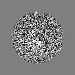
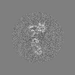
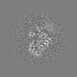
 Sample
 Keywords
 Function and homology information
 Human immunodeficiency virus type 1 BH10 /
  Authors
 United States, 3 items
  Citation
 Structure visualization
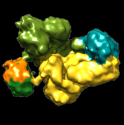
 Downloads & links
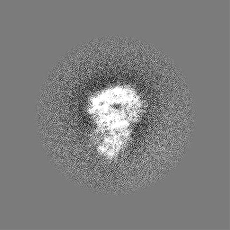
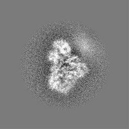
 emd_25165.png
 http://ftp.pdbj.org/pub/emdb/structures/EMD-25165


 Z (Sec.)
 Y (Row.)
 X (Col.)


 Sample components
 Processing
 Electron microscopy
 FIELD EMISSION GUN