Movie
Movie Controller

[English] 日本語
 Yorodumi
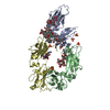
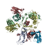
- EMDB-25136: Structure of a partially disrupted IgE high affinity receptor com... -
+ Open data

- Basic information
| Entry | Database: EMDB / ID: EMD-25136 | |||||||||
|---|---|---|---|---|---|---|---|---|---|---|
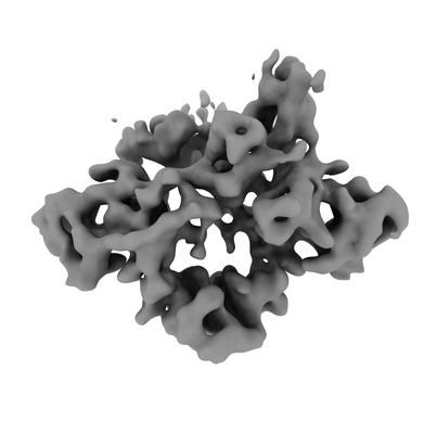
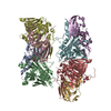
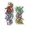
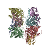
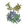
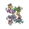
| Title | Structure of a partially disrupted IgE high affinity receptor complex bound to an omalizumab variant | |||||||||
 Map data Map data | ||||||||||
 Sample Sample |
| |||||||||
 Keywords Keywords | IgE / allergy / Xolair / Omalizumab / IMMUNE SYSTEM | |||||||||
| Function / homology |  Function and homology information Function and homology informationhigh-affinity IgE receptor activity / mononuclear cell differentiation / IgE B cell receptor complex / adaptive immune memory response / primary adaptive immune response / B cell antigen processing and presentation / type I hypersensitivity / Fc receptor-mediated immune complex endocytosis / eosinophil degranulation / IgE immunoglobulin complex ...high-affinity IgE receptor activity / mononuclear cell differentiation / IgE B cell receptor complex / adaptive immune memory response / primary adaptive immune response / B cell antigen processing and presentation / type I hypersensitivity / Fc receptor-mediated immune complex endocytosis / eosinophil degranulation / IgE immunoglobulin complex / macrophage activation / IgE binding / antibody-dependent cellular cytotoxicity / Fc epsilon receptor (FCERI) signaling / type 2 immune response / immunoglobulin complex, circulating / immunoglobulin receptor binding / mast cell degranulation / immunoglobulin mediated immune response / B cell proliferation / macrophage differentiation / B cell activation / Role of LAT2/NTAL/LAB on calcium mobilization / complement activation, classical pathway / antigen binding / FCERI mediated Ca+2 mobilization / B cell receptor signaling pathway / FCERI mediated MAPK activation / FCERI mediated NF-kB activation / antibacterial humoral response / Interleukin-4 and Interleukin-13 signaling / adaptive immune response / cell surface receptor signaling pathway / immune response / inflammatory response / external side of plasma membrane / cell surface / : / extracellular region / plasma membrane Similarity search - Function | |||||||||
| Biological species |  Homo sapiens (human) Homo sapiens (human) | |||||||||
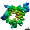
| Method | single particle reconstruction / cryo EM / Resolution: 7.29 Å | |||||||||
 Authors Authors | Pennington LF / Jardetzky TS | |||||||||
| Funding support |  United States, 2 items United States, 2 items
| |||||||||
 Citation Citation | Journal: Nat Commun / Year: 2021 Title: Directed evolution of and structural insights into antibody-mediated disruption of a stable receptor-ligand complex. Authors: Luke F Pennington / Pascal Gasser / Silke Kleinboelting / Chensong Zhang / Georgios Skiniotis / Alexander Eggel / Theodore S Jardetzky /  Abstract: Antibody drugs exert therapeutic effects via a range of mechanisms, including competitive inhibition, allosteric modulation, and immune effector mechanisms. Facilitated dissociation is an additional ...Antibody drugs exert therapeutic effects via a range of mechanisms, including competitive inhibition, allosteric modulation, and immune effector mechanisms. Facilitated dissociation is an additional mechanism where antibody-mediated "disruption" of stable high-affinity macromolecular complexes can potentially enhance therapeutic efficacy. However, this mechanism is not well understood or utilized therapeutically. Here, we investigate and engineer the weak disruptive activity of an existing therapeutic antibody, omalizumab, which targets IgE antibodies to block the allergic response. We develop a yeast display approach to select for and engineer antibody disruptive efficiency and generate potent omalizumab variants that dissociate receptor-bound IgE. We determine a low resolution cryo-EM structure of a transient disruption intermediate containing the IgE-Fc, its partially dissociated receptor and an antibody inhibitor. Our results provide a conceptual framework for engineering disruptive inhibitors for other targets, insights into the failure in clinical trials of the previous high affinity omalizumab HAE variant and anti-IgE antibodies that safely and rapidly disarm allergic effector cells. | |||||||||
| History |
|
- Structure visualization
| Movie |
Movie viewer |
|---|---|
| Structure viewer | EM map: SurfViewMolmilJmol/JSmol |
| Supplemental images |
- Downloads & links
-EMDB archive
| Map data | emd_25136.map.gz | 59.8 MB | EMDB map data format | |
|---|---|---|---|---|
| Header (meta data) | emd-25136-v30.xmlemd-25136.xml | 16.2 KB 16.2 KB | Display Display | EMDB header |
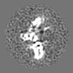
| Images |  emd_25136.png emd_25136.png | 44.3 KB | ||
| Filedesc metadata | emd-25136.cif.gz | 6.7 KB | ||
| Archive directory |  http://ftp.pdbj.org/pub/emdb/structures/EMD-25136ftp://ftp.pdbj.org/pub/emdb/structures/EMD-25136 http://ftp.pdbj.org/pub/emdb/structures/EMD-25136ftp://ftp.pdbj.org/pub/emdb/structures/EMD-25136 | HTTPS FTP |
-Related structure data
| Related structure data |  7shtMC  7shuC  7shyC  7shzC  7si0C M: atomic model generated by this map C: citing same article ( |
|---|---|
| Similar structure data |

-Links
| EMDB pages | EMDB (EBI/PDBe) / EMDataResource |
|---|---|
| Related items in Molecule of the Month |


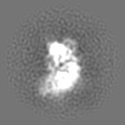
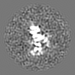
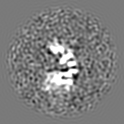
-Map
| File | Download / File: emd_25136.map.gz / Format: CCP4 / Size: 64 MB / Type: IMAGE STORED AS FLOATING POINT NUMBER (4 BYTES) | ||||||||||||||||||||||||||||||||||||||||||||||||||||||||||||
|---|---|---|---|---|---|---|---|---|---|---|---|---|---|---|---|---|---|---|---|---|---|---|---|---|---|---|---|---|---|---|---|---|---|---|---|---|---|---|---|---|---|---|---|---|---|---|---|---|---|---|---|---|---|---|---|---|---|---|---|---|---|
| Projections & slices | Image control
Images are generated by Spider. | ||||||||||||||||||||||||||||||||||||||||||||||||||||||||||||
| Voxel size | X=Y=Z: 0.8521 Å | ||||||||||||||||||||||||||||||||||||||||||||||||||||||||||||
| Density |
| ||||||||||||||||||||||||||||||||||||||||||||||||||||||||||||
| Symmetry | Space group: 1 | ||||||||||||||||||||||||||||||||||||||||||||||||||||||||||||
| Details | EMDB XML:
CCP4 map header:
| ||||||||||||||||||||||||||||||||||||||||||||||||||||||||||||
 Z (Sec.)
 Y (Row.)
 X (Col.)


-Supplemental data
- Sample components
-Entire : Locked complex of IgE-Fc (G335C) and alpha chain of the high affi...
| Entire | Name: Locked complex of IgE-Fc (G335C) and alpha chain of the high affinity IgE receptor (W156C) bound to clone_7 scFV |
|---|---|
| Components |
|
-Supramolecule #1: Locked complex of IgE-Fc (G335C) and alpha chain of the high affi...
| Supramolecule | Name: Locked complex of IgE-Fc (G335C) and alpha chain of the high affinity IgE receptor (W156C) bound to clone_7 scFV type: complex / ID: 1 / Parent: 0 / Macromolecule list: #1-#4 |
|---|---|
| Source (natural) | Organism: Homo sapiens (human) |
-Macromolecule #1: High affinity immunoglobulin epsilon receptor subunit alpha
| Macromolecule | Name: High affinity immunoglobulin epsilon receptor subunit alpha type: protein_or_peptide / ID: 1 / Number of copies: 1 / Enantiomer: LEVO |
|---|---|
| Source (natural) | Organism: Homo sapiens (human) |
| Molecular weight | Theoretical: 22.541799 KDa |
| Recombinant expression | Organism: Homo sapiens (human) |
| Sequence | String: APMAEGGGQN HHHHHHHHGG ENLYFQGGSP KVSLNPPWNR IFKGENVTLT CNGNNFFEVS STKWFHNGSL SEETNSSLNI VNAKFEDSG EYKCQHQQVN ESEPVYLEVF SDWLLLQASA EVVMEGQPLF LRCHGWRNWD VYKVIYYKDG EALKYWYENH N ISITNATV ...String: APMAEGGGQN HHHHHHHHGG ENLYFQGGSP KVSLNPPWNR IFKGENVTLT CNGNNFFEVS STKWFHNGSL SEETNSSLNI VNAKFEDSG EYKCQHQQVN ESEPVYLEVF SDWLLLQASA EVVMEGQPLF LRCHGWRNWD VYKVIYYKDG EALKYWYENH N ISITNATV EDSGTYYCTG KVCQLDYESE PLNITVIKA UniProtKB: High affinity immunoglobulin epsilon receptor subunit alpha |
-Macromolecule #2: Immunoglobulin heavy constant epsilon
| Macromolecule | Name: Immunoglobulin heavy constant epsilon / type: protein_or_peptide / ID: 2 / Number of copies: 2 / Enantiomer: LEVO |
|---|---|
| Source (natural) | Organism: Homo sapiens (human) |
| Molecular weight | Theoretical: 35.77316 KDa |
| Recombinant expression | Organism: Homo sapiens (human) |
| Sequence | String: ASFTPPTVKI LQSSCDGGGH FPPTIQLLCL VSGYTPGTIN ITWLEDGQVM DVDLSTASTT QEGELASTQS ELTLSQKHWL SDRTYTCQV TYQGHTFEDS TKKCADSNPR CVSAYLSRPS PFDLFIRKSP TITCLVVDLA PSKGTVNLTW SRASGKPVNH S TRKEEKQR ...String: ASFTPPTVKI LQSSCDGGGH FPPTIQLLCL VSGYTPGTIN ITWLEDGQVM DVDLSTASTT QEGELASTQS ELTLSQKHWL SDRTYTCQV TYQGHTFEDS TKKCADSNPR CVSAYLSRPS PFDLFIRKSP TITCLVVDLA PSKGTVNLTW SRASGKPVNH S TRKEEKQR NGTLTVTSTL PVGTRDWIEG ETYQCRVTHP HLPRALMRST TKTSGPRAAP EVYAFATPEW PGSRDKRTLA CL IQNFMPE DISVQWLHNE VQLPDARHST TQPRKTKGSG FFVFSRLEVT RAEWEQKDEF ICRAVHEAAS PSQTVQRAVS VNP GK UniProtKB: Immunoglobulin heavy constant epsilon |
-Macromolecule #3: clone_7 Variable fragment heavy chain
| Macromolecule | Name: clone_7 Variable fragment heavy chain / type: protein_or_peptide / ID: 3 / Number of copies: 2 / Enantiomer: LEVO |
|---|---|
| Source (natural) | Organism: Homo sapiens (human) |
| Molecular weight | Theoretical: 13.451814 KDa |
| Recombinant expression | Organism: Homo sapiens (human) |
| Sequence | String: ASEVQLVESG GGLVQPDGSL RLSCAVSGYN ITSGYSWNWI RQTPGKGLEW VASVTYDGST NYNPSVKGRI TISRDGSKNT FYLQMNSLR AEDTAVYYCA KGNNYFGHWH FAVWGQGTLV TVSS |
-Macromolecule #4: clone_7 Variable fragment light chain
| Macromolecule | Name: clone_7 Variable fragment light chain / type: protein_or_peptide / ID: 4 / Number of copies: 2 / Enantiomer: LEVO |
|---|---|
| Source (natural) | Organism: Homo sapiens (human) |
| Molecular weight | Theoretical: 14.641863 KDa |
| Recombinant expression | Organism: Homo sapiens (human) |
| Sequence | String: GSDIQLTQSP SSLSASVGDR VTITCRASKS VDSDGDSYMN WYQQKPGRAP KLLIYAASYL ESGVPSRFSG SGSGTHFTLT ISSLQPEDF ATYYCQQSHE DPYTFGQGTK VEIKGGSENL YFQGGSGHHH HHHHH |
-Macromolecule #8: 2-acetamido-2-deoxy-beta-D-glucopyranose
| Macromolecule | Name: 2-acetamido-2-deoxy-beta-D-glucopyranose / type: ligand / ID: 8 / Number of copies: 3 / Formula: NAG |
|---|---|
| Molecular weight | Theoretical: 221.208 Da |
| Chemical component information |  ChemComp-NAG: |
-Experimental details
-Structure determination
| Method | cryo EM |
|---|---|
 Processing Processing | single particle reconstruction |
| Aggregation state | particle |
-Sample preparation
| Concentration | 4.5 mg/mL |
|---|---|
| Buffer | pH: 7.5 |
| Grid | Model: Quantifoil R1.2/1.3 / Material: GOLD / Mesh: 200 / Support film - Material: CARBON / Support film - topology: HOLEY / Pretreatment - Type: GLOW DISCHARGE |
| Vitrification | Cryogen name: ETHANE |
- Electron microscopy
| Microscope | TFS KRIOS |
|---|---|
| Image recording | Film or detector model: GATAN K3 (6k x 4k) / Number grids imaged: 1 / Number real images: 523 / Average exposure time: 2.0 sec. / Average electron dose: 44.0 e/Å2 |
| Electron beam | Acceleration voltage: 300 kV / Electron source:  FIELD EMISSION GUN FIELD EMISSION GUN |
| Electron optics | Illumination mode: SPOT SCAN / Imaging mode: BRIGHT FIELD |
| Experimental equipment |  Model: Titan Krios / Image courtesy: FEI Company |
-Image processing
| Startup model | Type of model: OTHER Details: Ab initio from preliminary relion models used at template for final refinements in cryoSPARC |
|---|---|
| Final reconstruction | Resolution.type: BY AUTHOR / Resolution: 7.29 Å / Resolution method: FSC 0.143 CUT-OFF / Software - Name: cryoSPARC / Number images used: 24396 |
| Initial angle assignment | Type: MAXIMUM LIKELIHOOD |
| Final angle assignment | Type: MAXIMUM LIKELIHOOD |
-Atomic model buiding 1
| Initial model |
| ||||||||
|---|---|---|---|---|---|---|---|---|---|
| Details | Initial local fitting done in Chimera, followed by auto dock, and a single round of real space refinement. Carbohydrates were automatically built in phenix when possible, and a handful of flagged outliers in geometry were fixed manually in Phenix. | ||||||||
| Refinement | Protocol: FLEXIBLE FIT | ||||||||
| Output model | PDB-7sht: |