[English] 日本語
 Yorodumi
- EMDB-24741: Cryo EM analysis reveals inherent flexibility of authentic murine... -
+ Open data
- Basic information
| Entry | Database: EMDB / ID: EMD-24741 | |||||||||
|---|---|---|---|---|---|---|---|---|---|---|
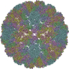
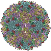
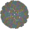
| Title | Cryo EM analysis reveals inherent flexibility of authentic murine papillomavirus capsids | |||||||||
 Map data Map data | Recombined Full Virus Capsid | |||||||||
 Sample Sample |
| |||||||||
 Keywords Keywords | MmuPV1 / papillomavirus / L1 / capsomer / subparticle / VIRUS | |||||||||
| Function / homology |  Function and homology information Function and homology informationT=7 icosahedral viral capsid / endocytosis involved in viral entry into host cell / host cell nucleus / virion attachment to host cell / structural molecule activity Similarity search - Function | |||||||||
| Biological species |  Mus musculus papillomavirus type 1 / Mus musculus papillomavirus type 1 /  Mouse papillomavirus 1 Mouse papillomavirus 1 | |||||||||
| Method | single particle reconstruction / cryo EM / Resolution: 3.3 Å | |||||||||
 Authors Authors | Hartmann SR / Hafenstein S | |||||||||
| Funding support |  United States, 2 items United States, 2 items
| |||||||||
 Citation Citation |  Journal: Viruses / Year: 2021 Journal: Viruses / Year: 2021Title: Cryo EM Analysis Reveals Inherent Flexibility of Authentic Murine Papillomavirus Capsids. Authors: Samantha R Hartmann / Daniel J Goetschius / Jiafen Hu / Joshua J Graff / Carol M Bator / Neil D Christensen / Susan L Hafenstein /  Abstract: Human papillomavirus (HPV) is a significant health burden and leading cause of virus-induced cancers. However, studies have been hampered due to restricted tropism that makes production and ...Human papillomavirus (HPV) is a significant health burden and leading cause of virus-induced cancers. However, studies have been hampered due to restricted tropism that makes production and purification of high titer virus problematic. This issue has been overcome by developing alternative HPV production methods such as virus-like particles (VLPs), which are devoid of a native viral genome. Structural studies have been limited in resolution due to the heterogeneity, fragility, and stability of the VLP capsids. The mouse papillomavirus (MmuPV1) presented here has provided the opportunity to study a native papillomavirus in the context of a common laboratory animal. Using cryo EM to solve the structure of MmuPV1, we achieved 3.3 Å resolution with a local symmetry refinement method that defined smaller, symmetry related subparticles. The resulting high-resolution structure allowed us to build the MmuPV1 asymmetric unit for the first time and identify putative L2 density. We also used our program ISECC to quantify capsid flexibility, which revealed that capsomers move as rigid bodies connected by flexible linkers. The MmuPV1 flexibility was comparable to that of a HPV VLP previously characterized. The resulting MmuPV1 structure is a promising step forward in the study of papillomavirus and will provide a framework for continuing biochemical, genetic, and biophysical research for papillomaviruses. | |||||||||
| History |
|
- Structure visualization
| Movie |
 Movie viewer Movie viewer |
|---|---|
| Structure viewer | EM map:  SurfView SurfView Molmil Molmil Jmol/JSmol Jmol/JSmol |
| Supplemental images |
- Downloads & links
-EMDB archive
| Map data |  emd_24741.map.gz emd_24741.map.gz | 771.7 MB |  EMDB map data format EMDB map data format | |
|---|---|---|---|---|
| Header (meta data) |  emd-24741-v30.xml emd-24741-v30.xml emd-24741.xml emd-24741.xml | 10.8 KB 10.8 KB | Display Display |  EMDB header EMDB header |
| Images |  emd_24741.png emd_24741.png | 113.3 KB | ||
| Filedesc metadata |  emd-24741.cif.gz emd-24741.cif.gz | 5.4 KB | ||
| Archive directory |  http://ftp.pdbj.org/pub/emdb/structures/EMD-24741 http://ftp.pdbj.org/pub/emdb/structures/EMD-24741 ftp://ftp.pdbj.org/pub/emdb/structures/EMD-24741 ftp://ftp.pdbj.org/pub/emdb/structures/EMD-24741 | HTTPS FTP |
-Related structure data
| Related structure data |  7ryjMC M: atomic model generated by this map C: citing same article ( |
|---|---|
| Similar structure data |
- Links
Links
| EMDB pages |  EMDB (EBI/PDBe) / EMDB (EBI/PDBe) /  EMDataResource EMDataResource |
|---|---|
| Related items in Molecule of the Month |
- Map
Map
| File |  Download / File: emd_24741.map.gz / Format: CCP4 / Size: 824 MB / Type: IMAGE STORED AS FLOATING POINT NUMBER (4 BYTES) Download / File: emd_24741.map.gz / Format: CCP4 / Size: 824 MB / Type: IMAGE STORED AS FLOATING POINT NUMBER (4 BYTES) | ||||||||||||||||||||||||||||||||||||||||||||||||||||||||||||||||||||
|---|---|---|---|---|---|---|---|---|---|---|---|---|---|---|---|---|---|---|---|---|---|---|---|---|---|---|---|---|---|---|---|---|---|---|---|---|---|---|---|---|---|---|---|---|---|---|---|---|---|---|---|---|---|---|---|---|---|---|---|---|---|---|---|---|---|---|---|---|---|
| Annotation | Recombined Full Virus Capsid | ||||||||||||||||||||||||||||||||||||||||||||||||||||||||||||||||||||
| Projections & slices | Image control
Images are generated by Spider. | ||||||||||||||||||||||||||||||||||||||||||||||||||||||||||||||||||||
| Voxel size | X=Y=Z: 1.1 Å | ||||||||||||||||||||||||||||||||||||||||||||||||||||||||||||||||||||
| Density |
| ||||||||||||||||||||||||||||||||||||||||||||||||||||||||||||||||||||
| Symmetry | Space group: 1 | ||||||||||||||||||||||||||||||||||||||||||||||||||||||||||||||||||||
| Details | EMDB XML:
CCP4 map header:
| ||||||||||||||||||||||||||||||||||||||||||||||||||||||||||||||||||||
-Supplemental data
- Sample components
-Entire : Mouse papillomavirus 1
| Entire | Name:  Mouse papillomavirus 1 Mouse papillomavirus 1 |
|---|---|
| Components |
|
-Supramolecule #1: Mouse papillomavirus 1
| Supramolecule | Name: Mouse papillomavirus 1 / type: virus / ID: 1 / Parent: 0 / Macromolecule list: all / NCBI-ID: 2171376 / Sci species name: Mouse papillomavirus 1 / Virus type: VIRION / Virus isolate: STRAIN / Virus enveloped: No / Virus empty: No |
|---|---|
| Host (natural) | Organism:  |
-Macromolecule #1: Major capsid protein L1
| Macromolecule | Name: Major capsid protein L1 / type: protein_or_peptide / ID: 1 / Number of copies: 6 / Enantiomer: LEVO |
|---|---|
| Source (natural) | Organism:  Mus musculus papillomavirus type 1 Mus musculus papillomavirus type 1 |
| Molecular weight | Theoretical: 57.623543 KDa |
| Sequence | String: MAMWTPQTGK LYLPPTTPVA KVQSTDEYVY PTSLFCHAHT DRLLTVGHPF FSVIDNDKVT VPKVSGNQYR VFRLKFPDPN KFALPQKDF YDPEKERLVW RLRGLEIGRG GPLGIGTTGH PLFNKLGDTE NPNKYQQGSK DNRQNTSMDP KQTQLFIVGC E PPTGEHWD ...String: MAMWTPQTGK LYLPPTTPVA KVQSTDEYVY PTSLFCHAHT DRLLTVGHPF FSVIDNDKVT VPKVSGNQYR VFRLKFPDPN KFALPQKDF YDPEKERLVW RLRGLEIGRG GPLGIGTTGH PLFNKLGDTE NPNKYQQGSK DNRQNTSMDP KQTQLFIVGC E PPTGEHWD VAKPCGALEK GDCPPIQLVN SVIEDGDMCD IGFGNMNFKE LQQDRSGVPL DIVSTRCKWP DFLKMTNEAY GD KMFFFGR REQVYARHFF TRNGSVGEPI PNSVSPSDFY YAPDSTQDQK TLAPSVYFGT PSGSLVSSDG QLFNRPFWLQ RAQ GNNNGV CWHNELFVTV VDNTRNTNFT ISQQTNTPNP DTYDSTNFKN YLRHVEQFEL SLIAQLCKVP LDPGVLAHIN TMNP TILEN WNLGFVPPPQ QSISDDYRYI TSSATRCPDQ NPPKEREDPY KGLIFWEVDL TERFSQDLDQ FALGRKFLYQ AGIRT AVTG RGVKRAASTT SASSRRVVKR KRGSK UniProtKB: Major capsid protein L1 |
-Experimental details
-Structure determination
| Method | cryo EM |
|---|---|
 Processing Processing | single particle reconstruction |
| Aggregation state | particle |
- Sample preparation
| Buffer | pH: 7.4 / Component - Concentration: 1.0 X / Component - Name: PBS |
|---|---|
| Grid | Model: Quantifoil R2/1 / Material: COPPER / Pretreatment - Type: GLOW DISCHARGE |
| Vitrification | Cryogen name: ETHANE / Chamber humidity: 100 % / Chamber temperature: 277 K / Instrument: FEI VITROBOT MARK IV |
- Electron microscopy
| Microscope | FEI TITAN KRIOS |
|---|---|
| Image recording | Film or detector model: FEI FALCON III (4k x 4k) / Detector mode: COUNTING / Number grids imaged: 1 / Number real images: 2859 / Average electron dose: 45.0 e/Å2 |
| Electron beam | Acceleration voltage: 300 kV / Electron source:  FIELD EMISSION GUN FIELD EMISSION GUN |
| Electron optics | Illumination mode: FLOOD BEAM / Imaging mode: BRIGHT FIELD / Cs: 0.01 mm |
| Sample stage | Cooling holder cryogen: NITROGEN |
| Experimental equipment |  Model: Titan Krios / Image courtesy: FEI Company |
 Movie
Movie Controller


 X (Sec.)
 Y (Row.)
 Z (Col.)


